Ruzibek T. Tolmasovich1, Utkir M. Mirsharapovich2, Akmal S. Khushbokovich1, Mirkomol P. Salimjon o‘g‘li1
1Assistant of the Department of Human Anatomy and OSTA, Tashkent Medical Academy, Uzbekistan
2Professor of the Department of Human Anatomy and OSTA, Tashkent Medical Academy, Uzbekistan
Correspondence to: Ruzibek T. Tolmasovich, Assistant of the Department of Human Anatomy and OSTA, Tashkent Medical Academy, Uzbekistan.
| Email: |  |
Copyright © 2025 The Author(s). Published by Scientific & Academic Publishing.
This work is licensed under the Creative Commons Attribution International License (CC BY).
http://creativecommons.org/licenses/by/4.0/

Abstract
To evaluate the morphological and morphometric changes in the gastric wall layers of white male laboratory rats taken for the study under the influence of palm oil. Materials and methods: For the study, we used white laboratory rats of different ages (21,60,90,120,150,180 days) with a body weight of 48 grams to 220 grams under the influence of palm oil. In this case, palm oil was additionally administered into the stomach using a probe after the daily special food given to the rats in the laboratory. In order to study the general structural structures of the stomach of experimental animals, it was stained using the Hematoxylin-Eosin and Van-Gieson methods. Results: When we gave white rats 1.7 g/kg of palm oil daily for 30 days, no toxicity was observed in their bodies. During the study, morphological and morphometric changes were detected in the body weight of rats and in the stomach wall. Conclusions: It was determined that the morphological and morphometric indicators of the layers of the stomach wall and morpho-functional changes in the experimental group of rats were significantly higher than in the control group.
Keywords:
Morphological, Morphometric, Stomach, Palm oil, Mucus, Submucosal, NanoZoomer, Hematoxylin-Eosin, Developmental dynamics, Morpho-functional, Microcirculatory basis, Experimental, Postnatal ontogenesis
Cite this paper: Ruzibek T. Tolmasovich, Utkir M. Mirsharapovich, Akmal S. Khushbokovich, Mirkomol P. Salimjon o‘g‘li, Evaluation of Morphological and Morphometric Changes in the Layers of the Stomach Wall under the Influence of Palm Oil During Postnatal Ontogenesis, American Journal of Medicine and Medical Sciences, Vol. 15 No. 5, 2025, pp. 1414-1420. doi: 10.5923/j.ajmms.20251505.21.
1. Introduction
In our country, comprehensive measures are being implemented to develop the medical field, in particular, to reduce the incidence of diseases of the functional and organic activity of the digestive organs and their complications, as well as to improve and prevent the treatment of diseases, and certain results are being achieved.Based on our scientific research, comparative morphological and morphometric analysis of the structure of the mucous, submucosal, muscular and serous layers of the stomach wall layers of rats fed palm oil, comparative analysis of the density of the mucous glands, and their morpho-functional changes in experimental conditions using laboratory tests and comparisons allow us to develop statistical analysis indicators of the stomach wall layers [1, 19].Research objective: To study the morphological and morphometric changes, developmental dynamics, microcirculatory vessels, and tissue structures in the layers of the stomach wall during postnatal ontogenesis and under the influence of palm oil.
2. Materials and Methodology
For the study, the stomachs of 150 white male rats, weighing from 48 to 220 grams, of 2 groups, of different ages: 21, 60, 90, 120, 150, 180 days, were taken. The experimental animals were kept under normal laboratory diet conditions. These rats were divided into two groups. The control group continued to be fed a daily constant diet. The diet of the second experimental group of white rats was restructured so that it consisted of red palm oil. For 30 days, 1.7 g/kg of Palm Oil Premium Carotino [2, 65] was additionally administered intragastrically via oral gavage after the special diet given to the rats in the laboratory during daytime feeding.The general morphology of the structural structures of the stomach of experimental animals was determined by histological sections of the stomach prepared on a microtome, stained with hematoxylin eosin and Van gizon.For morphometric examinations, the G.G. Avtandilov method and the NanoZoomer (REF C13140-21.S/N000198/HAMAMATSU PHOTONICS /431-3196 JAPAN) Hamamatsu (QuPath-0.4.0, NanoZoomer Digital Pathology Image) morphometric computer program were used. The obtained data were analyzed in the statistical section of Microsoft Excel 2010 to determine the arithmetic mean M, the average error of the relative sizes m and the coefficient of precision t. Micrographs of histological preparations were taken using a microscope with an OD400 camera on the CX40 model.
3. Results and Discussion
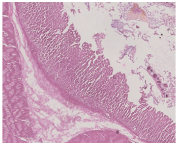
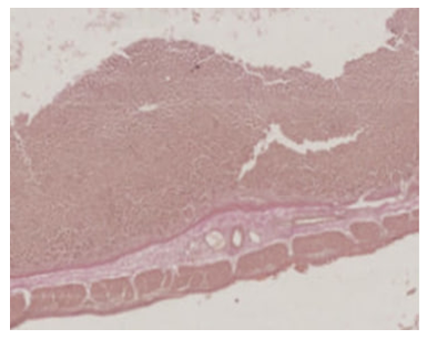
The following data were obtained regarding the structural components of the rat stomach wall. The gastric mucosa of rats in this group is covered with a multilayer epithelium. The epithelial layer consists of three rows of basal, round and oval cells.The basal cells are small, densely packed together and characterized by the location of the nucleus in the center of the cell. The cells of the middle and upper rows are oval in shape, larger in size, with cell nuclei located off-center. In the apical part of the cells there are granules indicating secretory properties. The cells of the proximal row of the epithelial tissue covering the stomach of rats are covered with cuticle.In the glandular tissue of the gastric mucosa of white rats involved in the experiment, unbranched simple and tubular glands can be seen. The glands consist of 3 parts (base, body, neck). The cellular elements in the base and body of the glands are mainly head and parietal cells. The neck of the glands is composed of parietal and mucous (accessory) cells.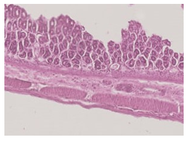 | Figure 1. Microscopic view of the increased gastric submucosa of a 21-day-old experimental group rat. Staining: hematoxylin-eosin. X: 10x40 |
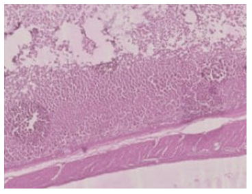
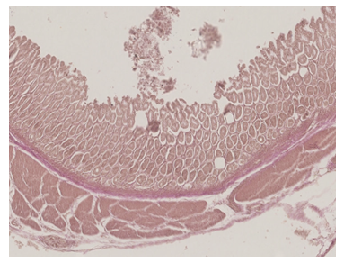
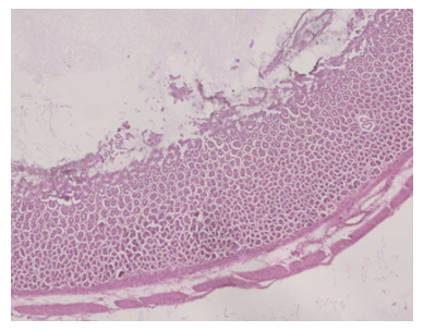 | Figure 2. Microscopic view of the stomach wall layers of a 60-day-old experimental group rat. Staining: hematoxylin-eosin. X: 10x40 |
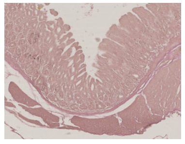
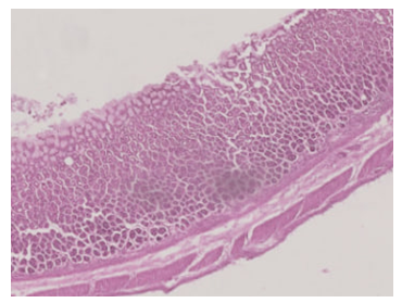 | Figure 3. Microscopic view of the layers of the stomach wall of a 90-day-old experimental group rat. Staining: hematoxylin-eosin. X: 10x40 |
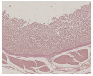
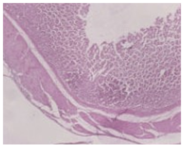 | Figure 4. Microscopic view of the layers of the stomach wall of a 120-day-old experimental group rat. Staining: hematoxylin-eosin. X: 10x40 |
 | Figure 5. Microscopic view of the layers of the stomach wall of a 150-day-old experimental group rat. Staining: hematoxylin-eosin. X: 10x40 |
 | Figure 6. Microscopic view of the layers of the stomach wall of a 180-day-old experimental group rat. Staining: hematoxylin-eosin. X: 10x40 |
As a result of the study of morpho-functional changes in the stomach of rats in the experimental group, the following data were obtained. A sharp increase in the mucoid layer on the surface of the gastric mucosa was detected (Figs. 2, 3, 4, 6). There were almost no lesions on the surface of the mucosa (Figs. 1, 2, 3, 4, 5, 6). [3, 65]In the submucosa of the stomach of rats, changes in the sparse fibrous connective tissue were observed (Figs. 1, 2, 3, 4, 5, 6).The structural elements of the special plate of the gastric mucosa of white rats consist of sparse connective tissue fibers. The submucosa of the stomach of rats consists of sparse fibrous connective tissue, the fibers of which are located in different directions in an irregular manner (Figs. 1, 4, 5).The muscularis mucosa of the stomach of white rats consists of smooth muscle fibers of internal longitudinal and external circular orientation, consisting of muscle layers. The outer serous membrane of the stomach of rats is replaced by a mesothelial lining with sparse fibrous connective tissue (Fig. 4).During the study of morpho-functional changes in the stomach of experimental rats at 21, 60, 90, 120, 150, 180 days, an increase in the mucous glands of the layers of the stomach wall and a fullness of small blood vessels in the submucosa, lymphocyte infiltration are detected (Figs. 7, 8, 9, 10, 11, 12).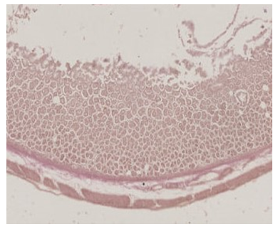 | Figure 7. Microscopic view of the layers of the stomach wall of a 21-day-old experimental group rat. Staining: Van-Gieson. X: 10x40 |
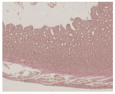 | Figure 8. Microscopic view of the layers of the stomach wall of a 60-day-old experimental group rat. Staining: Van-Gieson. X: 10x40 |
 | Figure 9. Microscopic view of the layers of the stomach wall of a 90-day-old experimental group rat. Staining: Van-Gieson. X: 10x40 |
 | Figure 10. Microscopic view of the layers of the stomach wall of a 120-day-old experimental group rat. Staining: Van-Gieson. X: 10x40 |
 | Figure 11. Microscopic view of the layers of the stomach wall of a 150-day-old experimental group rat. Staining: Van-Gieson. X: 10x40 |
 | Figure 12. Microscopic view of the layers of the stomach wall of a 120-day-old experimental group rat. Staining: Van-Gieson. X: 10x40 |
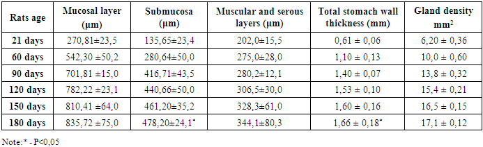
The following data were obtained as a result of studying the morpho-functional changes in the stomach of rats fed palm oil for 21, 60, 90, 120, 150, 180 days and 30 days. The structural elements of the gastric mucosa of rats are sparse connective tissue fibers, and the submucosal layer is sparse fibrous connective tissue, smooth muscle fibers of the muscularis mucosa, and thickening of small blood vessels of the gastric wall and submucosa (Figs. 7, 8, 9, 10, 11, 12).The mucous, muscular and outer layers of the stomach wall of white rats are supplied with blood by blood vessels. The lymphoid structures of the submucosa consist of several rows of chains of lymphocytes.Morphometry of the mucous, muscular and outer layers of the stomach wall of white rats G.G. The Avtandilov method and NanoZoomer (REF C13140-21.S/N000198/HAMAMATSU PHOTONICS/431-3196 JAPAN), NDP.viev2.0., QuPath 0.4.0. url., programs were widely used.The following data were obtained when the rats in the 21-day control group of the experiment were studied. The weight of the rats in the 21-day control group was 48-55 g, with an average of 51.5 ± 0.14 g.The thickness of the gastric mucosa of the 21-day male white rats in the control group was 270.81 ± 23.5 μm, the thickness of the submucosa was 135.65 ± 23.4 μm, the average thickness of the muscular and serous layers was 202.0 ± 15.5 μm, and the total thickness of the gastric wall was 0.61 ± 0.06 mm. The average distribution of the density of the mucous glands was 6.20 ± 0.36 mm2 (Table 1).The weight of 60-day-old male white rats in the control group ranged from 70 g to 82 g, with an average of 75.6 ± 2.08 g. The thickness of the gastric mucosa of the 60-day-old rats in the control group was 542.30 ± 50.2 μm, the thickness of the submucosa was 280.64 ± 50.0 μm, the average thickness of the muscular and serous layers was 275.0 ± 28.0 μm, and the total thickness of the gastric wall was 1.10 ± 0.13 mm. The average distribution of the density of the mucous glands was 10.0 ± 0.60 mm2 (Table 1).The weight of 90-day-old male white rats in the control group ranged from 100 g to 120 g, with an average of 110.2 ± 3.08 g. The thickness of the gastric mucosa of the 90-day-old rats in the control group was 701.81 ± 15.0 μm, the thickness of the submucosa was 416.71 ± 43.5 μm, the average thickness of the muscular and serous layers was 280.2 ± 12.1 μm, and the total thickness of the gastric wall was 1.40 ± 0.07 mm. The average distribution of the density of the mucous glands was 13.8 ± 0.32 mm2 (Table 1).The weight of 120-day-old male white rats in the control group ranged from 130 g to 150 g, with an average of 141.6 ± 2.23 g. The thickness of the gastric mucosa of the 120-day-old rats in the control group was 782.22 ± 23.1 μm, the thickness of the submucosa was 440.66 ± 50.0 μm, the average thickness of the muscular and serous layers was 306.5 ± 30.0 μm, and the total thickness of the gastric wall was 1.53 ± 0.10 mm. The average distribution of the density of the mucous glands was 15.4 ± 0.21 mm2 (Table 1).The weight of 150-day-old male white rats in the control group ranged from 165 g to 186 g, with an average of 175.5 ± 4.34 g. The thickness of the gastric mucosa of the 150-day-old rats in the control group was 810.41 ± 64.0 μm, the thickness of the submucosa was 461.20 ± 35.2 μm, the average thickness of the muscular and serous layers was 328.3 ± 61.0 μm, and the total thickness of the gastric wall was 1.60 ± 0.16 mm. The average distribution of the density of the mucous glands was 16.5 ± 0.15 mm2 (Table 1).The weight of 180-day-old male white rats in the control group ranged from 200 g to 220 g, with an average of 211.6 ± 3.54 g. The thickness of the gastric mucosa of the 180-day-old rats in the control group was 835.72 ± 75.0 μm, the thickness of the submucosa was 478.20 ± 24.1 μm, the average thickness of the muscular and serous layers was 344.1 ± 80.3 μm, and the total thickness of the gastric wall was 1.66 ± 0.18 mm. The average distribution of the density of the mucous glands was 17.1 ± 0.12 mm2 (Table 1).Table 1. Morphometric parameters of the gastric wall layers of control group rats of different ages (M±m)
 |
| |
|
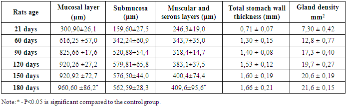
The following data were obtained when the 21-day experimental group of male white rats was studied. The weight of 21-day rats after 30 days of palm oil consumption was 73-80 g, an average of 76.5 ± 1.2 g. The thickness of the gastric mucosa of the 21-day rats in the experimental group was 300.90 ± 26.1 μm, the thickness of the submucosa was 159.60 ± 27.5 μm, the average thickness of the muscular and serous layers was 246.3 ± 19.0 μm, and the total thickness of the gastric wall was 0.71 ± 0.07 mm. The density of the mucous glands was 7.30 ± 0.42 mm2 on average, and the gastric mucosa of the control group rats changed by 21% from the density of the glands (Table 2).The morphometric parameters of the stomach wall of 60-day-old male white rats in the experiment were as follows: the weight of 60-day-old rats after 30 days of palm oil consumption was 100-112 g, an average of 107±2.5 g. The thickness of the gastric mucosa of the 60-day-old rats in the experimental group was 616.25 ±57.0 μm, the thickness of the submucosa was 342.24±60.9 μm, the average thickness of the muscular and serous layers was 343.7±35.0 μm, and the total thickness of the gastric wall was 1.30 ± 0.15 mm on average. The density of the mucous glands was 12.8 ± 0.77 mm2 on average, and the gastric mucosa of the control group rats changed by 21% from the density of the glands (Table 2).The following data were obtained from the morphometric parameters of the stomach wall in the 90-day experimental group of male white rats. The weight of 90-day rats after 30 days of palm oil consumption was 135-155 g, on average 146±1.7 g. The thickness of the gastric wall of the 90-day rats in the experimental group was 825.66 ±17.6 μm, the thickness of the submucosal layer was 520.88±54.4 μm, the average thickness of the muscular and serous layers was 318.4±14.7 μm, and the total thickness of the gastric wall was 1.40 ± 0.08 mm. The density of the mucous glands was on average 17.3 ± 0.40 mm2, and the gastric mucosa of the control group rats changed by 21% from the gland density (Table 2).When the 120-day-old male white rats in the experiment were studied, the following data were obtained from the morphometric parameters of the stomach wall.After 30 days of palm oil consumption, the weight of the 120-day-old rats was 168-188 g, on average 178 ± 1.4 g. The thickness of the gastric mucosa of the 120-day-old rats in the experimental group was 920.26 ± 27.2 μm, the thickness of the submucosa was 579.81 ± 65.8 μm, the average thickness of the muscular and serous layers was 383.1 ± 37.5 μm, and the total thickness of the gastric wall was 1.53 ± 0.12 mm. The density of the mucous glands was 19.7 ± 0.27 mm2 on average, and the gastric mucosa of the control group rats changed by 21% from the gland density (Table 2).When the 150-day-old male white rats in the experiment were studied, the following data were obtained from the morphometric parameters of the gastric wall.After 30 days of palm oil consumption, the weight of 150-day-old rats was 205-226 g, with an average of 215.5 ± 1.1 g. The thickness of the gastric mucosa of the 150-day-old rats in the experimental group was 920.92 ± 72.7 μm, the thickness of the submucosa was 576.50 ± 44.0 μm, the average thickness of the muscular and serous layers was 400.4 ± 74.4 μm, and the total thickness of the gastric wall was 1.60 ± 0.19 mm. The density of the mucous glands was 20.6 ± 0.19 mm2 on average, and the gastric mucosa of the control group rats changed by 20% from the gland density (Table 2).The following data were obtained from the morphometric parameters of the stomach wall in the 180-day experimental group of male white rats.The weight of 180-day rats after 30 days of palm oil consumption was 244-264 g, an average of 255±1.6 g. The thickness of the gastric wall of the 180-day rats in the experimental group was 960.60 ±86.2 μm, the thickness of the submucosal layer was 562.59±28.3 μm, the average thickness of the muscular and serous layers was 409.6±95.6 μm, and the total thickness of the gastric wall was 1.66 ± 0.21 mm. The density of the mucous glands averaged 21.6 ± 0.15 mm2, and the gastric mucosa of the control group rats changed by 21% of the gland density (Table 2).Table 2. Morphometric parameters of the stomach wall layers of experimental groups of rats of different ages (M±m)
 |
| |
|
When comparing the morphometric parameters of the stomach wall layers of rats in the control group at 21, 60, 90, 120, 150, and 180 days of age with rats in the experimental group, the differences were as follows (Figure 13): 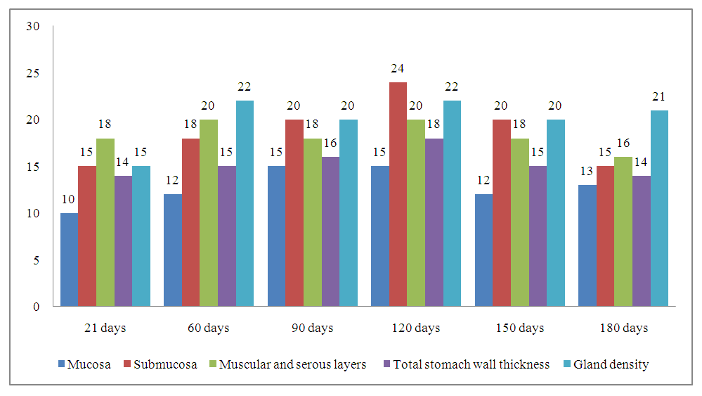 | Figure 13. Percentage (%) increase in morphometric parameters of the stomach wall layers of the experimental group of rats compared to the control group of different ages |
In the 21-day-old experimental animals studied, it was found that the total thickness of the stomach wall increased by 14% compared to the 21-day-old control group. The mucosa thickened by 10%, the submucosa by 15%, the muscular and serous layers by 18%, and the greatest increase was observed in the submucosa and the muscular-serous layers. The density of the mucous glands increased by 15% (Figure 13). In the 60-day-old experimental animals studied, it was found that the total thickness of the stomach wall increased by 15% compared to the 60-day-old control group. The mucosa thickened by 12%, the submucosa by 18%, the muscular and serous layers by 20%, and the greatest increase was observed in the muscular-serous layers. The density of the mucous glands increased by 22% (Figure 13).In the 90-day-old experimental animals studied, it was found that the total thickness of the stomach wall increased by 16% compared to the 90-day-old control group. In this case, the mucosa thickened by 15%, the submucosa by 20%, the muscular and serous layers by 18%, and the greatest increase was observed in the submucosa. The density of the mucous glands increased by 20% (Figure 13).In the 120-day-old experimental animals studied, it was found that the total thickness of the stomach wall increased by 18% compared to the 120-day-old control group. In this case, the mucosa thickened by 15%, the submucosa by 24%, the muscular and serous layers by 20%, and the greatest increase was observed in the submucosa. The density of the mucous glands increased by 22% (Figure 13).In the 150-day-old experimental animals studied, it was found that the total thickness of the stomach wall increased by 15% compared to the 150-day-old control group. In this case, the mucosa thickened by 12%, the submucosa by 20%, the muscular and serous layers by 18%, and the greatest increase was observed in the submucosa. The density of the mucous glands increased by 20% (Figure 13).In the 180-day-old experimental animals studied, it was found that the total thickness of the stomach wall increased by 14% compared to the 180-day-old control group. In this case, the mucosa thickened by 13%, the submucosa by 15%, the muscular and serous layers by 16%, and the greatest increase was observed in the muscular-serous layer. The density of the mucous glands increased by 21% (Figure 13). [10, 144]
4. Conclusions
Thus, as a result of studying the morpho-functional changes in the stomach of rats in the control group at 21, 60, 90, 120, 150, 180 days, the following data were obtained. Compared to all control groups (21, 60, 90, 120, 150, 180 days), it was found that the density of gastric mucosal glands in the 21-day experimental group of rats changed by 15%, the density of gastric mucosal glands in the 60-day experimental group of rats changed by 22%, the density of gastric mucosal glands in the 90-day experimental group of rats changed by 20%, the density of gastric mucosal glands in the 120-day experimental group of rats changed by 22%, the density of gastric mucosal glands in the 150-day experimental group of rats changed by 20%, and the total thickness of the gastric wall in the 180-day experimental group of rats changed by 21%. As a result of the study of morphometric changes in the layers of the gastric wall, the following data were obtained. It was found that compared to the control group at 21 days, the total thickness of the stomach wall of the experimental group rats changed by 14%, the total thickness of the stomach wall of the 60-day-old rats by 15%, the total thickness of the stomach wall of the 90-day-old rats by 16%, the total thickness of the stomach wall of the 120-day-old rats by 18%, the total thickness of the stomach wall of the 150-day-old rats by 15%, and the total thickness of the stomach wall of the 180-day-old rats by 14%.
References
| [1] | Shaykhova G.I., Ermatov N.J., Rustamov B.B. Hygienic Assessment of the Nutritional and Biological Value of Red Palm Oil // Monograph. – 2022. |
| [2] | Rustamov B.B., Ermatov N.J. Nutritional Value of Red Palm Oil // Medical News. – 2016. – No. 12 (267). – P. 65-67. |
| [3] | Bibik E.Yu., Romanenko D.V., Reshetilo N.V. [et al.] Excessive Consumption of Palm Oil as a Cause of Obesity in Different Periods of Ontogenesis // Ukrainian Morphological Almanac. – 2014. – Vol. 12. – No. 3. – P. 65-67. |
| [4] | Boikuziev Kh.Kh., Khamrayev A.Kh., Dzhurakulov B.I., Ismailova N.A. Morphology of Endocrine Cells in the Fundus of the Stomach in Mammals Depending on the Type of Nutrition // Questions of Science and Education. – 2020. – No. 13 (97). – P. 115-120. |
| [5] | Buvabekov M.M., Yuldashev A.Yu., Rakhmonov R.R. [et al.] Morphological Features of the Formation of the Mucous Membrane of the Fundal Part of the Stomach in Rats During Postnatal Ontogenesis // Medicine of Kyrgyzstan. – 2019. – No. 1. – P. 31-36. |
| [6] | Gaivoronskaya Yu.V. Structural and Functional State of the Adrenal Glands in White Rats of Different Ages with Excessive Palm Oil Consumption // Bulletin of Medical Internet Conferences. – 2018. – Vol. 8. – No. 7. – P. 274-276. |
| [7] | Ismailova K.R. The Effect of Excessive Palm Oil Consumption on the Ultrastructure of Dentin Biomineral at Different Stages of Postnatal Ontogenesis // Ukrainian Morphological Almanac named after Professor V.G. Koveshnikov. – 2017. – Vol. 15. – No. 2. – P. 14-19. |
| [8] | Kamoldinova R.A., Rakhmonov R.R., Ibragimova Kh.Z. Features of the Formation of the Gastric Mucosa in Rats During Postnatal Ontogenesis // Re-health Journal. – 2020. – No. 2(6). – P. 65-71. |
| [9] | Lyashchuk A.V. Morphofunctional State of the Proximal Epiphyseal Cartilage of Tibial Bones with Excess Palm Oil in the Diet of White Rats of Different Ages // Ukrainian Morphological Almanac. – 2015. – Vol. 13. – No. 3-4. – P. 81-85. |
| [10] | Mirzoyan E., Chekunkov V.V., Davidenko V.N., Markvo L.I. Histological Manifestations of Early Differentiation of Smooth Muscle Cells in the Wall of the Intestinal Tube of Rat Embryos During the Prenatal Period and in Postnatal Histogenesis in the Wall of the Stomach // International Student Scientific Bulletin. – 2015. – No. 2-2. – P. 144. |

 Abstract
 Reference
 Full-Text PDF
 Full-text HTML