American Journal of Medicine and Medical Sciences

p-ISSN: 2165-901X    e-ISSN: 2165-9036

2025;  15(4): 1006-1012

doi:10.5923/j.ajmms.20251504.33

Received: Mar. 9, 2025; Accepted: Mar. 25, 2025; Published: Apr. 10, 2025


The Prompt Identification and Management of Nephropathies Seen in Post-COVID Syndrome

Safarova Gulnoz A.1, Gadaev Abdugaffor G.2, Ismatova Mekhriniso N.1, Bakhronov Bekhruz B.1

1Bukhara State Medical Institute named after Abu Ali ibn Sino, Bukhara, Uzbekistan

2Tashkent Medical Academy, Tashkent, Uzbekistan

Correspondence to: Safarova Gulnoz A., Bukhara State Medical Institute named after Abu Ali ibn Sino, Bukhara, Uzbekistan.

Email:

Copyright © 2025 The Author(s). Published by Scientific & Academic Publishing.

This work is licensed under the Creative Commons Attribution International License (CC BY).
http://creativecommons.org/licenses/by/4.0/

Abstract

The outcomes of antioxidant therapy and dynamic observation in individuals with COVID-19 who did not have any comorbid conditions are presented in the article. Interleukin-6, collagen type IV, and nephrinuria levels in the urine significantly decreased in individuals with mild, moderate, and severe forms of COVID-19 and compromised renal function following six months of monitoring and antioxidant therapy.

Keywords: COVID-19, Interleukin-6, Cystatin-C, Creatinine, Collagen type IV, Nephrinuria, Glomerular filtration rate

Cite this paper: Safarova Gulnoz A., Gadaev Abdugaffor G., Ismatova Mekhriniso N., Bakhronov Bekhruz B., The Prompt Identification and Management of Nephropathies Seen in Post-COVID Syndrome, American Journal of Medicine and Medical Sciences, Vol. 15 No. 4, 2025, pp. 1006-1012. doi: 10.5923/j.ajmms.20251504.33.

1. Introduction

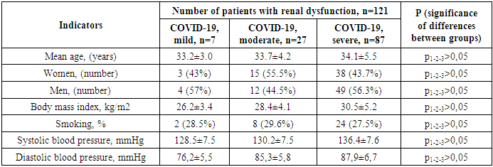
The COVID-19 infection that shook the world in 2019 poses a great threat to humanity. According to the latest official data, as of November 2022, 244,510 people have been infected in Uzbekistan, of whom 1,637 have died. According to Johns Hopkins University in America, these figures are 630,920,425 and 6,539,051 people worldwide, respectively. The World Health Organization reported that as of March 3, 2023, there were 758 million confirmed cases of COVID-19 worldwide [20]. Although most patients recover and return to their original condition, some have ongoing health problems, a process called post-acute COVID or long COVID. The World Health Organization defines long-term COVID as the persistence of symptoms for three months or more after the initial infection, as well as the appearance of new symptoms in the absence of other causes. The opinion of experts of the said authoritative organization is supported by a number of other observations [19,6].
Sometimes, even after a mild infection, persistent symptoms can be observed [2]. Such symptoms include fatigue, shortness of breath, chest tightness, cough, arthralgia, headache, and cognitive dysfunction. According to observers, a protracted course of COVID-19 is observed in 10-30% of patients and lasts more than a year [16,3] However, to date, no studies have been conducted to predict the mechanisms of the development of a long-term course of COVID.
Although the main target of the coronavirus is the respiratory system, as the virus spreads through the bloodstream, other organs can also be affected. Because it contains several transmembrane glycoproteins that create conditions for interaction with the cells of the human body. Thus, to date, numerous observations have proven that COVID-19 is a systemic disease that affects not only the respiratory system, but also other systems and causes severe complications [22,21].
Its extrapulmonary side effects and complications include disseminated thrombosis, myocardial dysfunction, various arrhythmias, acute coronary syndrome, acute kidney injury, various degrees of gastrointestinal changes, hypoglycemia, ketoacidosis, neurotoxic and neurological complications including cerebral ischemic changes, as well as various ocular and cutaneous reactions. It is known that angiotensin-converting enzyme II (ACE II) receptors serve as a gateway for the virus to enter cells. Since these receptors are present in all organs, the virus can spread through the blood, and its toxic effects can damage tissues and various organs. This, in turn, indicates that the disease becomes systemic. Of course, the direct action of the virus is primarily found in the epithelium of the respiratory tract, which is considered a target organ, and above all in the lungs, where type I cells involved in gas exchange and type II cells producing surfactant are located. Although the virus is found in type II cells of the lung parenchyma, in most cases it remains asymptomatic for 2-7 days after entering the body, after which it begins to spread rapidly throughout the body. Patients develop severe pneumonia and acute respiratory distress syndrome. Then, adjacent organs are damaged, in particular the gastrointestinal tract, renal tubules and liver capillaries, and a release of cytokines is observed. The latent course of the early stages of the disease is due to the fact that the virus hides in a number of cells in the body, creating reservoirs for latent replication. The squamous epithelium of the salivary glands and bronchi is a hidden incubator of viruses. A large number of viruses are released from them, which after a certain time cause viremia and serious changes in the body. Histopathological examination of COVID-19 shows that it has tropism for the epithelium of the kidneys, myocardium, trachea and gastrointestinal tract [10,5,12].
Following infection, a vicious cycle of endotheliitis that enhances thrombotic inflammation is maintained by increased ACE II levels on the surface of epithelial cells [1]. This is believed to signal the restoration of control over the renin-angiotensin-aldosterone system (RAAS), thereby triggering cardio- and visceroprotective mechanisms [9]. Of course, the rate and severity of COVID-19 progression are also partly related to the individual condition of each organism and the activation of inflammatory and immune-active cytokines, in particular interleukin (IL)-1β, IL-2, IL-6, IL-7, IL-8, tumor necrosis factor α (TNF-α), and others [4,11]. Patients with hyperactivity of cytokine genes have increased cytokine production. This cytokine storm leads to self-sustaining inflammatory processes, which in turn can lead to widespread endovascular thrombosis and, in extreme cases, to multiple organ failure [15]. The virus disrupts the regulation of RAAS by attaching to ACE II receptors. ACE II receptors are known to be present in large quantities in the epithelium of the heart and kidneys. COVID-19 negatively affects their functioning and leads to significant changes [17]. In addition, changes in RAAS under the influence of the virus cause a disruption of all areas of its control, including renal function, which is manifested by a number of clinical symptoms [8,13]. This is due to excessive activation of the kallikrein-kinin system, in particular bradykinin, in an inflammatory environment, which leads to the development of pulmonary edema. As a result of the disruption of the cardioprotective effect of this system under the influence of the virus, patients develop hypokalemia, decreased blood pressure, arrhythmia, which in turn leads to complications of cardiovascular and renal diseases [18]. The above conditions are detected in 20-30% of patients hospitalized due to the virus, and in people with existing heart and kidney problems, this figure reaches 50%. Although the pathophysiology of cardiovascular diseases, like other pathological processes, is multifactorial, it is associated with increased expression of ACE II receptors on cardiomyocytes and fibroblasts of cardiac tissue, as noted above. This is confirmed by the fact that the virus directly affects endothelial and smooth muscle cells, as well as the heart. The development of myocarditis is due to the viral load and inflammation or activation of immune system cells in the coronary arteries and myocardium due to infection. In addition, as the disease progresses, myocardial ischemia increases, and patients with comorbidities may develop myocardial infarction if such treatments are not used [14,7].
The above confirms that COVID-19 is a systemic disease that affects all organs. However, to date, no studies have been conducted in Uzbekistan to determine which internal organs, other than the lungs, are most often affected by this infection in relatively healthy people.
From this point of view, the study of kidney damage in patients with COVID-19 is also of great scientific and practical importance.
Purpose of the study: To improve the comprehensive assessment and treatment of renal dysfunction in patients with COVID-19 without comorbidities.
Materials and methods of the study: The source of the study was 121 patients who were treated for COVID-19 in the Bukhara Regional Infectious Disease Center and the Bukhara Regional Multidisciplinary Medical Center and were under subsequent observation, in whom micro- and macroalbuminuria (above 30 mg / l) was detected in daily urine, without concomitant diseases. Their average age was 33.1 ± 0.8 years, among them there were 65 men and 56 women. Of the study participants, 7 had a mild form of COVID-19, 27 had a moderate form, and 87 had a severe form.
Patients included in the study were under continuous observation for six months. Laboratory and instrumental studies were carried out during the first week of the study and after six months. Table 1 below presents a description of the patients included in the study.
Table 1. Description of patients with COVID-19 and renal impairment without comorbidities
     
As shown in the table, the average age in the groups was 33.2±3.0, 33.7±4.2 and 34.1±5.5 years, respectively, and they did not differ significantly from each other. When compared by gender, the first group consisted of 43% women and 57% men. In the second group, on the contrary, there were more women than men (55.5% versus 44.5%, respectively). In the third group, women accounted for 43.7%, and men – 56.3%. The body mass index in the first and second groups was 26.2±3.4 and 28.4±4.1 kg/m2, respectively, obesity was not revealed in the study participants. In the third group, the average body mass index was 30.5±5.2 kg/m2, they were obese. When comparing the body mass index in all groups, no reliable difference was observed (p>0.05). The number of smokers in all groups was 2 (28.5%), 8 (29.6%) and 24 (27.5%) people, respectively, the differences between them were not significant (p> 0.05). Systolic blood pressure in the first group was 128.5 ± 7.5 mm Hg, in the second group - 130.2 ± 7.5 mm Hg and in the third group 136.4 ± 7.6 mm. organized. When comparing them, no significant difference was found (p> 0.05). The average diastolic blood pressure between the groups was 76.2 ± 5.5, 85.3 ± 5.8 and 87.9 ± 6.7 mm Hg, respectively. (p> 0.05). The comparative analysis showed that the indicators in the three groups did not differ significantly from each other. Six months after hospital discharge, serum cystatin-C, interleukin-6, urinary nephrinuria, and antioxidant collagen IV levels were determined in patients before and after nephroprotective treatment using enzyme immunoassay. All patients also underwent ECG, radiography, and renal ultrasound.
Patients were prescribed quercetin in capsule form, 500 mg twice daily as an antioxidant and nephroprotective agent. This drug, along with antioxidant and nephroprotective effects, blocks the metabolism of arachidonic acid via the lipoxygenase pathway, suppressing the synthesis of leukotrienes, serotonin, and other inflammatory mediators. Blood biochemical parameters - serum alanine aminotransferase (ALT), aspartate aminotransferase (AST), bilirubin, glucose, urea, creatinine, total protein, potassium, sodium, coagulogram, blood lipid spectrum analysis were performed on a Mindry BA-88 biochemical analyzer using reagents from Human (Germany).
Enzyme immunoassays were performed in the laboratory of the Bukhara Regional Multidisciplinary Clinical Hospital on a Cobas-6000 device (Roche, Germany).
Serum cystatin C was determined by immunoturbidimetry using the DiaSys diagnostic kit (Germany). Normal values were considered to be 0.58–1.02 mg/ml (Hoek, 2003).
Calculation of capillary filtration rate. The rate of CKD development was determined using the formula recommended by Hook et al. based on the blood cystatin-C level: CKD [ml/min/1.73 m2] = (80.35/cystatin-C [mg/ml]) - 4.32.
The proinflammatory cytokine interleukin-6 was detected using the BEST 0 vector set. The test sensitivity is 0.13–0.3 ng/ml. The reference value is 1.7 [1.4; 4.8] ng/ml.
To determine the level of nephrin in urine (nephrinuria), the ELISA method was used using reagents from DiMediTec Diagnostic (Germany).
Analysis and discussion of the study results. It is known that regardless of the severity of the coronavirus infection, systemic inflammatory processes develop in the patient's body. Recent studies have shown that these processes continue in the body long after patients have recovered [10,6]. Considering the above, we assessed the level of interleukin-6, one of the main markers of inflammation, before and after treatment in the patients we observed. Figure 1 below shows the results.
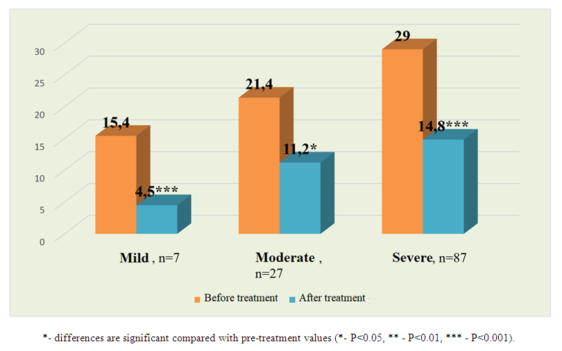
Figure 1. Changes in interleukin-6 levels (pg/ml) before and after treatment in patients with COVID-19 and varying degrees of renal impairment without comorbidities
As shown in the figure, pre-treatment interleukin-6 levels were 15.4±1.2, 21.4±2.7, and 29.0±2.8 pg/ml between the groups, respectively. When comparing, a significant difference (p<0.05) was found between the first and second groups, and a very significant difference (p<0.001) was found between the third group. There was no significant difference between the second and third groups (p>0.05). After treatment, the level of interleukin-6 in patients with a mild form of COVID-19 decreased by 3.42 times from 15.4 ± 1.2 pg / ml to 4.5 ± 0.8 pg / ml, which is a highly significant difference (p < 0.001). In patients with moderate and severe courses of the disease, its level was 21.4 ± 2.7 pg / ml before treatment and 11.2 ± 2.6 pg / ml after treatment, an improvement of 1.91 times, a significant (p < 0.05) difference was revealed. In patients with a severe form of coronavirus, the level of interleukin-6 improved by 1.97 times from 29.0 ± 2.8 pg / ml to 14.8 ± 2.5 pg / ml, which is a highly significant difference (p < 0.001). As is known, creatinine has been used for many years as the main marker for assessing renal impairment. However, rapid changes in its indicators in the body due to various reasons create a number of uncertainties in assessing renal impairment. Therefore, in recent years, cystatin C has been recommended to assess renal impairment, a marker that is more reliable than creatinine.
We studied renal impairment in patients included in our study using creatinine and cystatin C indicators before and after six months of treatment and compared the results. The results are presented in Table 2 below.
Table 2. Indices of renal impairment before and after treatment in patients included in the study
As shown in the table, the functional state of the kidneys in patients was initially assessed by the creatinine level before and after treatment. The creatinine level in patients with mild COVID-19 was 70.2±3.4 μmol/L, with moderate severity - 90.4±4.2 μmol/L, and with severe severity - 101.6±6.4 μmol/L. When comparing, a highly significant difference was noted in all groups before treatment compared to each other (p1-2-3<0.001). After nephroprotective treatment, the level in the first group changed from 70.2±3.4 to 60.2±2.1 μmol/L, and a significant difference was found (P<0.05). In patients with moderate COVID-19, after treatment, it decreased by 1.22 times from 90.4±4.2 to 73.6±3.4 μmol/L, the difference is statistically significant (P<0.01). In the third group, the difference between the indicators before and after treatment ranged from 101.6±6.4 to 88.3±4.5 μmol/L, and the differences were significant (P<0.01).
The creatinine-based SCF indicator was 120±2.8 ml, 99±2.5 ml and 86±2.1 ml per minute per 1.73 m2 of body surface area, respectively, in both groups before treatment. When comparing the results between the groups, a highly significant difference was noted (p1-2-3<0.001). After treatment, the cerebrospinal fluid flow rate increased to 126±3.1 ml, 118±2.8 ml, and 102±3.5 ml per minute per 1.73 m2 of body surface area in patients with mild, moderate, and severe COVID-19, respectively. When comparing the parameters before treatment, the differences in the first group were insignificant (p>0.05), while highly significant differences (p<0.001) were noted in the second and third groups.
The level of cystatin C in patients with mild, moderate, and severe COVID-19 before treatment was 0.95±0.05 mg/L, 1.1±0.05 mg/L, and 1.28±0.06 mg/L, respectively. The obtained parameters significantly (p<0.05) differed from each other. Although the first group improved from 0.95±0.05 to 0.86±0.02 mg/L after six months of treatment, the differences were insignificant. In the second group, it decreased by 1.14 times from 1.1±0.05 to 0.97±0.04 mg/L, with a significant difference (p<0.05). In patients with severe coronavirus infection, after treatment, the level improved by 1.22 times from 1.28±0.06 to 1.05±0.02 mg/L, with a significant difference (p<0.01).
The mean eGFR values calculated using cystatin C before and after treatment were 93±3.1 ml, 76±3.6 ml, and 62±4.9 ml per minute per 1.73 m2 of body surface area, respectively, between the groups. When these results were compared with the corresponding creatinine-based eGFR, a highly significant difference (p<0.001) was found in all groups. Therefore, it is reasonable to use cystatin C to detect renal impairment in patients who have recovered from COVID-19 but are considered relatively healthy. Compared with creatinine, it is more reliable in detecting existing latent renal failure. These differences are shown in Figure 2.
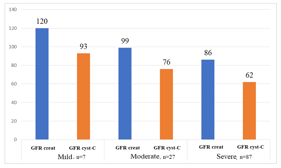
Figure 2. The estimated glomerular filtration rate (GFR) per minute per 1.73 m2 of body surface area in patients included in the study was determined using creatinine and cystatin C (mL)
Appendix: Glomerular filtration rate calculated using creatinine; Glomerular filtration rate is calculated using hCFTRcis-cystatin C.
After six months of observation and treatment, the estimated GFR per minute per 1.73 m2 of body surface area in patients with mild COVID-19 increased from 93±3.1 ml to 106±4.8, a significant difference (p<0.05) was noted. In moderate form, after six months, it increased from 76±3.6 to 90±3.4 ml, with a significant difference (p<0.01). In patients with severe disease, GFR was 62±4.9 ml before treatment and 81±4.1 ml after treatment, the differences are significant (p<0.01).
Also, long-term persistence of inflammatory processes in the body due to COVID-19 can lead to the development of fibrosis in this organ in patients with severe renal dysfunction. Taking this into account, we examined type IV collagen, one of the main markers of fibrosis, before and after treatment in patients included in our study. The results obtained are presented in Figure 3.
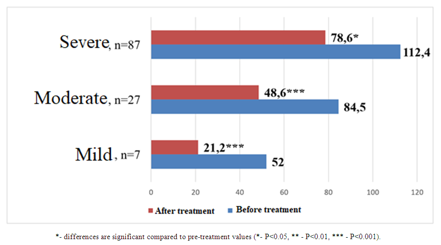
Figure 3. Comparative analysis of collagen type IV levels (ng/ml) before and after treatment in patients with COVID-19 and varying degrees of renal impairment without concomitant diseases
As shown in the figure, before treatment, the level of type IV collagen was 52.0±4.5, 84.5±7.6 and 112.4±10.2 ng/ml in patients with mild, moderate and severe COVID-19, respectively. When comparing, a highly significant (p<0.001) difference was found between the first group and the other two groups. No significant difference was found between the second and third groups (p<0.05).
After the treatment, the level of type IV collagen in patients with a mild form of COVID-19 decreased by 2.45 times from 52.0 ± 4.5 ng / ml to 21.2 ± 4.8 ng / ml, which is a highly reliable difference (p < 0.001). In patients with moderate and severe courses of the disease, its level was 84.5 ± 7.6 ng / ml before treatment and 48.5 ± 6.4 ng / ml after treatment, an improvement of 1.74 times, a significant (p < 0.05) difference was revealed. In patients with a severe form of coronavirus, the amount of type IV collagen improved by 1.43 times from 112.4 ± 10.2 ng / ml to 78.6 ± 8.4 pg / ml, which is a significant (p < 0.05) difference. The analysis conducted in this section confirmed the possibility of using cystatin C for the early detection of renal dysfunction in patients with coronavirus. In the examined patients, nephroprotective therapy demonstrated a positive effect on the glomerular filtration rate and the fibrosis marker type IV collagen, and reduced renal dysfunction.
Studies conducted in recent years have shown that kidney changes caused by various diseases begin with podocyte damage. Therefore, we studied the level of nephrinuria, an indicator of podocyte protein, in patients included in the study. Figure 4 below shows the results.
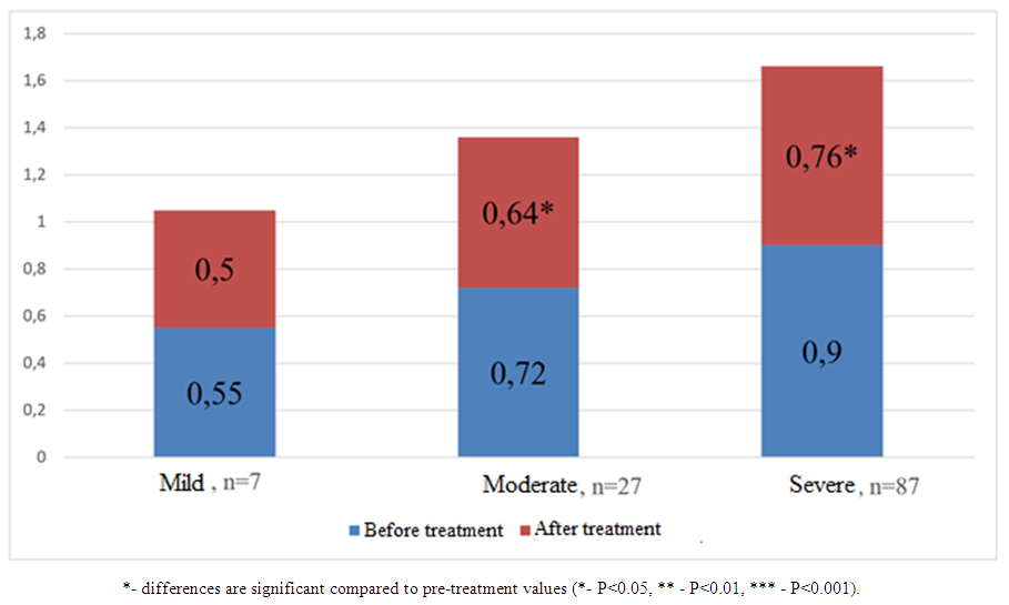
Figure 4. Comparative analysis of nephrinuria values before and after treatment (ng/ml) in patients with COVID-19 and varying degrees of renal impairment without comorbidities
As shown in the figure, before treatment, the urinary nephrin level was 0.55±0.03, 0.72±0.03, and 0.9±0.04 ng/ml in patients with mild, moderate, and severe COVID-19, respectively. When comparing between the groups, a highly significant (p<0.001) difference was found.
After treatment, the nephrinuria index in patients with mild COVID-19 decreased from 0.55±0.03 ng/ml to 0.5±0.02 ng/ml, without significant difference (p>0.05). In patients with moderate and severe disease, its level was 0.72±0.03 ng/ml before treatment and 0.64±0.02 ng/ml after treatment, a significant difference was noted (p<0.05). In patients with severe coronavirus, the level of nephrin in the urine improved by 1.18 times from 0.9±0.04 ng/ml to 0.76±0.03 pg/ml, which is a reliable (p<0.01) difference.

2. Conclusions

After six months of observation and antioxidant therapy, patients with mild, moderate and severe COVID-19 and renal impairment showed significant reductions in urinary interleukin-6, type IV collagen and nephrinuria.

References

[1]  Ackermann M. et al. Pulmonary vascular endothelialitis, thrombosis, and angiogenesis in Covid-19. N. Engl. J. Med. 2020; 383: 120–8. DOI: https://doi.org/10.1056/NEJMoa2015432.
[2]  Blomberg B, Mohn KG, Brokstad KA, Zhou F, Linchausen DW, Hansen BA, Lartey S, Onyango TB, Kuwelker K, Sævik M, et al. Long COVID in a prospective cohort of home-isolated patients. Nat Med. 2021; 27(9): 1607–1613. doi: 10.1038/s41591-021-01433-3.
[3]  Carfì A, Bernabei R, Landi F. Persistent symptoms in patients after acute COVID-19. JAMA. 2022; 324: 603–605. doi: 10.1001/jama.2020.12603.
[4]  Fagone P., Ciurleo R., Lombardo S.D., Iacobello C., et al. Transcriptional landscape of SARS-CoV-2 infection dismantles pathogenic pathways activated by the virus, proposes unique sex-specifi c differences and predicts tailored therapeutic strategies. Autoimmun Rev. 2020; 19 (7): 102571. DOI: https://doi.org/10.1016/j.autrev.2020.102571.
[5]  Giacosa A, Barrile GC, Gasparri C, Perna S, Rondanelli M. Positive Effect of Lecithin-Based Delivery Form of Curcuma and Boswellia Extracts on Irritable Bowel Syndrome After COVID-19 Infection. Nutrients. 2025 Feb 18; 17(4): 723. doi: 10.3390/nu17040723. PMID: 40005051; PMCID: PMC11858738.
[6]  Hurme A, Viinanen A, Teräsjärvi J, Jalkanen P, Feuth T, Löyttyniemi E, Vuorinen T, Kantele A, Oksi J, He Q, Julkunen I. Post-COVID-19 condition in prospective inpatient and outpatient cohorts. Sci Rep. 2025 Feb 26; 15(1): 6925. doi: 10.1038/s41598-025-90819-1. PMID: 40011519.
[7]  kint.2020.04.003, Sala S., Peretto G., Gramegna M., Palmisano A., et al. Acute myocarditis presenting as a reverse Tako-Tsubo syndrome in a patient with SARS-CoV-2 respiratory infec tion. Eur Heart J. 2020; 41 (19): 1861–1862. DOI: https://doi.org/10.1093/eurheartj/ehaa286.
[8]  Kuba K., Imai Y., Ohto-Nakanishi T., Penninger J.M. Trilogy of ACE2: a peptidase in the renin-angiotensin system, a SARS receptor, and a partner for amino acid transporters. Pharmacol Ther. 2010: 128 (1): 119–28. DOI: https://doi.org/10.1016/j.pharmthera.2010.06.00369.
[9]  Lax S.F., Skok K., Zechner P., et al. Pulmonary arterial thrombosis in COVID-19 with fatal outcome: results from a prospective, single-center, clinicopathologic case series. Ann Intern Med. 2020; 173 (5): 350–61. DOI: https://doi.org/10.7326/m20-2566.
[10]  Marques da Silva B, Gameiro J, Lei Teixeira J, Costa C, Branco C, Oliveira J, Bernardo J, Marques F, Agapito Fonseca J, Lopes JA. Long-term outcomes after AKI in hospitalized patients with COVID-19. Nefrologia (Engl Ed). 2025 Feb; 45(2): 150-158. doi: 10.1016/j.nefroe.2025.01.009. PMID: 39986713.
[11]  Mehta P., McAuley D.F., Brown M., Sanchez E., et al. COVID-19: consider cytokine storm syndromes and immunosuppression. Lancet. 2020; 395 (10229): 1033–4. DOI: https://doi.org/10.1016/S0140-6736(20)30628-0.
[12]  Stefania MN, Toma C, Bondor CI, Maria RV, Florin P, Adina MM. Long COVID and Lung Involvement: A One-Year Longitudinal, Real-Life Study. Medicina (Kaunas). 2025 Feb 10; 61(2): 304. doi: 10.3390/medicina61020304. PMID: 40005421; PMCID: PMC11857727.
[13]  Strawn W.B., Ferrario C.M., Tallant E.A. Angiotensin-(1-7) reduces smooth muscle growth after vascular in jury. Hypertension. 1999; 33: 207–11 DOI: https://doi.org/10.1161/01.hyp.33.1.207.
[14]  Su H., Yang M., Wan C., Yi L.X., et al. Renal histopathological analysis of 26 postmortem findings of patients with COVID-19 in China. Kidney Int. 2020; 98 (1): 219–27. DOI: https://doi.org/https://doi.org/10.1016/j.
[15]  Tang N., Li D., Wang X., Sun Z. Abnormal coagulation parameters are associated with poor prognosis in patients with novel coronavirus pneumonia. J Thromb Haemost. 2020; 18 (4): 844–7. DOI: https://doi.org/10.1111/ jth.14768.
[16]  Tran VT, Porcher R, Pane I, Ravaud P. Course of post COVID-19 disease symptoms over time in the ComPaRe long COVID prospective e-cohort. Nat Commun. 2022; 13: 1812. doi: 10.1038/s41467-022-29513-z.//
[17]  Vaduganathan M., Vardeny O., Michel T., McMurray J.J.V., et al. Renin-angiotensin-aldosterone system inhibitors in patients with Covid-19. N Engl J Med. 2020; 382 (17): 1653–9. DOI: https://doi.org/10.1056/NEJMsr2005760.
[18]  van de Veerdonk F., Netea M.G., van Deuren M., et al. Kinins and cytokines in COVID-19: a comprehensive pathophysiological approach. Preprints 2020. 2020040023. DOI: https://doi.org/10.20944/preprints202004.0023.v1.
[19]  World Health Organization (2021) A clinical case definition of post COVID-19 condition by a Delphi consensus. https://www.who.int/publications-detail-redirect.
[20]  Xiaoming Wu. And others. Damage to endothelial barriers and its contribution to long COVID Angiogenesis. 2024 Feb; 27(1): 5-22. doi: 10.1007/s10456-023-09878-5. Epub 2023 Apr 27.
[21]  Granovskaya M.V., Zaslavskaya K.Ya. COVID-19 - a set of symptoms or systemic pathology? Clinical lecture. Part 1. Features of multiple organ disorders // Infectious diseases: news, opinions, training. 2020. Vol. 9, No. 3. Appendix. P. 3–9. DOI: https://doi.org/10.33029/2305-3496-2020-9-3S-3-9.
[22]  N.G. Kulchenko. Epidemiology of kidney diseases in patients with covid-19. Research and practice in medicine 2020, Vol. 7, No. 3, pp. 74-82 Review https://doi.org/10.17709/2409-2231-2020-7-3-7.