-
Paper Information
- Next Paper
- Previous Paper
- Paper Submission
-
Journal Information
- About This Journal
- Editorial Board
- Current Issue
- Archive
- Author Guidelines
- Contact Us
Journal of Health Science
p-ISSN: 2166-5966 e-ISSN: 2166-5990
2015; 5(3A): 10-13
doi:10.5923/s.health.201501.04
Serum Ferritin Levels in Women with Telogen Effluvium: Do They Play a Role?
 Abstract
 Reference
 Full-Text PDF
 Full-text HTML
Mohammad I. Fatani1, Ahmed M. Bin Mahfoz2, Shahzana Naqqash1, Waleed A. Hussain1, Abdulmajeed S. Khan1, Abdulmohsen M. Zahrallayali3, Rogaiah Z. Sagr4
1Hera General Hospital, Makkah, KSA
2AL-Hada Armed Forces Hospital, Taif, KSA
3Al-Hada Military Hospital, Taif, Saudi Arabia
4AL-Hada Armed Forces Hospital, Taif, Saudi Arabia
Correspondence to: Mohammad I. Fatani, Hera General Hospital, Makkah, KSA.
| Email: |  |
Copyright © 2015 Scientific & Academic Publishing. All Rights Reserved.
Objective: To determine whether iron deficiency is more common in women with telogen effluvium than in control subjects. Methods: A cross-sectional study research design was used. The study included 160 Saudi women aged between 16 and 62 years with telogen effluvium seen in the outpatient Department of Dermatology, Hera General Hospital, Makkah, Saudi Arabia. A total of 425 Saudi women aged between 16 and 62 years without complaint of hair loss were included in the study as controls. All participants were tested for serum ferritin and hemoglobin level at the start of the study. Results: The mean serum ferritin level was 34.30 ng/ml among telogen effluvium patients compared with 75.57 ng/ml among controls. This difference was statistically significant (p=0.01). Mean hemoglobin was 11.56 g/dl among telogen effluvium patients compared with 11.26 g/dl hemoglobin among controls. This difference was statistically significant (p=0.000). Conclusions: Iron deficiency was found to be more common among women with telogen effluvium. The authors recommend that serum ferritin level be tested among women with excessive hair loss for better management.
Keywords: Telogen effluvium, Ferritin, Makkah, Saudi Arabia
Cite this paper: Mohammad I. Fatani, Ahmed M. Bin Mahfoz, Shahzana Naqqash, Waleed A. Hussain, Abdulmajeed S. Khan, Abdulmohsen M. Zahrallayali, Rogaiah Z. Sagr, Serum Ferritin Levels in Women with Telogen Effluvium: Do They Play a Role?, Journal of Health Science, Vol. 5 No. 3A, 2015, pp. 10-13. doi: 10.5923/s.health.201501.04.
1. Introduction
- More than 25% of women in developed countries are affected by hair loss. [1] Telogen effluvium (TE) is one of the most common causes of diffuse non-scarring alopecia in women. Although its exact prevalence rate is unknown, one study in Makkah, Saudi Arabia found it to be 1.74% among women. [2]TE is Characterized by diffuse hair shedding, often with acute onset. A chronic form with more insidious onset and a longer duration also exists. [3, 4] It is an abnormality of the hair cycle characterized by abrupt, generalized shedding of normal club hair. [5] It manifests after 2–3 months in reaction to various physical or mental stressors including childbirth, fever, crash dieting, major surgery, certain drugs, and vitamin and mineral deficiencies. [6]Some studies suggest a relationship between hair loss and iron deficiency in TE. [2, 7] Iron deficiency can also induce chronic diffuse telogen hair loss, cheilosis, and koilonychia. [7] Iron is involved in many critical physiologic processes within the hair follicle, suggesting that iron deficiency could disrupt hair synthesis. However, studies of iron as a cause of hair loss have produced conflicting results. Some of these discrepancies may relate to the limitations of assays for detecting iron deficiency, the most common nutritional deficiency in the world today. [2, 7]Iron in the body is divided into storage iron, transport iron, and functional iron compartments. Storage iron, which represents the body's iron (either ferritin or hemosiderin), is best measured by the concentration of serum ferritin. Transport iron is the iron bound to transferrin to transport iron to tissue. Functional iron, the iron bound to hemoglobin, is measured by the concentration of hemoglobin and hematocrit. [2, 7]Definitions of iron deficiency according to those compartments include iron depletion, iron-deficient erythropoiesis, and iron deficiency anemia. In iron depletion, functional and transport iron are normal but storage iron is decreased. In iron-deficient erythropoiesis, both storage and transport iron are decreased, while in iron deficiency anemia, all three iron compartments are decreased. [7, 8] Iron deficiency anemia can also be defined as absent bone marrow in iron stores (determined by bone marrow iron smears), an increase in hemoglobin concentration by more than 1.0 g/dL after iron supplementation therapy, or abnormal values of other biochemical tests such as serum ferritin level. [2, 7]Ferritin is a highly conserved protein complex that plays an important role in storage iron and is recognized as the main iron-binding protein in non-erythrothyroid cells. [7, 8] Only iron deficiency causes very low serum ferritin concentrations; therefore, a low serum ferritin level is specific to iron deficiency. [7, 8] The standard range of ferritin for women is between 12 and 150 nanograms per milliliter. However, normal does not mean the optimal level, and 70 ng/ml and above is considered to be optimal for both men and women. [9]Because there is controversy regarding the causative association of iron deficiency with TE, the aim of our study is to determine whether iron deficiency is more common in women that suffer hair loss compared with control women.
2. Methods
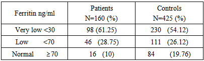
- This study employed a cross-sectional study. Ethical approval was obtained from the Institutional Ethical Committee before the commencement of the study. Enrolled in the study were 160 Saudi women aged between 16 and 62 years with TE seen in the outpatient Department of Dermatology, Hera General Hospital, Makkah, Saudi Arabia. The control group was selected from among patients who were treated at the study hospital for a condition other than TE. The control sample included 425 Saudi women aged between 16 and 62 years.All study participants including controls were enrolled after a detailed medical history and physical examination to rule out conditions that can cause hair loss. Subjects with acute fever, thyroid dysfunction, postpartum hair loss, and hemorrhagic disease, or those on certain medications, were excluded from the study. Similarly, subjects showing signs related to hyperandrogenism (menstrual dysfunction, hirsutism, acne) were also excluded.Participants were tested at least once for serum ferritin concentration and hemoglobin level. A serum ferritin level of <30 ng/ml was taken as very low, <70 as low, and ≥70 as normal in both cases and controls. Statistical analysis was performed by using SPSS. Values were reported as mean ± standard deviation. A Pearson correlation was carried out to determine the statistical relationships. Independent t-tests were then used to compare the means of the groups. A P value < 0.05 was considered to be significant.
3. Results
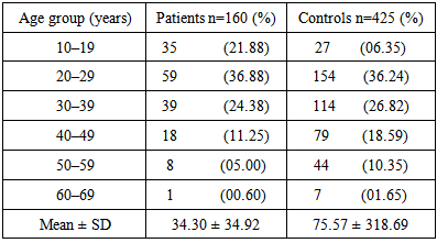
- The mean age was 28.94 ± 10.6 years in patients and 34.24 ± 11.4 years in controls (Table 1). The serum ferritin levels in patients and controls ranged 2.61–226.2 ng/ml and 0.79–5450 ng/ml, respectively (Table 2). The mean serum ferritin level was 34.30 ng/ml among TE patients compared with 75.57 ng/ml for controls. This difference was statistically significant (p=0.01).
|
|
4. Discussion
- A case-control study was conducted at the outpatient Department of Dermatology, Hera General Hospital, Makkah, Saudi Arabia. This study included only women aged 16–62, which is comparable with another study. [10] The mean age of patients in this study is also similar to those in other studies. [1, 11, 12] Most patients in our study with TE were aged 20–29 years (36.88%), perhaps because this age group is more concerned about hair loss.Mean serum ferritin levels were 34.30 ng/ml in patients with TE, which is comparable with other studies, [13, 14], and 75.57 ng/ml in controls, which is similar to another study. [15] However, the mean ferritin level for control patients in this study was double that presented in other studies. [12, 16, 17] This difference could be because our study sample was larger.The mean difference in serum ferritin level was statistically significant (p<0.01) between patients and controls according to an unpaired t-test, which is comparable with other studies. [1, 12, 14, 15, 16, 18] This finding showed a possible correlation between a low level of serum ferritin and the diagnosis of TE in female patients. About 10% of patients with TE had ferritin levels above 70 ng/ml. This lack of a lower ferritin level in patients with TE may be due to its multifactorial etiology. Medication, fever, rapid weight loss, and numerous other factors may cause TE. [5, 7]Most of the 585 subjects had low serum ferritin levels, while only 17.09% of the study population had ferritin levels above 70 ng/ml. This finding may be due to the high incidence of iron deficiency in the Saudi population as well as the fact that iron deficiency anemia is common among adult non-pregnant healthy Saudi women in Riyadh. [19] An inadequate iron intake and heavy consumption of tea and coffee by adults in Saudi Arabia during the day, especially after meals, inhibit the absorption of iron. [20]The mean hemoglobin level in our patient group was 11.56 g/dl, which is comparable with other studies. [11, 13] However, in the control group, the mean hemoglobin level was 11.26 g/dl, which showed a statistically significant difference of 0.3 g/dl from TE patients (p=0.0001). This finding could be because hemoglobin, hematocrit, and red blood cell indices are poor screens of iron deficiency.(21) Ferritin remains the most widely used test for iron deficiency but this is unreliable in patients with chronic inflammation, infection, neoplasia, or chronic kidney disease. Additional testing for soluble transferrin receptor, iron, and transferrin saturation, or a transferrin receptor-ferritin index, may increase accuracy. [21] Additional testing can be carried out in cases of chronic inflammatory diseases, as ferritin levels can be used to screen for iron deficiency and resultant disorders.Hair loss can have an emotional impact on patients, leading to anxiety and frustration. Therefore, diagnosing the underlying etiology is necessary for the better management of the disorder. Additional case-control studies with large sample sizes are needed to examine the potential role of iron deficiency during the normal hair cycle, as is a well-designed clinical research trial evaluating the effect of iron supplementation in women with TE that have iron deficiency.
5. Conclusions
- In our study, iron deficiency was found to be more common among women that have TE. The authors recommend that iron supplementation among women who suffer hair loss could address this problem.