-
Paper Information
- Paper Submission
-
Journal Information
- About This Journal
- Editorial Board
- Current Issue
- Archive
- Author Guidelines
- Contact Us
Research in Zoology
p-ISSN: 2325-002X e-ISSN: 2325-0038
2020; 10(1): 11-17
doi:10.5923/j.zoology.20201001.02

Comparative Study on the Morphological Blood Cell Profile of Hybrid Cattle Breeds in Pyinmapin Dairy Cattle Farm, Mingalardon Township, Yangon
 Abstract
 Reference
 Full-Text PDF
 Full-text HTML
Hsu Htoo1, Thida1, Nang Yee Mon Htoo2
1Department of Zoology, University of East Yangon, Yangon, Myanmar
2Department of Zoology, Mandalay University, Mandalay, Myanmar
Correspondence to: Hsu Htoo, Department of Zoology, University of East Yangon, Yangon, Myanmar.
| Email: |  |
Copyright © 2020 The Author(s). Published by Scientific & Academic Publishing.
This work is licensed under the Creative Commons Attribution International License (CC BY).
http://creativecommons.org/licenses/by/4.0/

This study was to compare the morphological blood cell profiles of hybrid bovines in Pyinmapin dairy farm. The study period started in October 2017 and ended in September 2018. Blood samples are taken every 2 months. After Dacie and Lewis (1975), basic experiments of conventional thin blood membrane preparation and staining methods were performed. Cell morphology will be identified based on cell morphology and average size, average cell morphology and size, and staining characteristics. Hematology analysis is performed in the Department of Zoology, University of East Yangon. Based on the results obtained, the morphological blood cell profiles of the three hybrid cows will be compared.
Keywords: Mammalogy, Hematology, Blood cell profiles, Hybrid cattle, Bos taurus
Cite this paper: Hsu Htoo, Thida, Nang Yee Mon Htoo, Comparative Study on the Morphological Blood Cell Profile of Hybrid Cattle Breeds in Pyinmapin Dairy Cattle Farm, Mingalardon Township, Yangon, Research in Zoology , Vol. 10 No. 1, 2020, pp. 11-17. doi: 10.5923/j.zoology.20201001.02.
Article Outline
1. Introduction
- Cattle are the word for a specific mammal in the genus Bos. The cow can be a cow, bull, cow, lamb, bull, bull or calf. Cows are the most tame hoofed animals. They are outstanding modern members of the Bovinae subfamily.Cattle were originally identified as three separate species. Bos taurus, the European or “taurine” cattle (including similar types from Africa and Asia); Bos indicus, the Zebu; and the extinct Bos primigenius, the aurochs (1627). The aurochs is ancestral to both Zebu and taurine cattle. These have been reclassified as one species, Bos taurus, with three subspecies, Bos taurus, primigenius, Bos taurus indicus and Bos taurus taurus.About 10,500 years ago, cattle were domesticated among some pioneers in southeastern Turkey. According to 2011 estimates, there are 1.4 billion cows in the world. In 2009, cows became one of the first livestock with a complete genome map.Many of the reference intervals commonly used in hematology are derived from studies conducted in the 1960s. A 2010 study compared the reference intervals of healthy North American cattle from 1957 to 2006 to account for changes in the number of whole blood cells (CBC) over the past 50 years. (George et al. 2010)Hematological analysis is helpful for the diagnosis of diseases of the blood system, as well as the diagnosis of many organs and systemic diseases. The diagnosis of disease can sometimes be based solely on the number of whole blood cells (CBC), but blood flow charts can provide useful information for the prognostic diagnosis, monitoring, and prognosis of an individual's future disease progression. Specific characteristics of bovine blood cells and diseases related to abnormal bovine hematological characteristics.In all vertebrates, blood contains (1) almost colorless plasma (2) several types of white blood cells or white blood cells (3) red blood cells or red blood cells stained with hemoglobin, and (4) smaller cells called platelets or platelets. Plasma carries dissolved food, waste, internal secretions and some gases. (Tibbo, 2004)Red blood cells are formed in the bone marrow, especially in the top of the femur and in vertebrates (Anderson, 2003). The main function of red blood cells is the transport of oxygen bound to hemoglobin. The life span of bovine red blood cells is relatively long, 130-160 days. (Wood and Quiroz-Rocha, 2010)White blood cells (WBC) or white blood cells play an important role in immune defense and include other subpopulations such as neutrophils, eosinophils and basophils, monocytes and lymphocytes. White blood cells are produced and matured in the bone marrow, whereas in the case of lymphocytes, white blood cells are produced in lymphoid tissue. (Webb and Latimer, 2011)Basophils have large particles, and the nucleus is twisted by two or more contractions. Basophils are the least common among white blood cells. Not present in many samples. Their function is unknown, but they are also produced by the bone marrow. (Science Diet Petco, 2018)It has the most neutrophils and many granules, but it is very thin, and is connected to the nucleus by three or more thin haired lines (Jordan, 1965). The normal range for mature neutrophils is 3000 to 12000 / μl. When the total number of neutrophils increases, this is usually a sign of bacteria or some form of extreme stress. (Science Diet Petco, 2018)There are two types of non-granular white blood cells. They are lymphocytes and monocytes. Lymphocytes have a large circular nucleus with a transparent cytoplasm and a thin circular border. They are found in lymphatic tissue, spleen, thymus and lymph glands. They do not move and are interested in antibody formation. A common cause of increased lymphocytes is leukemia, which is usually a fatal cancer that produces blood cells. (Science Diet Petco, 2018). Monocytes have a single round nucleus and many cytoplasms. They are found in the spleen and red bone marrow and have motility and phagocytosis (Jordan, 1965). When animals are weak due to chronic weakness and certain viral infections, the white blood cell count is lower than normal.Platelets play an important role in the formation of blood clots. Proteins called platelets and fibrinogen heal all damaged blood vessels. Decreased platelet counts can put the animal at risk because the body has exhausted a large number of cells that form clots or the platelet count is low. (Jordan, 2004).
2. Objectives
- This study was conducted to determine and compare the morphology of blood cells in hybrids. Therefore, this study is based on the following goals.1. Check the composition and morphology of the blood cells of the species under study2. Investigate and measure the average diameter of blood cells in the study species3. to compare morphometric measurements of male and female hybrid cattle blood cells
3. Materials and Methods
3.1. Study Area and Study Site
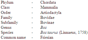
- The blood samples of twenty hybrid cattle species, namely Bos taurus (Linnaeus, 1758) were taken from Pyinmapin dairy cattle farm, Mingaladon Township (16°59'42" N 96°08'00" E), Yangon Region (Fig 3.1). The haematological analysis was done at laboratory of Zoology Department, East Yangon University.
3.2. Study Period
- The study period lasted from October, 2017 to September, 2018.
3.3. Specimen and Blood Sample Collection and Preparation
- This study was conducted in 20 crossbred cattle (Friesian x Local) to study the effect of breed on blood cell morphology. Blood samples were collected from 10 males and 10 females through the jugular vein with an extended neck. The left neck is fully extended and the medical surface of the neck is disinfected with cotton wool dipped in methylated alcohol. Use an 18 gauge needle to gently draw blood into a 3 ml syringe. The blood was collected in a vacuum tube containing ethylenediaminetetraacetic acid (EDTA). Each 1 ml blood sample was mixed in 3 ml EDTA in a vacuum tube. After gently rotating or inverting, blood should be mixed with anticoagulant as soon as possible. EDTA-treated samples are ideal for routine hematology examinations. Basic thin blood membrane preparation and staining were used for basic hematology experiments. This method is after Dacie and Lewis (1975).
3.4. Thin Blood Film Preparation
- Freshly drawn blood samples should be used to prepare thin blood films. I chose a few rails with smooth ends, clean and grease-free. Place the slide on a counter on another level. A drop of medium blood is about 2.4 centimeters from the right end, in the middle of the edge of the slide. Press the left side to grab this slide. Hold the other rail with your right hand, spreader, hold the right end and place it in the center of the flat rail to form an acute angle with the horizontal rail surface. Pull the spreader to the right until the tip touches the blood drop. Stop at this time, and the blood will spread along the acute angle by capillary action. Scan continuously to push the spreader to the left, and then spread the ink drops to the edge of the horizontal slide rail. This pulled a thin film. The smear should be thin and uniform, and the thickness depends on (1) the size of the droplets and (2) the spreader's angle. Sharp angles produce thin stains. Accelerate drying with a small amount of heat or air flow when coating. The quick drying of the membrane prevents red blood cells from being wrinkled and broken. Immediately stain and store the dust-free box so that flies, cockroaches, etc. do not swallow holes in the blood smear (Lewis, 1976).
3.5. Staining of Thin Blood Film
- The leishman stain was carefully dropped on the thin blood film to cover the entire thin blood film. Leave the stain on the thin blood film for about 2 minutes. Subsequently, the diluted blood is added to the stain on the diluted blood membrane. When it is a therapeutic pipette, the diluted stain will move up and down quickly and mix thoroughly. Diluting the liquid correctly will produce thin green garbage. Allow the diluted stain to act for 10 minutes, then wash with distilled water and dry the slide for two stains.After drying, a large amount of oil is immersed on the surface of the smear and then inspected by oil immersion. The diameter of blood cells was measured with a digital binocular microscope with an immersion magnification (x1000). (Model: NLCD-307B, Rated: AC 100-240V 50 HZ / 60 HZ, Lamp: S-LED WI, T 250V 500 mA, No. 1400040). When identifying most spots in the middle, from one edge to the middle, you need to include a smeared edge in this rough process. Continue this mode until the representative area is covered. This method follows Lewis (2000)
3.6. Formula of Leishman’s Stain
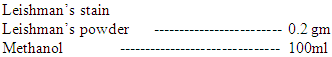 Directions0.2 gm of powdered dye is weighted out and transferred to a conical flask of 200-250 ml capacity. 100ml of methanol are added and the mixture is warmed to 50°C for 15 minutes with occasional shaking. The solution is filtered. It is them ready for use, but will improve on staining (Lewis, 2000).
3.7. Measurement of the Blood Cells
- Mature erythrocytes, leucocytes and platelets were measured for diameter in each sample of hybrid cattle and their mean diameter values in microns were calculated in each sample followed after Murry (1992).Formula for calculation of standard deviation

3.8. Identification of Hybrid Cattles and Blood Cells
- Morphological identification of erythrocytes, leucocytes and platelets were followed after Schermer (1958) and Thein Htut (1979). The classifications of the studied hybrid cattles were followed after Wilson, (1993).
3.9. Data Analysis
- Comparative analyses of morphometric measurements of blood cells between male and female hybrid cattle were carried out by applying student t – test at the 0.05 alpha level.
4. Results
- Morphological blood cell profile of hybrid cattle (Friesian x Local) were chosen to collect and study blood samples. From these samples, the characteristics of the morphology of the erythrocytes, leucocytes and platelets (Thrombocytes) were described (Figs 4.3, 4.4, 4.5, 4.6, 4.7). Table 4.1, 4.2, 4.3).
4.1. Systematic Position of the Study Species
4.2. The Blood Cells of Bos taurus (Male)
- Red blood cells, white blood cells and platelets of the Bos taurus (Male) were studied.
4.2.1. Red Blood Cells or Erythrocytes
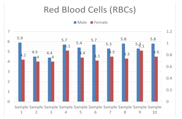
- The red blood cells were round and flat disc with little or no central depression. They do not contain a nucleus. The mean diameter of erythrocyte was 5.38 µm (Fig. 4.1, Table 4.1, 4.2, 4.3).
 | Figure 4.1. Comparison of mean diameter of red blood cells in the two collected cattles |
 | Figure 4.2. Comparison of mean diameter of platelet in the two collected cattles |
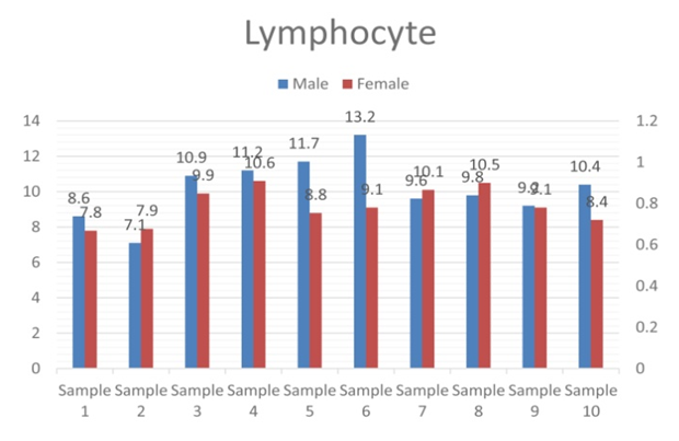 | Figure 4.3. Comparison of mean diameter of lymphocyte in the two collected cattles |
4.2.2. White Blood Cells or Leucocytes
- Two main types of leucocytes, granulocytes and non-granulocytes were investigated. Granulocytes:(1) The basophil had round granules and the nucleus was segmented with dark purple color. The cell had numerous small dark purple cytoplasmic granules obscuring part of the nucleus. The mean diameter of basophil was 11.44µm.(2) The eosinophil had round and very large. The nucleus had bi-lobed with dark purple. The eosinophil had round to oval reddish granule in the cytoplasm obscuring part of the nucleus. The mean diameter of eosinophil was 12.01µm.(3) The neutrophil had round (irregular shape) and cytoplasm with light pink granules. The nucleus had segmented with purple color. The mean diameter of neutrophil was 11.75µm (Fig 4.5, 4.6, 4.7, Table 4.1, 4.2 and 4.3).Non-granular Leucocytes:(1) The lymphocyte had round with rim of light blue cytoplasm. The nucleus had large round with purple color. The mean diameter of lymphocyte was 10.17µm.(2) The monocyte had large and round and its nucleus was bean-shaped with purple color. The cytoplasm had blue gray color. The mean diameter of monocytes was 12.45µm (Fig. 4.4, Table 4.1, 4.2 and 4.3).
4.2.3. Platelet
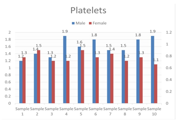
- The platelets were small round and pale blue color. Platelet have purple central granules in stained blood smears. The mean diameter of platelet was 1.59 µm (Fig. 4.2, Table 4.1, 4.2 and 4.3).
4.3. The Blood Cells of Bos taurus (Female)
- Red blood cells, white blood cells and platelets of the Bos taurus (Female) were studied.
4.3.1. Red Blood Cells or Erythrocytes
- The red blood cells were round and flat disc with little or no central depression. They do not contain a nucleus. The mean diameter of erythrocyte was 4.42µm (Fig. 4.1, Plate Table 4.1, 4.2 and 4.3).
4.3.2. White Blood Cells or Leucocytes
- Two main types of leucocytes, granulocytes and non-granulocyte were investigated. Granulocytes:(1) The basophil had round granules and the nucleus was segmented with dark purple color. The cell had numerous dark purple granules obscuring part of the cytoplasm. The mean diameter of basophil was 10.42µm.(2) The eosinophil had round and large. The nucleus had bi-lobed with dark purple. The cytoplasm had pale blue color. The mean diameter of eosinophil was 10.17µm.(3) The neutrophil had round and cytoplasm with light pink granules. The nucleus had segmented with purple color. The mean diameter of neutrophil was 10.13µm (Fig. 4.5, 4.6, 4.7 Table 4.1, 4.2 and 4.3).Non-granular Leucocytes:(1) The lymphocyte had round with rim of light blue cytoplasm. The nucleus had large round with purple color. The mean diameter of lymphocyte was 9.22µm (Fig.4.3, Table 4.1, 4.2 and 4.3).(2) The monocyte had large and round and its nucleus was bean-shaped with purple color. The cytoplasm had blue gray color. The mean diameter of monocyte was 10.9µm (Fig. 4.4. Table 4.1, 4.2 and 4.3).
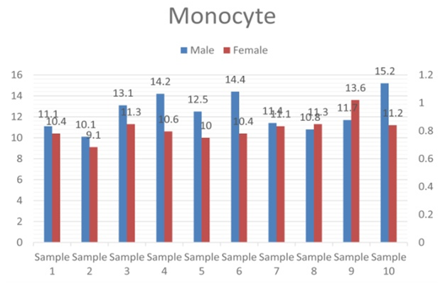 | Figure 4.4. Comparison of mean diameter of monocyte in the two collected cattles |
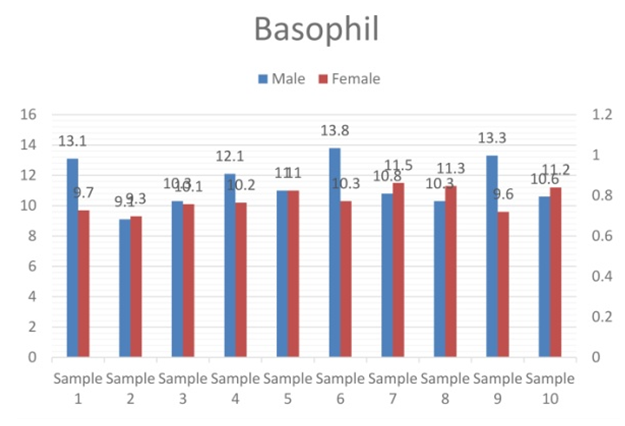 | Figure 4.5. Comparison of mean diameter of basophil in the two collected cattles |
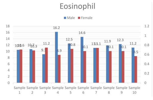 | Figure 4.6. Comparison of mean diameter of eosinophil in the two collected cattles |
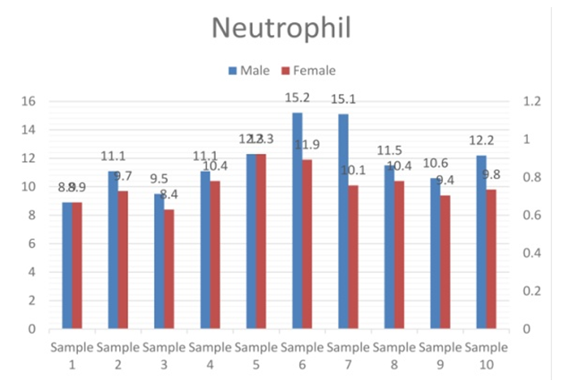 | Figure 4.7. Comparison of mean diameter of neutrophil in the two collected cattles |
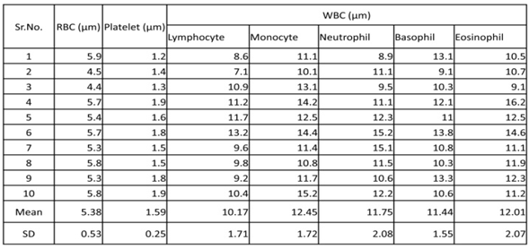 | Table 4.1. Morphometric measurement of male hybrid cattle blood cells |
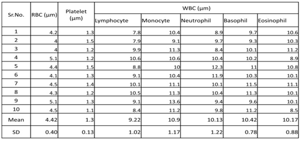 | Table 4.2. Morphometric measurement of female hybrid cattle blood cells |
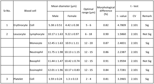 | Table 4.3. Comparison between morphometric measurement of male and female hybrid cattle blood cells |
4.3.3. Platelet
- The platelets were small round and pale blue color. The mean diameter of platelet was 1.3µm. (Fig. 4.2, Table 4.1, 4.2 and 4.3).
4.4. Comparison between Morphometric Measurements of Male and Female Hybrid Cattle Blood Cells
- The differences between morphometric measurement (diameter) of erythrocyte (t = 4.7809, cv = 2.101), monocyte (t = 2.4831, cv = 2.101), neutrophil(t = 2.2387, cv = 2.101), eosinophil(t = 2.7281, cv = 2.101) and platelet (t = 3.3965, cv = 2.101) cells in male and female hybrid cattle were significant while those of lymphocyte (t = 1.5860, cv = 2.101) and basophil (t = 1.9594, cv = 2.101)were not significant (Table 4.1, 4.2 and 4.3).
5. Discussions and Conclusions
- Morphological studies of red blood cells, white blood cells or white blood cells and platelets or platelets have an important effect on determining the type of cow. (Adili et al., 2016) In this study, blood samples from 20 cattle, 10 from adult males and the others 10 from adult females, were collected. From these samples, the morphological characteristics of red blood cells, white blood cells and platelets are described. The dog's red blood cells are large (7 µm), uniform in size, pale in the center (double concave disc). In cats, the average diameter of cells is 6 microns, and in cattle, the average diameter of cells is 6 microns. The average diameter of the sheep is 4.5 microns, flat and almost without central depression. In goats, red blood cells are over 4 microns in size and have the most different shapes. In horses, red blood cells have an average diameter of 5.7 microns, uniform size, and central paleness without skewer formation.The width of bovine red blood cells is 5-6 μm (Wood and Quiroz-Rocha, 2010), and the diameter of sheep red blood cells is 4.5 μm. However, compared to other species, goats have the smallest red blood cells, with a width of 2.5 – 3.9 µm (Byers and Kramer, 2010). Puppies have larger red blood cells than other species, and have a diameter of 6 to 8 µm. (Rizzi, et al., 2010)In previous reports, mature red blood cells in adult cows were double concave with a width of 5.6 µm, which is smaller than other species. (Roland et al., 2014). However, in Myanmar, livestock red blood cells are round, flat discs and have no central depression. The average diameter is 6 µm. (Khin Sabae Htut, et al., 2014)The red blood cells of mammals are spherical, biconcave and nucleated. The largest red blood cell was observed in Bandicota bengalensis (8 µm), and the smallest red blood cell was observed in Rattus rattus (6 µm). (Thein Htut, 1979)In this study, red blood cells in Taurus, both males and females, were rounded, flat discs with little or no central depression. The largest red blood cells (5.38 µm) were observed in Taurus males and the smallest (4.42 µm) in females. The average diameters of red blood cells in both species were almost the same, with 5.38 µm and 4.42 µm respectively.In the granular leucocytes, basophils, eosinophils and neutrophils are consisted. Basophils are rarely seen in the peripheral blood of all common family species. However, in dairy cows that we studied, as basophils are less than 1% in total leukocyte count, it is difficult to find in blood smears. The largest basophil was observed in Anser cygnoides (11.7 µm) and the smallest one was observed in Anas platyrhynchos (10.7 µm). (Kyi Kyi Mar, 2016)The study also found that the neutrophils of both species have round and cytoplasmic, light pink particles. The average diameter of the male Taurus neutrophil (11.75 µm) is greater than the average diameter of the female Taurus neutrophil (10.13 µm). According to the data, the average diameters of basophils are 11.44 µm in males and10.42 µm in females respectively, eosinophils (12.01 µm male, 10.17 µm female) and neutrophils (11.75 µm male, 10.13 µm female) are approximately the same. Lymphocytes are a non-granular and are common in ruminants, birds and rodents and their size ranges from 6 to 15 μm. Most lymphocytes in predators and horses are small while larger cells occur more frequently in ruminants. The largest lymphocytes were observed in Anas platyrhynchos (9.7 µm) and the smallest lymphocytes in Anser cygnoides (9.6 µm), Kyi Kyi Mar, 2016. In chicken-backed chickens, the largest lymphocyte was 9.7 µm and the smallest lymphocyte was 8.7 µm. (Wut Yee Nandar, 2016) In this study, Bos Taurus, both males and females, lymphocytes are rounded and have a light blue cytoplasm on their edges. In both cows, the nucleus has a large purple circle. The mean lymphocyte diameter (10.17μm) in Bos taurus (male) is greater than the mean diameter (9.22μm) in Taurus (female). In cows, monocytes are the largest white blood cells (15-20 μm in diameter). The shape of the nucleus is very diverse and can be oval, irregular, kidney-shaped or horseshoe-shaped.The largest monocytes were observed in Anser cygnoides (10.5 µm), and the smallest monocytes were observed in Anas platyrhynchos (9.8 µm) (Kyi Kyi Mar, 2016). The largest monocytes in C. gallinarum was 11.7 µm and the smallest was (10.8 µm) (Wut Yee Nandar, 2016). In this study, monocytes were large and round. The nuclei of both cows are purple and bean-shaped. The cytoplasm of both men and women is blue-gray. The maximum average diameter of monocytes (12.45 μm) was studied in Bos taurus (male), and the minimum average diameter (10.9 μm) of monocytes was studied in Bos taurus (female). It was found that the two monocytes studied had bean-shaped nuclei.In cattle, platelets are fragments of non-nuclear cells 2-4 μm in diameter. The main role of platelets is to promote blood clotting. Khin Sabae Htut (2014) of Myanmar explained that the average platelet diameter of livestock was (2.13 μm). In this study, male Taurus platelets are small, round, and sky blue. Female Taurus platelets are small, round, light blue. The average platelet diameters for both species are approximately the same with males (1.59 µm) and female (1.3 µm) respectively.Cows have been domesticated for thousands of years and play an important role in serving food to people all over the world, dairy farm for dairy farmers and at the present the dairy industry has become a major occupation in the center of large cities with high milk demand. Therefore, healthy and clean farms require food and milk safety. As Hematology is the study of blood health and disease, blood cell circumference and surface measurements appear to be more suitable for comparative studies. In addition, these two parameters are more representative in the study of blood cell morphology and morphological changes (Adili et al., 2016).Finally, it was concluded that the study could explain the morphological parameters of blood cells in a drop of blood. However, if pathological or abnormal morphology or impaired blood cell functions are suspected, other microscopic blood smear assessments are strongly recommended. (Roland et al., 2014) According to this study, it was found that the morphology of blood cells are almost similar to other researchers in previous cattle studies, but monocytes are the largest of white blood cells. It should also be a new morphological study of different species and varieties. Attention should be paid to hematologic changes and pathological conditions in bovine blood cells.The research was conducted in the laboratory of the Department of Zoology, University of East Yangon. The study period is from October 2017 to September 2018. Blood samples of hybrid cows were collected from the Pyinmapin Dairy Farm in Mingaladon Township, Yangon. In the boss bull male and female, the red blood cells are round and do not contain nuclei. It may appear as a flat disc with almost no central depression. The average red blood cell diameter of males is 5.38 μm, which is larger than the average diameter of females (4.42 μm). In leukocytes, basophils have round particles. The nucleus is dark purple. The cytoplasm has dark purple particles. Eosinophils are dark purple nuclei with round and double lobes. The cytoplasm has red-brown (male) and light blue (dark) particles. Neutrophils are rounded and divided into purple nuclei. The cytoplasm has light pink particles. The average diameters of basophils, eosinophils and neutrophils were observed in Bos Taurus (males and females).In non-granular white blood cells, lymphocytes are rounded and have a light blue edge. The nucleus is large, round, purple. Mononuclear cells are large, round and have a bean-shaped purple nucleus. The cytoplasm is blue-gray. The average diameter of male lymphocytes and monocytes is greater than that of women. Platelets are small, light blue. In male and female species, the average diameter of platelets is about the same.
ACKNOWLEDGEMENTS
- The author would like to thank Prof. Dr. Thet Thet Myaing and Dr. Thida for her invaluable insights into blood cell taxonomy and comments on the working draft.