-
Paper Information
- Paper Submission
-
Journal Information
- About This Journal
- Editorial Board
- Current Issue
- Archive
- Author Guidelines
- Contact Us
Research in Zoology
p-ISSN: 2325-002X e-ISSN: 2325-0038
2017; 7(1): 7-10
doi:10.5923/j.zoology.20170701.02

Absence of Hemoparasites in Wildlife Snakes, Located in the Ecological Reserves Cota 70, Cotacachi-Cayapas and Sumaco-Napo-Galeras in Ecuador
 Abstract
 Reference
 Full-Text PDF
 Full-text HTML
Erika Medrano-Tupiza1, Samantha Morales-Arciniega1, Silvana Santander-Parra2, Luis Núñez-Naranjo1, 2, Byron Puga-Torres1
1College of Veterinary Medicine, Central University of Ecuador, Quito, Ecuador
2Department of Pathology, College of Veterinary Medicine, University of Sao Paulo, São Paulo, Brazil
Correspondence to: Byron Puga-Torres, College of Veterinary Medicine, Central University of Ecuador, Quito, Ecuador.
| Email: |  |
Copyright © 2017 Scientific & Academic Publishing. All Rights Reserved.
This work is licensed under the Creative Commons Attribution International License (CC BY).
http://creativecommons.org/licenses/by/4.0/

Ecuador is one of the most mega diverse countries in the world, covering a large amount of flora and fauna in all its geographical regions. The aim of this study was to determine the presence or absence of blood parasites in wildlife snakes in the ecological reserves Cota 70, Cotacachi - Cayapas and Sumaco - Napo - Galeras, located in the continental Ecuador. A total of 14 individuals were captured: 9 Colubrids (Dipsa elegans, Atractus dunni, Sibonne bulata, Mastigodryas heathii, Oxyrhopus petola digitalis, Atractus major, Oxyrhopus petolarius, Phrynonax polylepis, Mastigodryas pulchriceps), 3 boids (Corallus hortolanus) 2 Boa constrictor, 1 viperid (Bothrops asper) and 1 Elapid (Micrurus surinamensis surinamensis). Samples of blood were collected from the caudal vein or by cardiac puncture depending on the size of the snake. The blood smears were dried, fixed with methanol and stained with Giemsa solution. Samples were examined by light microscopy using a 100 × magnification lens in search for hemoparasites. The present study shows that in the snakes analyzed there is no evidence of the presence of hematic parasites showing that the areas where the ecological reserves are located could be free of blood parasites. No ectoparasites were found in the captured animals.
Keywords: Snakes, Haemoparasites, White blood cells, Giemsa
Cite this paper: Erika Medrano-Tupiza, Samantha Morales-Arciniega, Silvana Santander-Parra, Luis Núñez-Naranjo, Byron Puga-Torres, Absence of Hemoparasites in Wildlife Snakes, Located in the Ecological Reserves Cota 70, Cotacachi-Cayapas and Sumaco-Napo-Galeras in Ecuador, Research in Zoology , Vol. 7 No. 1, 2017, pp. 7-10. doi: 10.5923/j.zoology.20170701.02.
Article Outline
1. Introduction
- Medicine in wild species is relatively new in Ecuador, however, the interest in its practice and learning has been growing in the country, so it is necessary to know more about these animals, their characteristics, the threats to which they are exposed and their development with the environment [1]. According to Carrillo et al. (2005) [1] in his book " Lista Roja de Reptiles del Ecuador ", presents 16 species of snakes considered endangered in the country, 21 vulnerable species and 4 in almost critical situation. There are several causes for the increase of species at risk of extinction, among which we can mention diseases, many of which have gained space due to human intervention [2, 3]. In recent years important findings have been made regarding the life cycles of the parasites. This has provided information about the biosecurity that must be had to prevent disease transmission [4]. In reptiles, due to morphological variability and high diversity of hemoparasites, there are not as many reports as there are in mammals and birds, probably related to the large size of reptile erythrocytes and to the presence of a prominent nucleus within them, Which can strongly influence the appearance of the parasite within the cell and also because the diversity of hemoparasites is greater in reptiles, both in genus and in species [5].Within the hemoparasites, in the genus Hepatozoon, family Hepatozoidea we find more than 50 species of intracellular hemoparasites which occur frequently in groups of vertebrates and some invertebrates; Representatives of this genus can parasite leukocytes of birds and mammals (including dogs and cats), also red blood cells in reptiles, amphibians and fish; All vertebrate animals infected with hemoparasites are considered intermediate hosts [6].Hemoparasites in snakes and reptiles in general affect the organism in different ways, either by forming cysts in various organs such as liver, spleen, kidney and brain, which may contain pigment deposits or may be surrounded by inflammatory cells, causing variation in the Hemoglobin, alterations in serum or plasma proteins [3]. In cases of generalized infection or immunosuppression, hemolytic anemia may occur; in natural hosts these changes mentioned are rare or absent. In young animals, the presence of hemoparasites, added to stress situations or other pathologies, can lead to the death of the animal [3].The data obtained in the present research are of importance for their originality and refer to a population not studied, which sets precedents for further research. It is expected to continue the study of wildlife snakes in the country, as the populations of these animals have declined considerably in recent years, and more attention is required from the scientific community to address such a worrying situation. The main objective of this study was to determine the presence or absence of hemoparasites in wildlife snakes in Ecuador’s natural reserves: Cota 70, Cotacachi - Cayapas and Sumaco - Napo - Galeras.It seems that the presence of hemoparasites in reptiles is due to their infestation with ectoparasites (like ticks) as it happens in green iguanas in the Guayaquil city [7]. The transmission of Haemoproteus in snakes require the presence of a natural vector (Crhysops fly), for the transmission of Haemoproteus metchnikovi [2].
2. Materials and Methods
2.1. Study Area
- The study, of a descriptive nature, was carried out in three natural reserves of Ecuador, located in the coast, east and mountain region: Reserve Cota 70, Portoviejo - province of Manabí, Reserve Sumaco Napo Galeras, Napo - province of Tena [8], Reserve Cotacachi Cayapas, Cotacachi - Province of Imbabura [9] (Table 1).
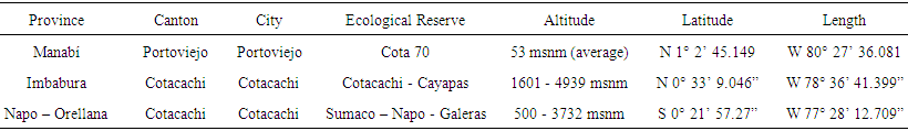 | Table 1. Location and characteristics of the studied reserves. Source: Ministry of the Environment - Ecuador, 2016 |
2.2. Size and Target Population
- The target population for this study was all snakes that were located within the natural reserves. The search for snakes was by means of sampling by transects (technique of observation and recording of data along a line that crosses the zones), in anthropic and non-anthropic areas, with emphasis on sectors with dry leaves, fallen trees, hollow logs, holes in the ground, branches of trees, and every place a snake could hide, besides to paths to which they come out to warm themselves. The size of the sample was based on the availability of individuals in wildlife throughout the project, for four months. In total, 14 snakes were collected, distributed as follows: 6 in the Sumaco - Napo - Galeras reserve, 5 in the Cotacachi - Cayapas reserve and 3 in the Cota 70 reserve. The individuals were captured in situ, by means of search and direct capture. It was also shown if there was presence or absence of ectoparasites on snakes.
2.3. Collection of Blood Samples
- The method of manipulation and collection of blood samples were performed according to the standards of preservation and animal ethics established by the Ministry of the Environment in the "Codification of forest law and conservation of natural areas and wildlife (Law No. 2004-017, Official Register No. S-418 of September 10, 2004). Data on weight (gr), size (cm), sex and species were taken from all individuals captured. Heparinized syringes were used to control the blood clotting process. Approximately 1 mL of blood sample was collected by puncture of the caudal vein, in case of boids and large viperids, and by cardiac puncture in colubrids and small elapids. The samples obtained were stored in properly identified heparinized microtubes for the subsequent blood smear. When the blood sample was insufficient, the blood smear was performed immediately using a drop of blood.
2.4. Examination of Blood Smears
- The blood smears of the captured individuals were withdrawn for a period of 10 minutes and then fixed for 4 minutes in 95% methanol. Once the blood smears were fixed, they were stained with Giemsa solution (belonging and prepared by the Laboratory of Parasitology of the Faculty of Veterinary Medicine and Animal Science of the Central University of Ecuador) for four minutes, according to protocol already established [10]. Examination of the smears was performed by light microscopy with a 100X magnification, using 10 quadrants in search of haemoparasites.
3. Results and Discussion
- A total of 14 individuals were captured: 9 Colubrids (Dipsa elegans, Atractus dunni, Sibonne bulata, Mastigodryas heathii, Oxyrhopus petola digitalis, Atractus major, Oxyrhopus petolarius, Phrynonax polylepis, Mastigodryas pulchriceps), 3 boids (Corallus hortolanus) 2 Boa constrictor, 1 viperid (Bothrops asper) and 1 Elapid (Micrurus surinamensis surinamensis). Of the 14 snakes, 6 belong to the reserve Sumaco - Napo - Galeras (%), 5 in the Cotacachi -. Cayapas (%) and three in reserve Cota 70 (%). Of the total captured snakes, 28.57% were males, while the remaining 71.43% were females. The 14 samples taken, showed negative results, did not evidence the presence of hemoparasites in the blood cells of the captured individuals. Neither vector (ticks, mosquitoes) were found on the individuals found in the sample (Table 2). Most of the specimens found in the field belong to the Colubridae family, a fact that coincides with the research carried out by Moreno and Bolaños in 1977, in this study, none of the 28 specimens of colubrids were found to be parasitized. In the same study, it was also evidenced that in the snakes where ticks were found, after a histopathological analysis, all of these animals were found to be positive for Haemogregarinidae hemoparasites [11].
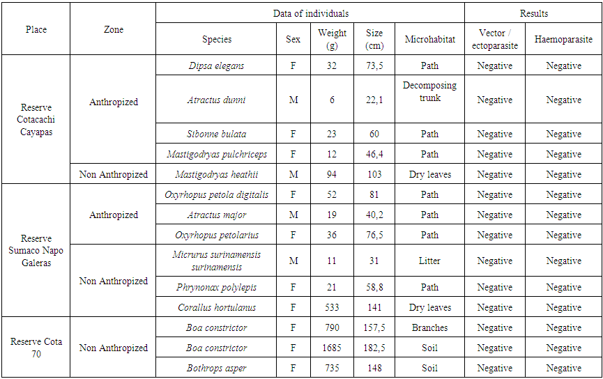 | Table 2. Results of research |
4. Conclusions
- The present study shows that there are no hemoparasites present in the ophidians analyzed, suggesting that the ecological reserves at Ecuador where the animals were captured could be free of hemoparasites, however more studies should be performed in order to corroborate these results.
ACKNOWLEDGEMENTS
- To Andy Proaño and Jonathan Proaño, for their great help in the field phase of this investigation. To the personnel of the Laboratory of Parasitology of the Faculty of Veterinary Medicine and Animal Science of the Central University of Ecuador, for facilitating the use of equipment and supplies.