-
Paper Information
- Paper Submission
-
Journal Information
- About This Journal
- Editorial Board
- Current Issue
- Archive
- Author Guidelines
- Contact Us
Research in Zoology
p-ISSN: 2325-002X e-ISSN: 2325-0038
2016; 6(3): 33-36
doi:10.5923/j.zoology.20160603.01

Prevalence of Liver Flukes in Cattle, and Small Ruminants at Slaughter in Zaria, Nigeria
 Abstract
 Reference
 Full-Text PDF
 Full-text HTML
Ieren I. I.1, Ajanusi O. J.2, Mbaya P. Y.1
1Department of Veterinary Public Health and Preventive Medicine, Ahmadu Bello University, Zaria, Nigeria
2Department of Veterinary Parasitology and Entomology, Ahmadu Bello University, Zaria, Nigeria
Correspondence to: Ieren I. I., Department of Veterinary Public Health and Preventive Medicine, Ahmadu Bello University, Zaria, Nigeria.
| Email: |  |
Copyright © 2016 Scientific & Academic Publishing. All Rights Reserved.
This work is licensed under the Creative Commons Attribution International License (CC BY).
http://creativecommons.org/licenses/by/4.0/

The study was conducted to determine the prevalence of liver flukes (Fasciola gigantica and Dicrocoelium hospes) in cattle, and small ruminants (sheep and goats) slaughtered in Zaria, Nigeria. A total of 500 gall bladders were harvested from 227 (45.4%) cattle, 10 (2.0%) sheep and 263 (52.6%) goats after slaughter from the abattoir and slaughter slabs within Zaria metropolis. Out of the animals sampled, 240 (48.0%) were positive for eggs of liver flukes. There was a significantly higher level of infection in cattle (39.2%) than small ruminants (8.8%). Among the infected animals, 99 (50.51%) of the infected cattle had mixed infections with eggs of F. gigantica and D. hospes(p<0.05). A non-statistically significant higher rate of infection was observed in cows 47 (88.68%) than bulls 149 (85.63%) (p> 0.05). Season-specific prevalence showed a significantly (p<0.05) higher rates of infections during the rainy season than during the dry season. This study therefore confirms a high prevalence of liver fluke infections among animals slaughtered in Zaria for consumption, therefore adequate measures needs be taken to control the infection in order to reduce the economic losses accruing to this infection.
Keywords: Abattoir, Fasciola gigantica, Dicrocoelium hospes, Prevalence, Zaria
Cite this paper: Ieren I. I., Ajanusi O. J., Mbaya P. Y., Prevalence of Liver Flukes in Cattle, and Small Ruminants at Slaughter in Zaria, Nigeria, Research in Zoology , Vol. 6 No. 3, 2016, pp. 33-36. doi: 10.5923/j.zoology.20160603.01.
Article Outline
1. Introduction
- Liver fluke diseases of ruminants are caused by trematodes from the genera Fasciola, Fascioloides and Dicrocoelium [1]. Diseases caused by the genus Fascioloides may occur in domestic ruminants, though the incidence is low. Where these diseases occur, they are as a result of grazing domestic ruminants on the same pasture utilized by deer which are the natural hosts [2].In the temperate region, these diseases are caused by Fasciola hepatica and Dicrocoelium dendriticum, but in the tropics and Asia, as well as the pacific islands, the diseases are caused by Fasciola gigantica and Dicrocoelium hospes which cause fasciolosis and dicrocoeliosis respectively [2]. The trans-human system which involves movement of animals to areas of wet regions during the dry season when the grass for grazing is scarce encourages the spread of these liver flukes as result of the eggs being passed out in faeces as the animals graze. Liver flukes infections in animals may result to huge economic losses associated with decreased productivity, loss of weight and poor carcass quality, cost of antihelminthics for treatment, and condemnation of offals, particularly the liver at slaughter [8, 14]. Liver flukes infections may also pose public health infections as the infectious forms of these helminthes (metacercaria) may be found on the surface waters and on vegetables through the use of manure produced from already contaminated faces. Humans drinking fresh untreated water or consuming raw vegetables which may not be properly washed or peeled containing metacercaria, a situation that is not uncommon in some parts of rural Africa, may become infected [15]. In Nigeria such infections have been reported in HIV patients who reported consuming partially cooked liver in order to meet their dietary needs. This could be due to consumption of liver infected with juvenile flukes [3].Limited information exists in Nigeria on the situation of liver flukes infection in cattle and small ruminants. Where such reports exist, they are based mainly on F. gigantica in faeces of cattle with little attention given to small ruminants and gall bladder examination, which often under reports the real situation. This study therefore aims at determining the prevalence of liver flukes (D. hospes and F. gigantica) cattle, small ruminants at slaughter in Zaria, Nigeria to provide epidemiological information that may contribute to the control of the infection.
2. Materials and Methods
2.1. Study Area
- This study was carried out in Zaria located in Kaduna state, within latitudes 11° 7’, 11° 12’N and longitudes 07° 41’E. It is a medium sized city with an estimated population of 547,000 and a growth rate of 3.5% per annum. It is divided administratively into Zaria and Sabon Gari LGAs [13] and about 40-75% of its working population derives their principal means of livelihood from agriculture [4].Agricultural activity in Zaria can be divided into two types: rain-fed (May to October) and irrigation farming in the dry season (November to April). It is characterized by a tropical climate, a monthly mean temperature ranging from 13.8 to 36.7°C and an annual rainfall of 1092.8mm [5]. Horticulture is the second most prevalent agricultural activity in Zaria with vegetables being mostly produced, but in some cases fruits are inter-cropped among cereal crops.
 | Figure 1. Map of Kaduna state showing the Study area [6] |
2.2. Sampling and Sampling Technique
- A cross-sectional study design was used and convenience sampling technique was adopted based on informed consent of the abattoir/slaughter slab operators and butchers, and only those who accepted to participate in the study had their animals sampled. A total of 500 gall bladder samples were collected from 227 (45.4%) cattle, 10 (2.0%) sheep and 263 (52.6%) goats. The samples were packaged in sterile polythene bags and labelled appropriately. The sex, breed and species of the animals were also recorded. The gall bladder samples were then packed into ice pack containers and transported to the Helminthology laboratory of the Department of Veterinary Parasitology and Entomology, Ahmadu Bello University, Zaria.
2.3. Sample Processing
- Each of the gall bladder samples was cut open and washed into clean containers and allowed to sediment as described by Ulayi, et al. [7]. The supernatant was then decanted off and the sediments were pipetted and drops were placed on clean glass slide for examination under the microscope at ×10 magnification. Where it was anticipated that the sediments could not be examined over a 24-hour period, drops of formaldehyde were added to prevent the eggs from hatching prior to the time they will be viewed. Eggs identification for these flukes was based on morphological characteristics and size of the eggs as described by Soulsby [8].
2.4. Data Analysis
- Data was presented in the form of frequency distribution tables and Chi Square was also used, where necessary to show degree of association using the Statistical Package for Social Sciences (SPSS) version 20.0 and STATA 12. Values of p<0.05, where used, were considered to be statistically significant.
3. Results
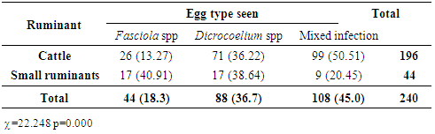
- Among the 500 animals sampled, 240 (48.0%) were positive for eggs of liver flukes. Distribution of eggs of the parasites showed a statistically significant difference (p<0.05) in the occurrence of eggs of liver flukes in cattle 196 (39.2%) and 44 (8.8%) for small ruminants (sheep and goats) sampled at slaughter (Table 1).
|

|
|
|
4. Discussion
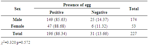
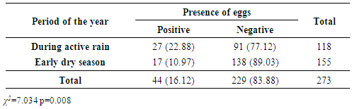
- The high incidence of liver fluke infection observed in cattle is similar to 31.7% and 37.2% obtained by Ozung et al. in a retrospective study over a 5-year period [8] and Ulayi et al in abattoir study [7] respectively which shows that the prevalence of liver fluke is still high in Zaria, Nigeria. The prevalence rate of F. gigantica (86.34%) obtained in cattle is however higher than 37.2% reported by Ulayi and others [7]. This could be attributed to the fact that this study was carried out for a longer period of time (5 months) including period rainfall was almost at its peak (July- September) and early dry season (October-November) as compared to that obtained by Ulayi et al. [7] which was for 30 days in between December and January which is about the peak of dry harmattan season. This further shows that seasons may have influence on the occurrence these parasites [7] and also confirmed in this study where a statistically significantly (p=0.008) higher infection rate was observed in the rainy season than in the dry season (Table 4). The significantly higher prevalence of liver flukes in cattle (86.34%) could be due to the fact that most of the herds of cattle in this region are reared using the pastoral system where animals move to different places in search of pasture thereby increasing their chance of exposure to sources of infection which include stagnant water points and lush pasture that have been identified as containing the infectious forms of the parasites [15] as compared to the small ruminants that are mostly reared around the homestead and fed by food from sources other than pasture and provided with fairly clean water for drinking, thereby reducing their chances of exposure to infection.The prevalence of D. hospes infection was higher in cattle (36.20%) agrees with the findings by Ulayi et al. [7]. The occurrence of mixed infection with eggs of both D. hospes and F. gigantica was significantly higher (50.51%) in cattle than pure infections with eggs of the individual helminthes which agrees with the finding by Olusegun-Joseph et al. [10]. This finding could be attributed to the environmental and climatic conditions that favour the development of these helminthes are similar [11] and it is therefore not uncommon to have the two parasites occurring together hence potentiating the chance of having mixed infections.The prevalence of eggs of liver flukes was lower in bulls (85.63%) than cows (88.68%) which disagree with the findings by Elkannah [12] and Olusegun-Joseph et al. [10] who reported a higher prevalence in bulls; however, this difference was not statistically significant (p=0.572) (Table 3). This shows that bulls and cows present the same chance of being infected if they are exposed to the same management system.A higher prevalence of the liver fluke infection during the period of active rainfall (22.88%) than during the dry season (10.97%) could be due to the abundance of moisture and water collection points which are present in the rainy season. These conditions have been reported to favour the availability of the snail intermediate hosts [11] which in-turn favours the occurrence of these helminthes.The findings in small ruminants represent one of the rare reports of these parasites in these species as there as hardly reports of liver flukes infections in sheep and goats in this environment.
5. Conclusions
- The study has established that the prevalence of liver flukes in animals slaughtered in Zaria for consumption is high and this could result to reduction in the production capacity of these animals as well as present potential threat to public health for those who consume vegetables produced from manure of such animals. Adequate control measures should therefore be put in place to reduce the occurrence of these diseases especially during the rainy season so as to reduce economic losses due to these infections.
ACKNOWLEDGMENTS
- The authors wish to acknowledge the technical support of Mr. D. Gimba and Mr. K. Amadi, all of the Department of Veterinary Parasitology and Entomology, Ahmadu Bello University, Zaria, Nigeria.