-
Paper Information
- Paper Submission
-
Journal Information
- About This Journal
- Editorial Board
- Current Issue
- Archive
- Author Guidelines
- Contact Us
Research in Zoology
p-ISSN: 2325-002X e-ISSN: 2325-0038
2016; 6(1): 11-16
doi:10.5923/j.zoology.20160601.03

Differential Mites Infestation of Domesticated Animals and Handlers Dermatitis in Ijumu Nigeria
 Abstract
 Reference
 Full-Text PDF
 Full-text HTML
Foluke Helen Ajobiewe 1, Olu Joseph Ajobiewe 2
1University of Jos Nigeria, Department of Zoology, Nigeria
2National Veterinary Research Institute Vom Plateau State of Nigeria, Nigeria
Correspondence to: Foluke Helen Ajobiewe , University of Jos Nigeria, Department of Zoology, Nigeria.
| Email: |  |
Copyright © 2016 Scientific & Academic Publishing. All Rights Reserved.
This work is licensed under the Creative Commons Attribution International License (CC BY).
http://creativecommons.org/licenses/by/4.0/

Principally, Mites infestation of domesticated animals, viz:- cattle, sheep, and goats were examined. In like manner, other ectoparasites viz, Lice, Fleas, Ticks, and Flies were also examined. This was aimed at verifying the exact species of mites and other ectoparasites responsible for dermatitis and lesions on human handlers of these infested animals. Random sampling technique was adopted in the collection of the ectoparasites from Ijumu local government area of Kogi state, Nigeria. Result revealed that mites infestations on cattle, goats and sheep were 0%, 1.90, 4.79% respectively when compared with other parasites. Dermatitis on human handlers of cattle, sheep and goats were 0%, 98.1%, and 95.21% respectively. (as caused by mites when compared with other ectoparasites) There is a significant correlation (P&0.05) between mites infestation and dermatitis lesions on the human handlers.
Keywords: Dermatitis, Differential Infestation, Psoroptes, Sarcoptes, Mites
Cite this paper: Foluke Helen Ajobiewe , Olu Joseph Ajobiewe , Differential Mites Infestation of Domesticated Animals and Handlers Dermatitis in Ijumu Nigeria, Research in Zoology , Vol. 6 No. 1, 2016, pp. 11-16. doi: 10.5923/j.zoology.20160601.03.
Article Outline
1. Study Background/Literature Reviews
- Mites were described as very minute arthropods belonging to the class Arachnida and order Acarina [1]. They are medically important because they are source of great discomfort to man and domestic animals, due mainly to their biting and burrowing habit. According to Becks et al, [2] mites are small and have hypostome hidden and unarmed (without hooks). The female is bigger than male in size and it is found between the fingers toes, in the groin, external genitalia and axillary regions. They excavates tunnels in the epidermis and sometimes even the dermal tissue of their domestic hosts. They suck blood and tissue fluids, cause irritations and set up inflammatory and hypersensitivity reactions that present clinical pathogenic lesion, follicular papules, intense itching, hence scabies is acquired. The bites and the pruritis due to the presence of mites can cause restlessness and lack of sleep [3]. Gordon et al (1962) [4] indicated that the sarcoptic mites cause diseases in almost all species of animals and sarcoptic mange is common in domestic animals such as horses, cattle, dogs and pigs. Sarcoptes canis is from the dog, Sarcoptes ovis is from the sheep, Sarcoptes equi from the horse. The mite causing scabies in man is known as Sarcoptes scabiei var canis. Sarcoptesscabiei is a small flattened disc-shaped creature, whitish in colour with the hypostome devoid of teeth and the chelicerae are of the pincer type. The adult possesses eight short equal legs. In the female, the two first pairs of legs have suckers situated at the end of an unjointed pedicel whereas the last two pairs have no suckers. The male is similar but smaller and last pair of legs also possess suckers. The individuals of both sexes have the dorsum armed with backwardly directed spines, which facilitate the mites progress down the burrow.Nelson et al (1975) [5] divided mites into the following families:-Family Psoroptidae:- e.g. Chorioptes bovis which cause chorioptic mange primarily indomestic herbivores. Family Sarcoptidae:- e.g. Sarcoptes scabiei which cause sarcoptes mange or scabies in man, domestic and wild animals throughout the world. Family Demodicidae:- e.g. Demodex spp which spend the entire life cycle upon the host and it takes about 24 days. Family Trombiculidae:- e.g. Chiggera which parasitize all vertebrates. They cause severe dermatitis in humans and produce lesions on horses. The suborder Mesostigmata is a large group of active mites. Lung mites (Pneumonyssus) are found in the lungs of mammals, where they cause nodules which resembles tubercular lesions. Dermanyssid mites are common in poultry, rats and mice, and are quite important because they affect man incidentally but frequently [6]. The mites of the family dermanyssidae when numerous cause irritation and restlessness. The “apartment mite” Allodermanyssus sanguineus lives in house –mouse nests and often man. This mite is a vector of the micro-organism, Rickettsia akari which causes a disease similar to chicken pox. Cheyletiella and Cheyletus spp. parasitize other species of mites. These mite –inhabiting mites can attack man. The genus Demodex include very minute mites which inhabit the oily skin glands of man and other mammals. Demodectic invasions are accompanied by bacterial infection which are usually mild or symptomless [6].Families of Sarcoptidae and Psoroptidae consist of parasites which cannot survive away from integumentary structures or tissue of vertebrates (Askew 1971). The genus Psoroptes, Otodectes, Notoedres, Chorioptes and Sarcoptes have various species which cause “itch”, “scab” and mange in sheep, cattle, horses, dogs, cats and other animals including man. The feeding process of mites in particular have been extensively studied and reviewed by [5]. They have been observed to cause the following:- - They cause dermatitis or other tissue damages to man and his domestic animals.- They help in transmitting or transferring pathogenic agents either as vectors or developmental hosts.- They also cause strong allergic reactions in man, pets and livestock.- They help in the loss of blood or other tissue fluid [8].Gray (1961) [9] discovered mites to be an irritating and parasitic ectoparasite likewise other forms of mites like chiggers. Recently, Yerubam (1984) [10] discovered that out of 30 herds of local (black) goats monitored throughout 1983 in different part of Israel, ten (10) were infested with mites. He also stated that the infested goats were aged 2 years or more and were in good physical condition. Steelman (1976) [11] observed large numbers of nodules caused by Demodex bovis on the side of the body, head, back, hip, legs and abdomen of cow aged 2-14 years in July. A heifer was infested with mites of the genus Demodex, and was found inside the external ear, also Psoroptes ovis was identified in the lesions of cattle [12]. Mixed infestations by Chorioptes, Psoroptes, and Sarcoptes spp. were found on cattle [13]. Enemalah (1976) [14] also observed that S. scabiei var suis were found on growing pigs under commercial conditions. Kerkut (1961) [15] reported that sheep scab caused by Psoroptes ovis was eradicated from sheep in united kingdom. Gordon (1962) [4] reported that a herd of 14 local breed goats were infested with S. scabiei var coprae. However, Hall (1977) [16] added that red mite Dermanyssus gallinae attacks only at night and hide in crevices during the day which might be a reason for low incidence of mites reported by many authorities. Furthermore, Iwuala and Okpala (1977) [17] indicated that mites species Psoroptes ovis were only found restricted to the trunk region of sheep sampled to be infested. Puccini et al (1986) [12] stated Sarcoptes scabiei var suis was found on 108 wormed pigs in USA. Mohr (1961) [18] reported that 4 species of mites that cause mange in cattle are Chorioptes bovis, Demodex bovis, Psoroptes ovis and Sarcoptes scabiei var bovis.
2. Recent Trends in Mites Infestation in Nigeria
- Age and sex prevalence of infectious dermatoses among primary school children in a rural South-Eastern Nigerian community.The main ectoparasitic dermatoses include scabies and pediculosis [19]. Examples include eczema herperticum, due to superimposition of herpes simplex lesions on eczematous lesions; and the various secondary pyodermas usually superimposed on atopic dermatitis, dermatophytosis, scabies and papular [20]. Most of the skin lesions exhibit known typical clinical morphological patterns, along with characteristic sites of predilection [21]. The typical primary school child is aged between six years to twelve years [22]. Statistics indicate that this age group may constitute about 44% of the entire Nigerian population; and up to 60% of this population reside in the rural areas [23]. Children in the primary school age group are not “small adults [24]. They are yet physically, physiologically and immunologically immature; and so, they are vulnerable to injuries from the environment [24]. Specific characteristics of these children, therefore, include rapid physical and mental development [22]. These result in high nutritional need and rapid development of nutritional deficiencies if they are persistently underfed [25]. Inadequate feeding is, in turn, associated with immunodeficiency and enhanced susceptibility to infection [23]. Furthermore, children in this age group are survivors of the tropical environmental risk factors of high early childhood mortality; and many of these risk factors remain relevant in the primary school age [26]. These risk factors include poverty, male sex, low maternal education, low maternal age, shorter birth intervals, large family size, malnutrition, incomplete immunization and low standards of sanitation [27]. The primary school children are also exposed to the typical school hazards: physical injuries, emotional problems and infection [28]. The commonly overcrowded school environment, in developing countries, is a strong dissemination factor as the infectious dermatoses have a high chance of spreading among this group of people who may not have learnt hygiene skills and who tend to be inherently careless about their health [29]. This proneness to infections call for special attention to these children in relation to their health, including their skin health. Furthermore, various dermatoses, due to their morbidity characteristics, have been shown to constitute a serious setback to the education of the child [30]. Although these diseases are not common causes of mortality, they may be common causes of morbidity and may interfere with learning [31]. Among children, the most epidemiologically important of these dermatoses seem to be the infectious types because of their high prevalence and transmissibility [32]. In the study on the prevalence of parasitic skin diseases in Benin, Nigeria, most of the cases were found among children [33]. Infectious dermatoses were the greatest indications of primary health care clinic attendance among children in Enugu, Nigeria, in another study [34]. Reports from several studies in this sub-region show that, due to the physical and socioeconomic environments, the clinical types of most significant prevalence in children include the dermatophytoses, scabies, pediculosis and the pyodermas [35]. Different authors studying specific infectious skin diseases have found high prevalences of various infectious skin diseases among school children in different parts of Nigeria [36]; and several factors, including age and sex, have been shown to be associated [37]. There is paucity of data on this subject in eastern Nigeria. The few related studies were hospital-based, and not community-based [38] and so assessed only the expressed needs, rather than the real needs of the people [39]. As a result of the amenability of these infectious dermatoses to simple public health control efforts, their control can be incorporated into the school health programme, in line with the Nigerian school health policy in 2006 [40]. Adequate epidemiologic database on infectious dermatoses in the reference population is necessary to ascertain the need and mode of interventions. This survey is set to determine the age- and sex-prevalence of dermatoses of infectious origin, among children attending primary schools in NdiUduma Awoke community of Ohafia Local Government Area (LGA) of Abia State, Nigeria.
3. Methods
- Random sampling technique was adopted in the collection of the mites from Ilorin and oyi local government areas of Kwara state as at the time this study was conducted. Skin scrapings were collected with one or two drops of mineral oil randomly to a suspected lesion, which was then scraped or shaved with a scalpel blade. The specimens were examined directly under a low-power light microscope.
4. Result
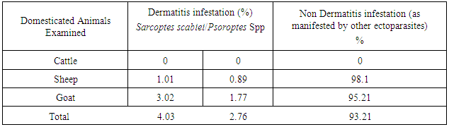
- Result revealed that mites infestations on cattle, goats and sheep were 0%, 6.75%, 14.37% respectively. Sarcoptes scabiei infestation rate in cattle, sheepand goat were 0%, 60.22% and 39.78% respectively. Ticks infestation rate taken in the same order was 47.88%, 42.56% and 21.81% respectively. Fleas infestation rate on cattle, sheep and goat was 0%, 8.92% and 22.25% respectively. Flies infestation rate on the domesticated animals exemplified by cattle, sheep and goat was 36.15%, 24.48% and 16.20% respectively. Lice infestation rate similarly taken in the same order was 15.71%, 16.32% and 25.27%. Psoroptes spp. infestation rate in cattle, sheep and goat were respectively 0%, 0.89%, and 1.77% respectively. Sarcoptes scabiei infestation rate in the same was 0%, 1.01%, and 3.02% respectively. Being the two types of mite, we suggested that its infestation rate for the purpose of this research was 0%. The analysis of the result has shown that cattle were not infested by mites (Table 1). Mites were noted on only goats and sheep. It accounted for about 6.79% of the total number of ectoparasites scored (Table 2). In Sheep, Sarcoptes scabiei were more abundant than Psoroptes spp being 1.01% as against 0.89% respectively. While in Goat, Sarcoptes scabiei were more abundant than Psoroptes spp. Being 3.02% as against 1.01 % respectively. These results are as shown in Table 3.
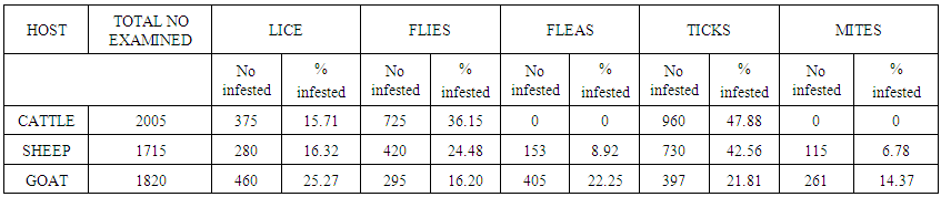 | Table 1. Infestation rates of ectoparasites on domestic animals |
|
|
5. Discussion
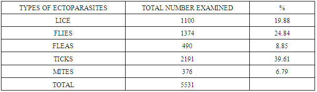
- The ten month investigation revealed that all the animal types had ectoparasites. The ectoparasites were lice, flies, fleas, ticks and mites. It was observed that infestation rates of the arthropods varied amongst the animals examined. Among a total five thousand five hundred and five hundred and thirty one ectoparasites examined, three hundred and seventy six had mites (376/5,531) translating to 6.79 %. In like manner, those that had ticks were Two thousand one hundred and thirty one (2,131/5,571) translating to 39.61%. Those examined for fleas were four hundred and ninety (490/5,531) translating to 8.85%. Those for lice and flies were respectively One thousand three hundred and seventy four (1374/5,531) translating to 24.84% and one thousand one hundred (1,100/5,531) translating to 19.88 % respectively. Overall, mites infestation rates were the lowest when compared to all the other ectoparasites investigated. Details of the methods adopted and the medical and economic importance and implications of these other ectoparasites of these domesticated animals had already been cited and published elsewhere. The presence of Sarcoptes scabiei on goats and sheep were equally reported by [16]. More so, Psoroptes spp. was also noted on goats and sheep. The analysis of the result showed that cattle were not infested by mites in agreement with the work of [19]. Mites were noted only on goats and sheep. Despite this observation, mixed infestations were prominent features of the domesticated animals examined as shown in tables one (1) and two (2). This was similarly cited by [20]. It is therefore suggested that the irritation and dermatitis usually observed in handlers of domesticated animals principally could be associated with those who rear, provide medication, care or farm sheep and goats. Being the two types of mite, we suggested that its infestation rate for the purpose of this research was 0% on cattle. The analysis of the result has shown that cattle were not infested by mites (Table 1). Mites were noted on only goats and sheep. It accounted for about 6.79% of the total number of ectoparasites scored (Table 2). In Sheep, Sarcoptes scabiei were more abundant than Psoroptes spp. being 1.01% as against 0.89% respectively. While in goat, Sarcoptes scabiei were more abundant than Psoroptes spp. being 3.02% as against 1.01 % respectively. These results are as shown in Table 3. We suggest that these ectoparasites rate of causing dermatitis have preferential tropisms in their various domesticated animal hosts. This agreed with the work of Horsefall in 1962. As they were not found in cattle at all.Thus Zoonotic disease associated with mite infestation of domesticated animals could be very rare if not impossible in, custodians, farmers, or veterinary doctors of cattle; as there was significant correlation (P < 0.05) between mites infestation and dermatitis lesions found only on the human handlers of sheep and goats but never on the human handlers of cattle.
6. Conclusions
- While dermatitis on human handlers of cattle, sheep and goats were 0%, 1.89%, and 4.79% respectively. There was significant correlation (P < 0.05) between mites infestation and dermatitis lesions on the human handlers of sheep and goat. Thus Sarcoptes scabiei and Psoroptes spp. that were the only examples of mites studied in this work, infested sheep and goats only and as such were responsible for the dermatitis found in their handlers.