Research in Zoology

p-ISSN: 2325-002X    e-ISSN: 2325-0038

2014;  4(2): 29-42

doi:10.5923/j.zoology.20140402.01

Comparative Osteology of the Jaws in Representatives of the Eurypterygian Fishes

Yazdan Keivany

Department of Natural Resources (Fisheries Division), Isfahan University of Technology, Isfahan, 84156-83111, Iran

Correspondence to: Yazdan Keivany, Department of Natural Resources (Fisheries Division), Isfahan University of Technology, Isfahan, 84156-83111, Iran.

Email:

Copyright © 2014 Scientific & Academic Publishing. All Rights Reserved.

Abstract

The osteology of the jaws in representatives of 49 genera in 40 families of eurypterygian fishes, including: Aulopiformes, Myctophiformes, Lampridiformes, Polymixiiformes, Percopsiformes, Mugiliformes, Atheriniformes, Beloniformes, Cyprinodontiformes, Stephanoberyciformes, Beryciformes, Zeiformes, Gasterosteiformes, Synbranchiformes, Scorpaeniformes (including Dactylopteridae), and Perciformes (including Elassomatidae) were studied. Generally, in this group, the upper jaw consists of the premaxilla, maxilla, and supramaxilla. The lower jaw consists of the dentary, anguloarticular, retroarticular, and sesamoid articular. In higher taxa, the premaxilla bears ascending, articular, and postmaxillary processes. The maxilla usually bears a ventral and a dorsal articular process. The supramaxilla is present only in some taxa. The dentary is usually toothed and bears coronoid and posteroventral processes. The retroarticular is small and located at the posteroventral corner of the anguloarticular.

Keywords: Acanthopterygii, Bone, Eurypterygii, Osteology, Percomprpha

Cite this paper: Yazdan Keivany, Comparative Osteology of the Jaws in Representatives of the Eurypterygian Fishes, Research in Zoology , Vol. 4 No. 2, 2014, pp. 29-42. doi: 10.5923/j.zoology.20140402.01.

1. Introduction

Despite the introduction of modern techniques such as DNA sequencing and barcoding, osteology, due to its reliability, still plays an important role in the systematic study of fishes and comprises a major percent of today’s works [e.g., 1-6]. Eurypterygian fishes composed of several orders including Aulopiformes, Myctophiformes Lampridiformes, Polymixiiformes, Percopsiformes, Mugiliformes, Atheriniformes, Beloniformes, Cyprinodontiformes, Stephanoberyciformes, Beryciformes, Zeiformes, Gasterosteiformes, Synbranchiformes, Scorpaeniformes, Perciformes, Pleuronectiformes and Tetraodontiformes [7]. The diversity of jaw skeleton as the main feeding apparatus is described in this study. Each section describes the jaw osteology of a family. The aims of these sections are to describe and illustrate the variable and significant features of the taxa at higher levels. The systematic of the taxa follows reference [7]. For alternative views on the systematic of this group, also see references [3, 8-17].

2. Materials and Methods

Representatives of the taxa were prepared for dissection following method for clearing and staining bone and cartilage provided in reference [18]. A camera lucida attached to a Wild M5 dissecting stereomicroscope was used to prepare the drawings. The bones in the first figure of each anatomical section are arbitrarily shaded and labeled and in the others are shaded in a consistent manner (dark, medium, and clear) to facilitate comparison among the taxa. Forty nine genera representing 41 families were studied. The number of genera studied in each order are: Aulopiformes (1), Myctophiformes (1), Lampridiformes (2), Polymixiiformes (1), Percopsiformes (1), Mugiliformes (1), Atheriniformes (2), Beloniformes (2), Cyprinodontiformes (2), Stephanoberyciformes (2), Beryciformes (2), Zeiformes (2), Gasterosteiformes (18) Synbranchiformes (2), Scorpaeniformes (including Dactylopteridae) (5), and Perciformes (including Elassomatidae) (5).
Most of the specimens were obtained from the University of Alberta Museum of Zoology (UAMZ). Some were obtained from the Smithsonian Institution (United States National Museum) (USNM), California Academy of Sciences (CAS), and Australian Museum at Sydney (AMS). All the specimens were fixed in 10% formalin and preserved in 70% ethanol. Numbers in parentheses indicate the standard length in millimeters of specimens; those in bold are specimens used for figuring osteology of the taxa. The scale bars in all the figures are representatives of 1 mm.
Aulopiformes
Synodontidae (lizardfishes)
Synodus synodus UAMZ 1806 (147).
Synodus intermedius UAMZ 4889 (78).
Myctophiformes
Myctophidae (lanternfishes)
Myctophum sp. UAMZ 2689 (60, 77, 80).
Lampridiformes
Veliferidae
Velifer hypselopterus AMS 21839005 (101, 115).
Trachipteridae (ribbonfishes)
Trachipterus altivelis CAS 24297 (85), CAS 51177 (105).
Polymixiiformes
Polymixiidae (beardfishes)
Polymixia lowei USNM 159300 (81, 115).
Percopsiformes
Percopsidae (trout-perches)
Percopsis omiscomaycus UAMZ 2048 (17, 19, 27, 55, 60).
Mugiliformes
Mugilidae (mullets)
Mugil sp. UAMZ 5125 (55, 63, 63, 66, 91).
Atheriniformes
Melanotaeniidae (rainbowfishes)
Melanotaenia sp. UAMZ 3526 (40, 42, 51).
Atherinidae (silversides)
Allanetta harringotonensis UAMZ 2673 (47, 58, 59, 60, 61).
Beloniformes
Belonidae (needlefishes)
Pseudotylosurus sp. UAMZ 8165 (173).
Hemiramphidae (halfbeaks)
Arrahamphus sclerolepis UAMZ 3523 (103).
Cyprinodontiformes
Aplocheilidae (rivulines)
Rivulus hartii UAMZ 6660 (44, 47).
Cyprinodontidae (pupfishes)
Cyprinodon nevadensis UAMZ 3114 (24, 34, 41).
Stephanoberyciformes
Stephanoberycidae (pricklefishes)
Stephanoberyx monae USNM 304353 (85, 92).
Rondeletiidae (redmouth whalefishes)
Rondeletia loricata AMS 21141001 (88), AMS 20523001 (34, 37), AMS 18813001 (53).
Beryciformes
Monocentridae (pinecone fishes)
Monocentris sp. UAMZ 7854 (92).
Holocentridae (squirrelfishes)
Sargocentron vexillarium UAMZ 5075 (34, 34, 40, 44, 84).
Zeiformes
Grammicolepididae
Xenolepidichthys dalgleishi USNM 322673 (68, 75).
Caproidae (boarfishes)
Antigonia sp. USNM 266901 (37, 41).
Gasterosteiformes
Hypoptychidae (sand eel)
Hypoptychus dybowskii UAMZ 5550 (75, 80, 81).
Aulorhynchidae (tubesnouts)
Aulorhynchus flavidus: UAMZ 3783 (104, 105, 109).
Aulichthys japonicus UAMZ 5542 (47, 48).
Gasterosteidae (sticklebacks)
Spinachia spinachia UAMZ 6582 (25, 53, 55).
Apeltes quadracus UAMZ 7958 (34, 37, 39).
Gasterosteus aculeatus UAMZ 3894 (32, 57, 58, 58, 73, 77, 83).
Culaea inconstans UAMZ 3797 (21, 30, 49, 56, 57, 58, 60).
Pungitius pungitius UAMZ 3049 (39, 43, 46, 55).
Pegasidae (seamoths)
Pegasus volans UAMZ 4616 (99, 104).
Solenostomidae (ghost pipefishes)
Solenostomus paradoxus AMS 17111002 (51), AMS 17160002 (50), AMS 18852002 (52), AMS 18314001 (59).
Syngnathidae (pipefishes and seahorses)
Syngnathus scovelli UAMZ 3782 (117).
Syngnathus griseolineatus UAMZ 3469 (225, 240, 272).
Hippocampus ingens UAMZ 3594 (170).
Indostomidae (nailfishes)
Indostomus paradoxus UAMZ 6700, CAS 64017 (23, 23, 24, 25).
Aulostomidae (trumpetfishes)
Aulostomus valentini CAS 11979 (139).
Aulostomus sp. CAS 145550 (109, 125).
Aulostomus maculatus CAS 145176 (213), CAS 145549 (171).
Fistulariidae (cornetfishes)
Fistularia petimba UAMZ 6348 (125, 158, 171).
Macroramphosidae (snipefishes)
Macroramphosus scolopax USNM 344398 (94, 99, 99, 100).
Centriscidae (shrimpfishes)
Centriscus scutatus UAMZ 3480 (94, 107).
Aeoliscus strigatus UAMZ 4048 (79, 89).
Synbranchiformes
Synbranchidae (swamp-eels)
Monopterus albus USNM 192939 (193, 245).
Mastacembelidae (spiny eels)
Macrognathus aculeatus UAMZ 1625 (120), UAMZ 1855 (119).
Scorpaeniformes
Dactylopteridae (flying gurnards)
Dactylopterus volitans UAMZ 2633 (61, 74).
Dactyloptena sp. UAMZ 7519 (65).
Scorpaenidae (rockfishes)
Sebastes caurinus UAMZ 3142 (71, 75).
Hexagrammidae (greenling)
Hexagrammos decagrammus UAMZ 3190 (47, 50).
Agonidae (poachers)
Xeneretmus latifrons UAMZ 3196 (90, 95, 143).
Perciformes
Percidae (perches)
Perca flavescens UAMZ 1244 (50, 51, 52, 54, 55).
Cirrhitidae (hawkfishes)
Amblycirrhitus pinos UAMZ 3640 (24, 25, 26, 45, 61).
Elassomatidae (pygmy sunfishes)
Elassoma zonatum UAMZ 6920 (26, 29, 30).
Pomacentridae (damselfishes)
Stegastes partitus UAMZ 3640 (12, 12, 13, 14, 25, 34, 47).
Centrarchidae (sunfishes)
Lepomis gibbosus UAMZ 7715.4 (23, 24, 25, 31, 40).

3. Results

The upper jaw composed of the premaxilla, maxilla, and supramaxilla and The lower jaw consists of the dentary, anguloarticular, retroarticular, and sesamoid articular [19]. In higher taxa, the premaxilla bears ascending, articular, and postmaxillary processes [19]. The maxilla usually bears a ventral and a dorsal articular process. The supramaxilla is present only in some taxa. The dentary is usually toothed and bears coronoid and posteroventral processes. The retroarticular is small and located at the posteroventral corner of the anguloarticular. The terminology of reference [19] for the jaw skeleton was followed, except that the term anguloarticular was used for the angular. In atherinomorphs, what is called the ascending process of premaxilla might be homologous to the articular process of other acanthomorphs, because the ascending process in other acanthomorphs is usually long, thin, pointed, and the rostral cartilage is tightly attached to it. In atherinomorphs the ascending process of the premaxilla is short, thick, round or blunt, like the articular process of other acanthomorphs, and the rostral cartilage is separate from the process. Mugiliformes (Figure 4) have the same upper jaw configuration as atherinomorphs, except that the rostral cartilage is attached to the premaxilla.
Aulopiformes
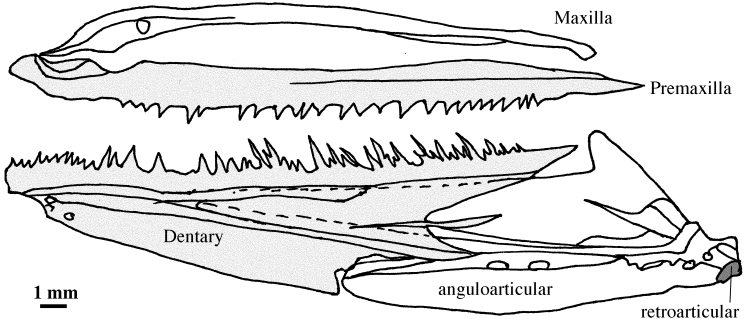
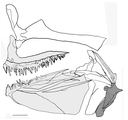
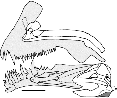
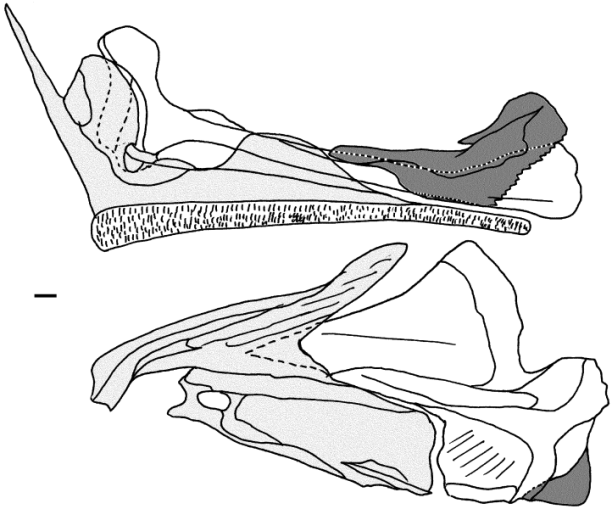
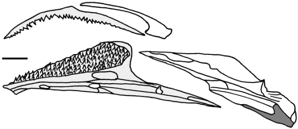
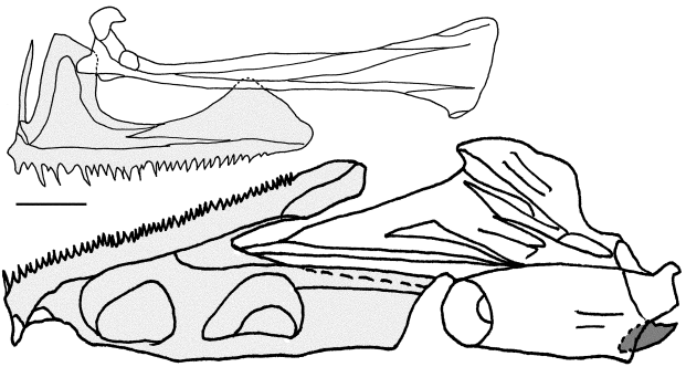
Synodontidae (Figure 1). The premaxilla is toothed and lacks the ascending and postmaxillary processes, but a small knob-like articular process is present. The maxilla is narrow, excluded from the gape by premaxilla, and lacks the articular processes. The dentary is toothed, triangular, well developed, and with a long coronoid process. The anguloarticular is bifurcated anteriorly and the dorsal fork is inserted into the dentary. Based on the literature, in Saurida [20] and Bathysaurus [21], a small supramaxilla is present. In Pseudotrichonotus, the ascending and articular processes of the premaxilla, and the articular and posterior processes of the maxilla are present [20].
Figure 1. Lateral view of the jaws in Synodus synodus (Synodontidae) (UAMZ 1806, 147 mm)
Myctophiformes
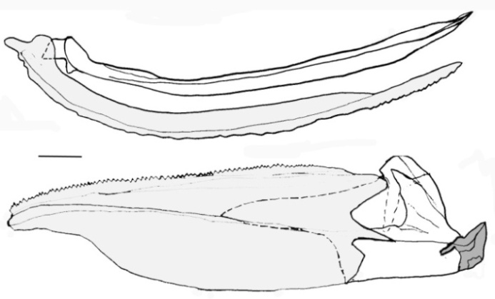
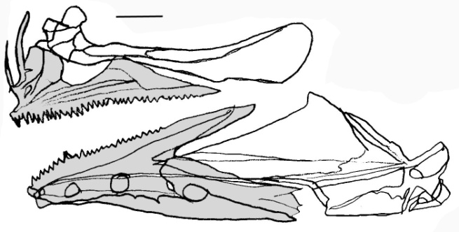
Myctophidae (Figure 2). The premaxilla bears tiny teeth and small bud-like ascending and articular processes. The maxilla is narrow, excluded from the gape by premaxilla, and bears small ventral and dorsal articular processes. The dentary is thinner anteriorly and bears tiny teeth and equal coronoid and posteroventral processes. The anguloarticular is not bifurcated anteriorly. Also see reference [11].
Figure 2. Lateral view of the jaws in Myctophum sp. (Myctophidae) (UAMZ 2689, 60)
Lampridiformes
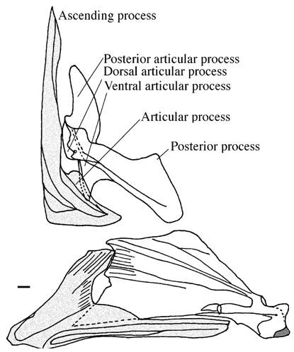
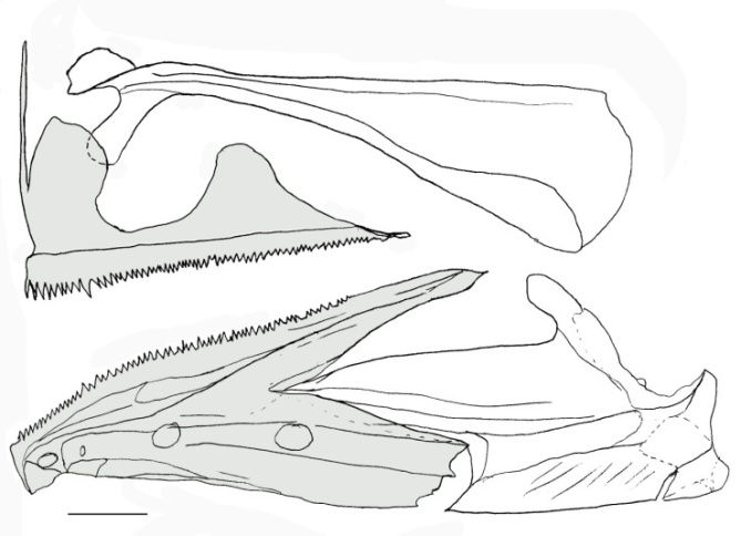
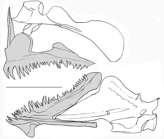
Veliferidae (Figure 3). The premaxilla is toothless and bears well developed ascending and articular processes. The maxilla is broad, not fully excluded from the gape, and bears well developed ventral, dorsal, and posterior articular processes. The dentary is toothless and bears a long posteroventral process. The anguloarticular is not bifurcated anteriorly. Also see reference [22].
Figure 3. Lateral view of the jaws in Velifer hypselopterus (Veliferidae) (AMS 21839005, 101 mm)
Polymixiiformes
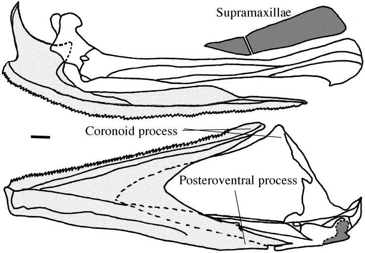
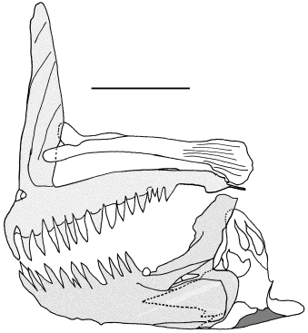
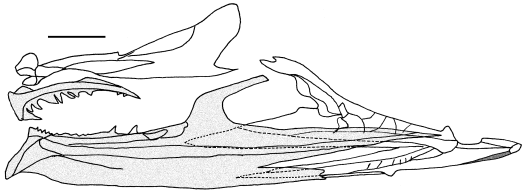
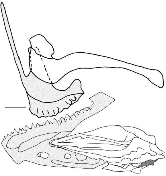
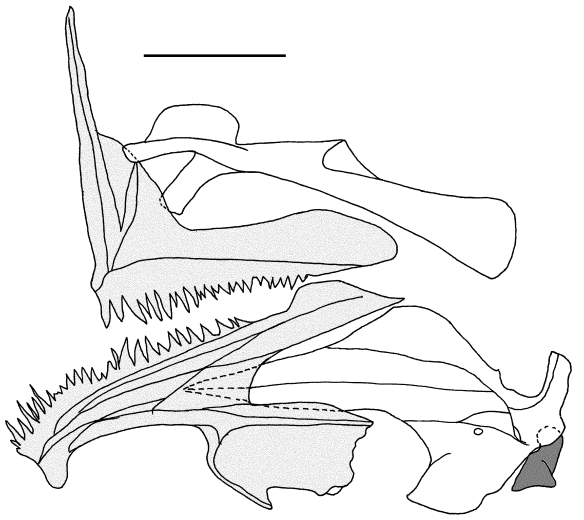
Polymixiidae (Figure 4). The premaxilla bears tiny teeth and short ascending and articular processes. The maxilla is broad and hook-shaped distally, not excluded from the gape, and bears small ventral and dorsal articular processes. One large and one small supramaxilla are present on the dorsal margin of maxilla. The dentary bears tiny teeth and equal coronoid and posteroventral processes. The anguloarticular is not bifurcated anteriorly.
Figure 4. Lateral view of the jaws in Polymixia lowei (Polymixiidae) (USNM 159300, 115 mm)
Percopsiformes
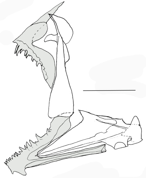
Percopsidae (Figure 5). The premaxilla is toothed and bears an ascending process. The maxilla is narrow, bears ventral and posterior articular processes, a posterior process, and contributes to the gape. The dentary is toothed, highly deep posteriorly, and bears small coronoid and posteroventral processes. The anguloarticular is bifurcated and weakly articulated with the dentary. Also see reference [23].
Figure 5. Lateral view of the jaws in Percopsis omiscomaycus (Percopsidae) (UAMZ 2048, 55 mm)
Mugiliformes
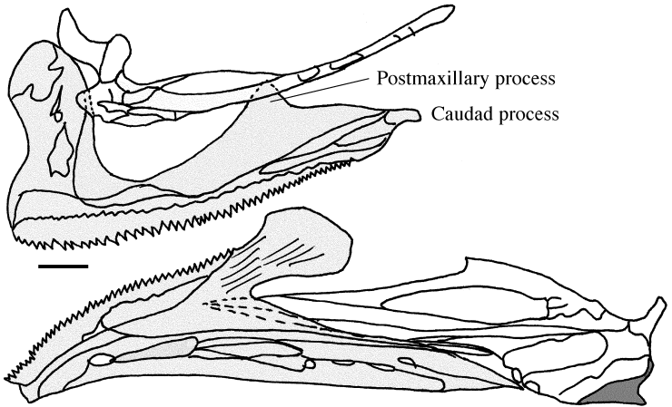
Mugilidae (Figure 6). The premaxilla is toothed and bears the articular and postmaxillary processes and a small caudad process. The maxilla is narrow, not fully excluded from the gape, and bears ventral and dorsal articular processes. The dentary is toothed and its posteroventral process is much longer than the coronoid process. The anguloarticular is not bifurcated and is weakly articulated with the dentary. For further studies on this order, see also reference [24].
Figure 6. Lateral view of the jaws in Mugil sp. (Mugilidae) (UAMZ 5125, 66 mm)
Atheriniformes
Melanotaeniidae (Figure 7). The premaxilla is toothed and bears the articular and postmaxillary processes. The maxilla is narrow, not fully excluded from the gape, and bears ventral and dorsal articular processes and a posterior process. The dentary is toothed and bears equal coronoid and posteroventral processes. The anguloarticular is not bifurcated and is weakly articulated with the dentary. Also see references [25-27].
Figure 7. Lateral view of the jaws in Melanotaenia sp. (Melanotaeniidae) (UAMZ 3526, 51 mm)
Atherinidae (Figure 8). The premaxilla is toothed and bears the articular and postmaxillary processes. The maxilla is narrow, not fully excluded from the gape, and bears ventral and dorsal articular processes. The dentary is toothed and with equal coronoid and posteroventral processes. The anguloarticular is not bifurcated and is weakly articulated with the dentary. For further studies on this order, also see references [28-32].
Figure 8. Lateral view of the jaws in Allanetta harringotonensis (Atherinidae) (UAMZ 2673, 58 mm)
Beloniformes
Belonidae (Figure 9). The premaxilla is elongated, toothed, fused to its counterpart along most of its length, and bears no processes, but is expanded posteriorly. The maxilla is sutured to the premaxilla. The dentary is toothed and with a longer posteroventral process. The anguloarticular is not bifurcated and is tightly articulated with the dentary. Also see references [33, 34]
Figure 9. Lateral view of the jaws in Pseudotylosurus sp. (Belonidae) (UAMZ 8165, 173 mm)
Hemiramphidae (Figure 10). The premaxilla bears tiny teeth and a triangular articular process. The maxilla is narrow, not excluded from the gape, and bears a long ventral articular process and a dorsal articular process. The dentary is toothed and with a longer posteroventral process. The anguloarticular is not bifurcated and is tightly articulated with the dentary. Also see reference [35].
Figure 10. Lateral view of the jaws in Arrhamphus sclerolepis (Hemiramphidae) (UAMZ 3523, 103 mm)
Cyprinodontiformes
Aplocheilidae (Figure 11). The premaxilla is toothed and bears the articular, postmaxillary, and caudad processes. The maxilla is narrow, not excluded from the gape, and bears ventral and dorsal articular processes. The dentary is toothed and with a longer coronoid process. The anguloarticular is bifurcated and weakly articulated with the dentary.
Figure 11. Lateral view of the jaws in Rivulus hartii (Aplocheilidae) (UAMZ 6660, 47 mm)
Cyprinodontidae (Figure 12). The premaxilla is sinusoid, with weakly attached teeth, and bears the articular and caudad processes. The maxilla is narrow, not excluded from the gape, and bears ventral and dorsal articular processes. The dentary is toothed, short and deep, and bears a short coronoid and posteroventral processes. The anguloarticular is slightly bifurcated and weakly articulated with the dentary. For other species of this family, also see reference [36, 37].
Figure 12. Lateral view of the jaws in Cyprinodon nevadensis (Cyprinodontidae) (UAMZ 3114, 34 mm)
Stephanoberyciformes
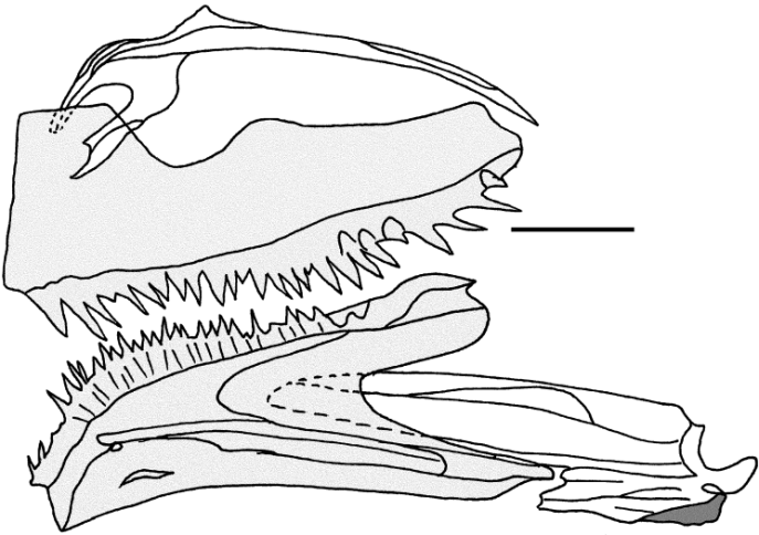
Stephanoberycidae (Figure 13). The premaxilla bears tiny teeth and a short ascending, small articular, and deep postmaxillary process. The maxilla is broad posteriorly, not excluded from the gape, and bears ventral and dorsal articular processes. A broad supramaxilla is present. The dentary is toothed and bears a slightly longer coronoid process. The anguloarticular is not bifurcated and is weakly articulated with the dentary. Also see references [38,39].
Figure 13. Lateral view of the jaws in Stephanoberyx monae (Stephanoberycidae) (USNM 304353, 92 mm)
Rondeletiidae (Figure 14). The premaxilla bears very tiny teeth and a small bud-like articular process. The maxilla is broader posteriorly, not excluded from the gape, and bears short ventral and dorsal articular processes and a posterior process. A relatively large supramaxilla is present. The anguloarticular is highly incorporated into the dentary and hardly distinguishable from that. Also see reference [40].
Figure 14. Lateral view of the jaws in Rondeletia loricata (Rondeletiidae) (AMS 20523001, 37 mm)
Beryciformes
Monocentridae (Figure 15). The premaxilla bears very tiny teeth and the ascending, articular, and postmaxillary processes. The maxilla is broader posteriorly and bears short ventral, dorsal, and posterior articular processes and a small posterior process. A large supramaxilla is present. The anguloarticular is not bifurcated anteriorly. Also see reference [41].
Figure 15. Lateral view of the jaws in Monocentris sp. (Monocentridae) (UAMZ 7854, 92 mm)
Holocentridae (Figure 16). The premaxilla is toothed and bears the ascending, articular, and postmaxillary processes. The maxilla is broad posteriorly, much longer than the premaxilla, and bears ventral and elongated dorsal and posterior articular processes. A large and a small supramaxilla are present. The dentary bears a longer coronoid process. The anguloarticular is not bifurcated anteriorly. Also see reference [42, 43].
Figure 16. Lateral view of the jaws in Sargocentron vexillarium (Holocentridae) (AMS 5075, 44 mm)
Zeiformes
Grammicolepididae (Figure 17). The premaxilla is short, bears tiny teeth and a long ascending, an articular, and a posterior process. The maxilla is broad, longer than the premaxilla and bears a ventral and an enlarged dorsal and a posterior process. The dentary is triangular, toothed, and not distinctly bifurcated. The anguloarticular is not bifurcated and is fully inserted in the dentary.
Figure 17. Lateral view of the jaws in Xenolepidichthys dalgleishi (Grammicolepididae) (USNM 322673, 68 mm)
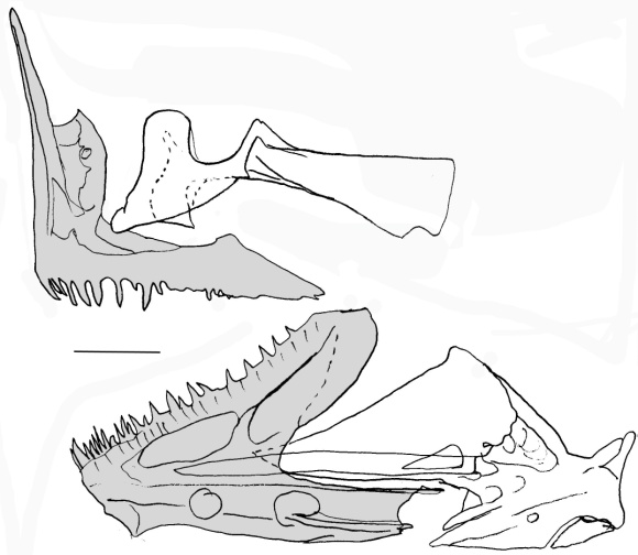
Caproidae (Figure 18). The premaxilla is short, toothed proximally, and bears the ascending, articular, and postmaxillary processes. The maxilla is broad and bears the ventral and dorsal articular and posterior processes. The dentary bears a longer posteroventral process. The anguloarticular is not bifurcated and is fully coupled with the dentary. In Capros aper, the articular process of the premaxilla is not distinct [10]. For this order, see also references [44-46].
Figure 18. Dorsal view of the upper jaw and lateral view of the lower jaw in Aulorhynchus flavidus (Caproidae) (UAMZ 3783, 109 mm)
Gasterosteiformes
Hypoptychidae (Figure 19). The premaxilla is toothed in males, but toothless in females, and bears a long cylindrical ascending and a postmaxillary process. The maxilla is longer than the premaxilla and bears the ventral and dorsal articular processes. The anguloarticular is not bifurcated and is fully inserted in the dentary. This species is also studied by reference [47].
Figure 19. Lateral view of the jaws in Hypoptychus dybowskii (Hypoptychidae) (UAMZ 5550, 80 mm)
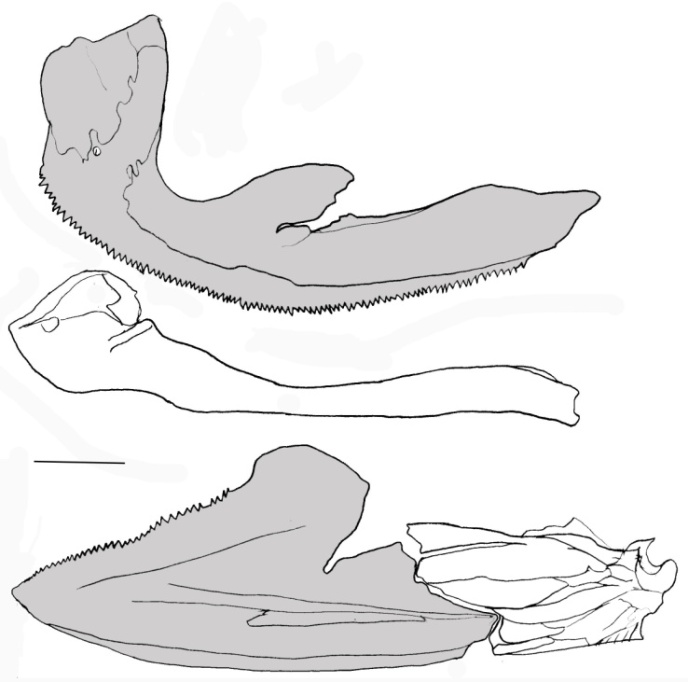
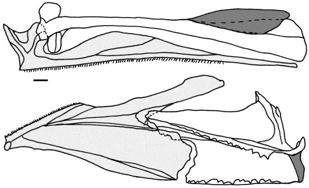
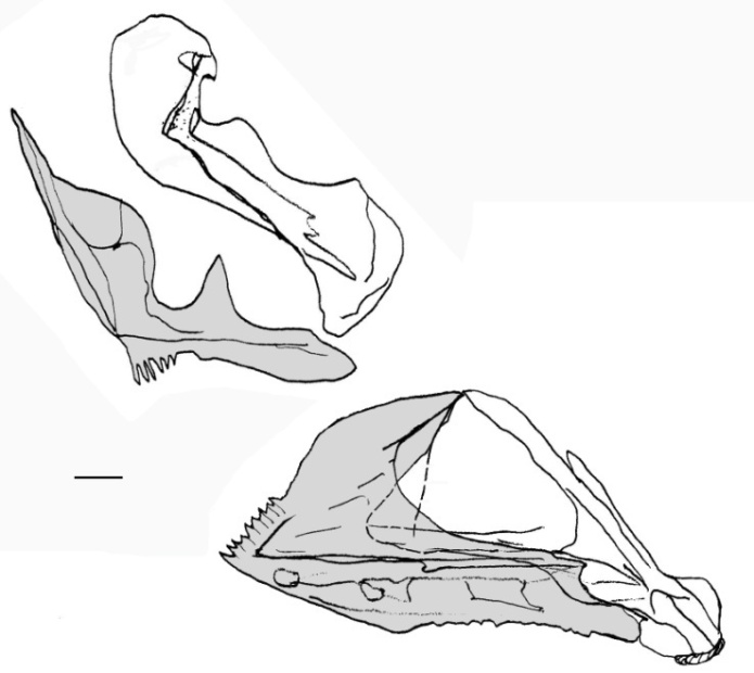
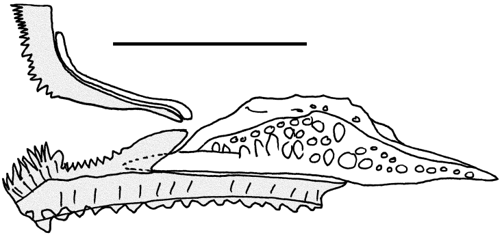
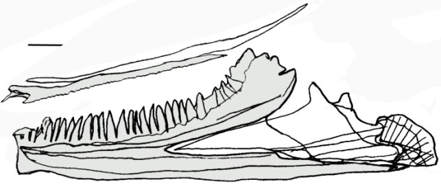
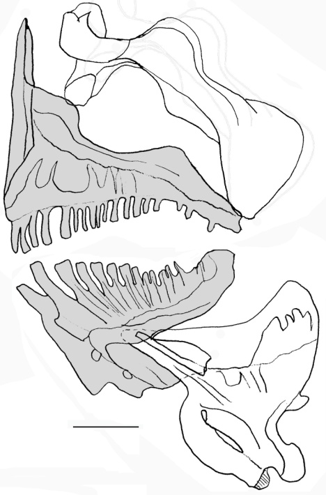
Aulorhynchidae (Figure 20). The premaxilla is well developed and toothed in males, but toothless or poorly toothed in females [40], and bears a long, strong, and pointed ascending process. The postmaxillary process of the premaxilla is present in Aulichthys japonicus, but absent in Aulorhynchus flavidus. The maxilla is longer than the premaxilla and bears the ventral and dorsal articular processes. The anguloarticular is not bifurcated and is fully inserted in the dentary. For this family, also see reference [52]
Figure 20. Dorsal view of the upper jaw and lateral view of the lower jaw in Aulorhynchus flavidus (Aulorhynchidae) (UAMZ 3783, 109 mm)
Gasterosteidae (Figure 21). The premaxilla is toothed and bears a long and strong ascending process. In the ascending process of juveniles there is a line that might indicate the fusion of the articular and ascending processes, but there is no distinct articular process. The maxilla bears the ventral and dorsal articular processes. The anguloarticular is not bifurcated and is fully inserted in the dentary. For this family, also see references [4-6, 48-54].
Figure 21. Lateral view of the jaws in Apeltes quadracus (Gasterosteidae) (UAMZ 7958, 37 mm)
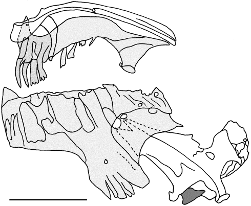
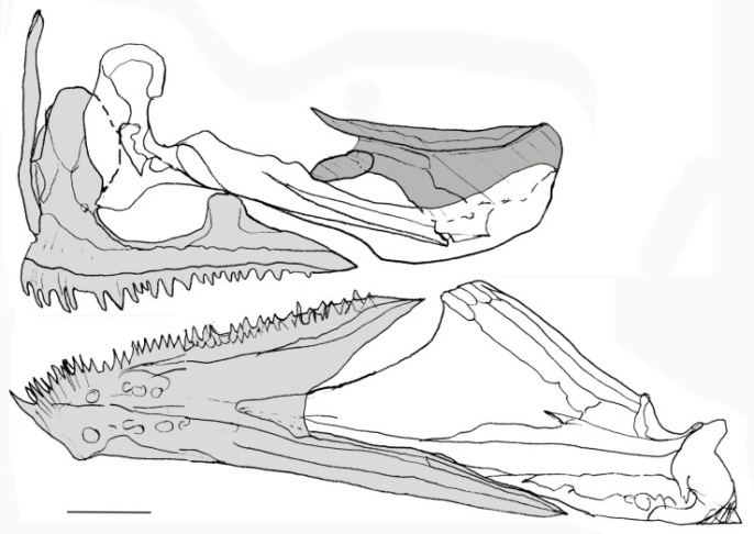
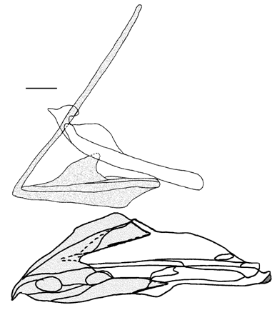
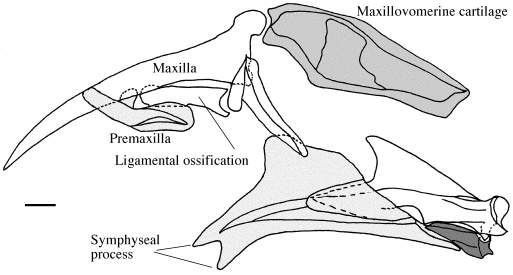
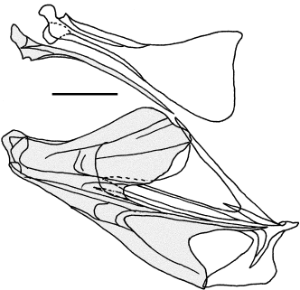
Pegasidae (Figure 22). The terminology for the unusual jaws of Pegasidae follows reference [55] that studied the osteology of this family. The premaxilla is toothless and bears a small articular process that connects it to the maxilla by a ligamental ossification. The maxilla is triradiate and its posterior process connected to the vomer by the ossified maxillovomerine cartilage and is longer than premaxilla. The dentary is not bifurcated posteriorly, the coronoid process is not distinct, the posteroventral process is long, and the symphyseal process is well developed. The anguloarticular is not bifurcated, but bears a large coronoid process and is tightly connected to the dentary. The retroarticular is relatively large.
Figure 22. Lateral view of the jaws in Pegasus volans (Pegasidae) (UAMZ 4616, 99 mm)
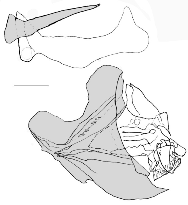
Figure 23. Lateral view of the jaws in Solenostomus paradoxus (Solenostomidae) (AMS 17111002, 51 mm)
Solenostomidae (Figure 23). The premaxilla is toothless and bears a small articular process. The maxilla is much broader posteriorly, longer than the premaxilla, and bears small ventral and dorsal articular processes. The dentary is trifurcated posteriorly. The anguloarticular is bifurcated and tightly connected to the dentary.
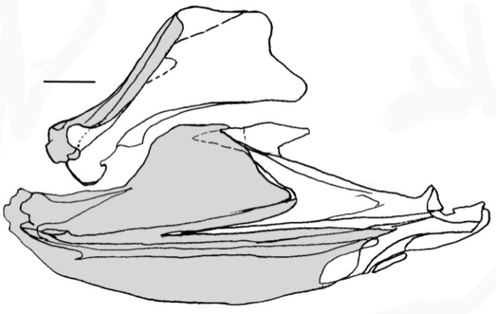
Syngnathidae (Figure 24). The premaxilla is toothless and bears a small articular process. The maxilla is broad, longer than the premaxilla, and bears a small articular process. The dentary is much broader posteriorly. The anguloarticular is not bifurcated and is tightly connected to the dentary.
Figure 24. Lateral view of the jaws in Hippocampus ingens (Syngnathidae) (UAMZ 3594, 170 mm)
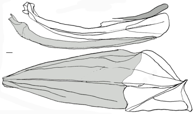
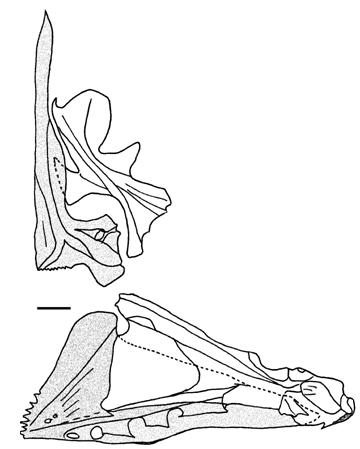
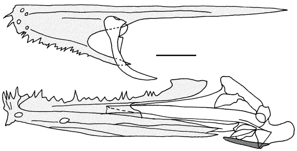
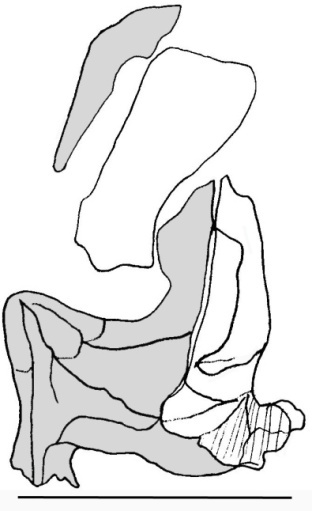
Indostomidae (Figure 25). The premaxilla is toothed and bears no distinct processes. The maxilla is thread-like and broader posteriorly, shorter than the premaxilla, not reaching the midline, and with a tiny ventral articular process. The dentary bears a longer posteroventral process. The anguloarticular is not bifurcated anteriorly. The osteology of this species was also studied by reference [56, 57].
Figure 25. Dorsal view of the upper jaw and lateral view of the lower jaw in Indostomus paradoxus (Indostomidae) (CAS 64017, 25 mm)
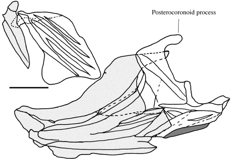
Aulostomidae (Figure 26). The premaxilla is toothless and poorly developed and bears a small articular process. The maxilla is broader distally and bears small dorsal and ventral articular processes. The anguloarticular is bifurcated and tightly connected to the dentary and bears a posterocoronoid process.
Figure 26. Lateral view of the jaws in Aulostomus valentini (Aulostomidae) (CAS 11979, 139 mm)
Fistulariidae (Figure 27). The premaxilla is toothed and poorly developed and bears small articular and postmaxillary processes. The maxilla is broad, much longer than the premaxilla, and bears small dorsal and ventral articular processes. The dentary is trifurcated posteriorly. The anguloarticular is bifurcated and tightly connected to the dentary.
Figure 27. Lateral view of the jaws in Fistularia petimba (Fistulariidae) (UAMZ 6348, 158 mm)
Macroramphosidae (Figure 28). The premaxilla is toothless, poorly developed, and bears a small articular process. The maxilla is broad, longer than the premaxilla, and bears small articular processes. The dentary is deep and bears a well developed coronoid process. The anguloarticular is bifurcated and tightly connected to the dentary and bears a posterocoronoid process.
Figure 28. Lateral view of the jaws in Macroramphus scolopax (Macroramphosidae) (USNM 344398, 99 mm)
Centriscidae (Figure 29). The premaxilla is toothless and poorly developed and bears a small articular process. The maxilla is broad, longer than the premaxilla, and bears no distinct processes. The dentary bears an upright coronoid and a posteroventral process. The anguloarticular is upright, not bifurcated and is tightly connected to the dentary. The osteology of this family is also studied by reference [58]. For this order, also see references [47-59].
Figure 29. Lateral view of the jaws in Aeoliscus strigatus (Aulostomidae) (UAMZ 4048, 89 mm)
Synbranchiformes
Synbranchidae (Figure 30). The premaxilla is toothed, narrow, and bears only a small articular process. The maxilla is broader posteriorly, longer than the premaxilla, and bears no processes. The anguloarticular is not bifurcated, but fully coupled with the dentary.
Figure 30. Lateral view of the jaws in Monopterus albus (Synbranchidae) (USNM 192939, 193 mm)
Mastacembelidae (Figure 31). The premaxilla is toothed but poorly developed and bears no processes. The maxilla is broader and hook shaped posteriorly, much shorter than the premaxilla, and bears no processes. The anguloarticular is not bifurcated and not fully coupled with the dentary. For this order, also reference [60].
Figure 31. Lateral view of the jaws in Macrognathus aculeatus (Mastacembelidae) (UAMZ 1855, 119 mm)
Scorpaeniformes
Dactylopteridae (Figure 32). The premaxilla is toothed and bears long ascending and articular processes. The maxilla is much longer than premaxilla and bears the ventral and dorsal articular processes. The coronoid and posteroventral processes of the dentary are widely separated from each other. The anguloarticular is not bifurcated anteriorly and does not bear a distinct coronoid process. Also see references [61, 62].
Figure 32. Lateral view of the jaws in Dactylopterus volitans (Dactylopteridae) (UAMZ 2633, 74 mm)
Scorpaenidae (Figure 33). The premaxilla is toothed and bears the ascending, articular, and postmaxillary processes. The maxilla is broader posteriorly, longer than the premaxilla, and bears the ventral and dorsal articular processes. The anguloarticular is not bifurcated and not fully coupled with the dentary.
Figure 33. Lateral view of the jaws in Sebastes caurinus (Scorpaenidae) (UAMZ 3142, 75 mm)
Hexagrammidae (Figure 34). The premaxilla is toothed and bears the ascending, articular, and postmaxillary processes. The maxilla is broader posteriorly, much longer than the premaxilla, and bears ventral and dorsal articular processes. The anguloarticular is not bifurcated and not fully coupled with the dentary. Also see reference [63].
Figure 34. Lateral view of the jaws in Hexagrammos decagrammus (Hexagrammidae) (UAMZ 3190, 50 mm)
Agonidae (Figure 35). The premaxilla is toothed and bears a short ascending and a well developed articular process. The maxilla is broader posteriorly, much longer than the premaxilla, and bears ventral and dorsal articular processes. The anguloarticular is not bifurcated and not fully coupled with the dentary and bears a large tube. Also see reference [64].
Figure 35. Lateral view of the jaws in Xeneretmus latifrons (Agonidae) (UAMZ 3196, 95 mm)
Perciformes
Percidae (Figure 36). The premaxilla is toothed and bears a small ascending, an articular, and a postmaxillary process. The maxilla is broader posteriorly, much longer than the premaxilla, and bears ventral and dorsal articular processes. The anguloarticular is not bifurcated and not fully coupled with the dentary. Also see reference [65-68].
Figure 36. Lateral view of the jaws in Perca flavescens (Percidae) (UAMZ 1244, 54 mm)
Cirrhitidae (Figure 37). The premaxilla is toothed and bears the ascending, articular, and postmaxillary processes. The maxilla is broader posteriorly, longer than the premaxilla, and bears the ventral and dorsal articular processes and a posterior process. The anguloarticular is not bifurcated and not fully coupled with the dentary.
Figure 37. Lateral view of the jaws in Amblycirrhitus pinos (Cirrhitidae) (UAMZ 3640, 45 mm)
Elassomatidae (Figure 38). The premaxilla is toothed and bears the ascending and articular processes. The maxilla is broader posteriorly, longer than the premaxilla and bears the ventral and dorsal articular processes and a posterior process. The anguloarticular is bifurcated but not fully coupled with the dentary. This species was also studies by others [69, 70].
Figure 38. Lateral view of the jaws in Elassoma zonatum (Elassomatidae) (UAMZ 6920, 30 mm)
Pomacentridae (Figure 39). The premaxilla is toothed and bears the ascending, articular, and postmaxillary processes. The maxilla is broader posteriorly and bears the ventral and dorsal articular processes and a posterior process. The dentary bears a longer posteroventral process. The anguloarticular is large and forms most of the ventral jaw, is not bifurcated and not fully coupled with the dentary. Also see referenes [71-73].
Figure 39. Lateral view of the jaws in Stegastes partitus (Pomacentridae) (UAMZ 3640, 34 mm)
Centrarchidae (Figure 40). The premaxilla is toothed and bears a long ascending, an articular, and a postmaxillary process. The maxilla is broad posteriorly, longer than the premaxilla, and bears ventral and dorsal articular processes and a posterior process. The anguloarticular is bifurcated anteriorly and not fully coupled with the dentary. For osteology of other perciform fishes, also see references [74-76].
Figure 40. Lateral view of the jaws in Lepomis gibbosus (Centrarchidae) (UAMZ 7715.4, 40 mm)

4. Discussion

Euteleosts are characterized by a protrusible jaw, which is intensified by development of the premaxillary process in members of higher taxa [10]. However, the ascending process is secondarily reduced or lost in several groups including Syngnathoidei. Most Syngnathoidei have an elongated snout and a small mouth at the end of the tube-like snout. The elongated vomer extends forward superficially and fills most of the space in the ethmoid region and leaves little or no space for a well developed ascending process. This modification of the jaws caused a secondary reduction or loss of the process in this group. In Mugiliformes and Atherinomorpha, probably due to the mode of feeding and shortening of the snout, the ascending process is lost or fused to the articular process. However, there is a disagreement on the homology of the premaxillary process. The ascending process is generally defined as the process that is in contact with rostral cartilage and the articular process as the one in contact with the maxilla [36]. Reference [17] believed that atherinomorphs do not bear a true ascending process and alternatively, reference [29] believed that the process is the ascending process. Reference [36], based on presence of distinct ascending and articular processes in some primitive atherinomorphs such as Bedotiidae and Melanotaeniidae, argued that the process is the ascending process and the articular process is secondarily lost in other atherinomorphs. In Melanotaeniidae (Figure 7) and Bedotiidae [28], there are large postmaxillary and articular processes but not any other distinct process. In Phallostethidae [36], there is a relatively distinct long process but as in Atherinopsinae [30], it is not positioned at the proximal end of the premaxilla (unlike in other acanthopterygians) and it is in contact with the maxilla, not the rostral cartilage. Besides, this process in Phallostethidae, as derived atheriniforms [31], might be secondary. Although differentiated ascending and articular process are not found in the ontogeny of atherinomorphs [32], it is conceivable that the ascending and articular processes are fused together in some atherinomorphs; however, the configuration of the upper jaw in Mugiliformes and Atherinomorpha is different from that of other acanthopterygians.

5. Conclusions

Generally, in this group, the upper jaw consists of the premaxilla, maxilla, and supramaxilla. The lower jaw consists of the dentary, anguloarticular, retroarticular, and sesamoid articular. In higher taxa, the premaxilla bears ascending, articular, and postmaxillary processes. The maxilla usually bears a ventral and a dorsal articular process. The supramaxilla is present only in some taxa. The dentary is usually toothed and bears coronoid and posteroventral processes. The retroarticular is small and located at the posteroventral corner of the anguloarticular.

ACKNOWLEDGEMENTS

I would like to express my gratitude to my late supervisor, Dr. J.S. Nelson, Dr. P.R. Willoughby, Dr. M.V.H. Wilson, Dr. B.S. Heming, Dr. G. Goss and Dr. L.R. Parenti for providing helpful comments and advice during the course of this study. I wish to extend my sincere appreciation to Mr. W.E. Roberts for lending museum materials and Mr. R. Mandryk for preparing stains and other chemical solutions, the curators and staff of the Smithsonian Institution, California Academy of Sciences, and Australian Museum at Sydney for lending their fish specimens. This study was financially supported by a MCHE scholarship to the author and an NSERC grant to Dr. J.S. Nelson and Isfahan University of Technology.

References

[1]  S. Eagderi and D. Adriaens, 2014, Cephalic morphology of Ariosoma gilberti (Bathymyrinae: Congridae). Iran. J. Ichthyol., 1(1): 39-50.
[2]  Y. Keivany, 2014, Comparative osteology of the suspensorial and opercular series in representatives of the eurypterygian fishes. Iran. J. Ichthyol., 1(2): 73-89.
[3]  M. L. J. Stiassny and J. A. Moore, 1992, A review of the pelvic girdle of acanthomorph fishes, with comments of hypotheses of acanthomorph intrarelationships. Zool. J. Linn. Soc., 104: 209-242.
[4]  Y. Keivany, and J. S. Nelson, 2004, Phylogenetic relationships of sticklebacks (Gasterosteidae), with emphasis on ninespine sticklebacks (Pungitius spp.). Behaviour, 141(11/12): 1485-1497.
[5]  Y. Keivany, and J. S. Nelson, 2006. Interrelationships of Gasterosteiformes (Actinopterygii, Percomorpha). J. Ichthyol., 46(suppl. 1): S84-S96.
[6]  Y. Keivany, J. S. Nelson, and P. S. Economidis, 1997, Validity of Pungitius hellenicus, a stickleback fish from Greece. Copeia, 1997(3): 558-564.
[7]  J. S. Nelson, 2006, Fishes of the world. Second edition. John Wiley & Sons, New York. 600 pp.
[8]  C. Patterson, and G. D. Johnson, 1995. The intermuscular bones and ligaments of Teleostean fishes. Smithson. Contrib. Zool., 1-83.
[9]  G. V. Lauder, 1982, Patterns of evolution in the feeding mechanism of actinopterygian fishes. Amer. Zool., 22(2): 275-285.
[10]  D. E. Rosen, 1985, An essay on Euteleostean classification. Am. Mus. Nov., (2827): 1-57.
[11]  M. L. J. Stiassny, 1996, Basal ctenosquamate relationships and the interrelationships and the interrelationships of the myctophiform (Scopelomorph) fishes, p. 405-426. In: Interrelationships of fishes. M. L. J. Stiassny, L. R. Parenti, and G. D. E. Johnson (eds.). Academic Press, San Diego.
[12]  G. J. Nelson, 1969, Gill arches and the phylogeny of fishes, with notes on the classification of vertebrates. Bull. Am. Mus. Nat. Hist., 141: 475-552.
[13]  G. V. Lauder, and K. F. Liem, 1983, The evolution and interrelationships of the actinopterygian fishes. Bull. Mus. Comp. Zool., 150: 95-197.
[14]  D. E. McAllister, 1968, The evolution of branchiostegals and classification of teleostome fishes. Bull. Natl. Mus. Can., (221): 1-239.
[15]  H. K. Mok, 1983, Osteology and phylogeny of Squamipinnes. Taiwan Mus. Spec. Publ. Ser., (1): 1-87.
[16]  G. D. Johnson, and C. Patterson. 1993. Percomorph phylogeny: A survey of Acanthomorphs and a new proposal. Bull. Mar. Sci., 52: 554-626.
[17]  P. H. Greenwood, D.E. Rosen, S. H. Weitzman, and G. S. Myers, 1966, Phyletic studies of teleostean fishes, with a provisional classification of living forms. Bull. Am. Mus. Nat. Hist., 131: 339-456.
[18]  W. R. Taylor, and G. C. Van Dyke, 1985, Revised procedures for staining and clearing small fishes and other vertebrates for bone and cartilage study. Cybium, 9: 107-119.
[19]  A. L. Rojo, 1991, Dictionary of evolutionary fish osteology. CRC Press, London. pp.
[20]  G. D. Johnson, C.C. Baldwin, M. Okiyama, and Y. Tominaga. 1996, Osteology and relationships of Pseudotrichonotus altivelis (Teleostei: Aulopiformes: Pseudotrichonotidae). Ichthyol. Res., 43: 17-45.
[21]  C. C. Baldwin, and G. D. Johnson, 1996, Interrelationships of Aulopiformes, p. 355-404. In: Interrelationships of fishes. M. L. J. Stiassny, L. R. Parenti, and G. D. Johnson (eds.). Academic Press, San Diego.
[22]  Carnevale, G., 2004, The first fossil ribbonfish (Teleostei, Lampridiformes, Trachipteridae). Geol. Mag., 141(05), 573-582.
[23]  H. D. Reed, 1903, The Cranial Osteology and Systematic Position of the Family Percopsidae. PhD dissertation, Cornell University.
[24]  M. L. J. Stiassny, 1993, What are grey mullets? Bull. Mar. Sci., 52: 197-219.
[25]  J. M. Patten, and W. Ivantsoff, 1983, A new genus and species of atherinid fish, Dentatherina merceri from the western Pacific. Jpn. J. Ichthyol., 29: 329-339.
[26]  G. R. Allen, (1983). Kiunga ballochi, a new genus and species of rainbowfish (Melanotaeniidae) from Papua New Guinea. Trop. Fish Hobb., 32(2): 72-77.
[27]  G. R. Allen, 1998, A new genus and species of Rainbowfish (Melanotaeniidae) from fresh waters of Irian Jaya, Indonesia. Rev. Fr. Aquariol., 25(1-2): 11-16.
[28]  M. L. J. Stiassny, 1990. Notes on the anatomy and relationships of the Bedotiid fishes of Madagascar, with a taxonomic revision of the genus Rheocles (Atherinomorpha: Bedotiidae). Am. Mus. Nov., (2979): 1-34.
[29]  R. M. Alexander, 1967, Mechanisms of the jaws of some atheriniform fish. J. Zool., 151: 233-255.
[30]  B. S. Dyer, 1997, Phylogenetic revision of Atherinopsinae (Teleostei, Atherinopsidae), with comments on the systematics of the South American freshwater fish genus Basilichthys Girard. Misc. Publ. Mus. Zool. Univ. Mich., (185): 1-64.
[31]  B. S. Dyer, and B. Chernoff, 1996, Phylogenetic relationships among atheriniform fishes (Teleostei: Atherinomorpha). Zool. J. Linn. Soc., 117: 1-69.
[32]  R. M. Langille, and B. K. Hall, 1987, Development of the head skeleton of the Japanese medaka, Oryzias latipes (Teleostei). J. Morphol., 193: 135-158.
[33]  S. Kužir, Z. Kozarić, E. Gjurčević, B. Baždarić, Z. Petrinec, 2009, Osteological development of the garfish (Belone belone) larvae. Anat. Histol. Embryo., 38(5): 351-354.
[34]  B. B. Collette, 1966, Belonion, a new genus of fresh-water needlefishes from South America. Amer. Mus. Nov., (2274).
[35]  P. Vandewalle, V. Lambert and E. Parmentier, 2002, Particularities of the bucco-pharyngeal apparatus in Zenarchopterus kampeni (Pisces: Hemiramphidae) and their probable significance in feeding. Belg. J. Zool., 132(2).
[36]  L. R. Parenti, 1984, On the relationships of Phallostethid fishes (Atherinomorpha), with notes on the anatomy of Phallostethus dunckeri Regan, 1913. Am. Mus. Nov., (2779): 1-12.
[37]  L. R. Parenti, 1981, A phylogenetic and biogeographic analysis of Cyprinodontiform fishes (Teleostei, Atherinomorpha). Bull. Am. Mus. Nat. Hist., 168: 335-557.
[38]  Y. Keivany, 2003, Superfacial skull bones in Zagros toothcarp, Aphanius vladykovi (Cyprinodontidae). Iran. J. Biol., 15(3): 25-30.
[39]  A. N. Kotlyar, 1996, The osteology, intraspecific structure, and distribution of Rondeletia loricata (Rondeletiidae). J. Ichthyol., 36: 207-221.
[40]  A. N. Kotlyar, 1991, Osteology of fish of the suborder Stephanoberycoidei. 2. Melamphaidae. J. Ichthyol., 31: 100-116.
[41]  A. N. Kotlyar, 1991b, Osteology of the suborder Stephanoberycoidei. Communication 1. The Stephanoberycidae and Gibberichthyidae. J. Ichthyol., 31: 18-32.
[42]  S. J. Zehren, 1979, The comparative Osteology and Phylogeny of the Beryciformes (Pisces, Teleostei). Evol. Monog., 1–389.
[43]  A. N. Kotlyar, 1992, Osteology of Sorosichthys ananassa and its position in the systematics of Beryciform fishes (Beryciformes). J. Ichthyol., 32: 56-68.
[44]  D. E. Rosen, 1984, Zeiforms as primitive plectognath fishes. Am. Mus. Nov., (2782): 1-45
[45]  J. C. Tyler, B. O'toole, and R. Winterbottom, 2003, Phylogeny of the genera and families of zeiform fishes, with comments on their relationships with tetraodontiforms and caproids. Smithsonian Institution Press.
[46]  F. Santini, J. C. Tyler, A. F. Bannikov, and D. S. Baciu, 2006, A phylogeny of extant and fossil buckler dory fishes, family Zeidae (Zeiformes, Acanthomorpha). Cybium, 30(2): 99-107.
[47]  H. Ida, 1976, Removal of the family Hypoptychidae from the suborder Ammodytoidei, order Perciformes, to the suborder Gasterosteoidei, order Syngnathiformes. Jpn. J. Ichthyol., 23: 33-42.
[48]  P. S. Bowne, 1985, The systematic position of Gasterosteiformes. PhD dissertation. Department of Zoology. University of Alberta, Edmonton, 461 pp.
[49]  P. S. Bowne, 1994, Systematics and morphology of the Gasterosteiformes, p. 28-60. In: Evolutionary biology of the threespine stickleback. M. A. Bell and S. A. Foster (eds.). Oxford University Press, Oxford.
[50]  Y. Keivany, 1996, Taxonomic revision of the genus Pungitius with emphasis on P. hellenicus. MSc. thesis. Department of Biological Sciences. University of Alberta, Edmonton. 98 pp.
[51]  Y. Keivany, 2000, Phylogenetic relationships of Gasterosteiformes (Teleostei, Percomorpha). PhD. dissertation. Department of Biological Sciences. University of Alberta, Edmonton. 201 pp.
[52]  J. S. Nelson, 1971, Comparison of the pectoral and pelvic skeleton and of some other bones and their phylogenetic implications in the Aulorhynchidae and Gasterosteidae (Pisces). J. Fish. Res. Bd. Can., 28: 427-442.
[53]  Y. Keivany, and J.S. Nelson, 1998. Comparative osteology of the Greek ninespine stickleback, Pungitius hellenicus (Teleostei, Gasterosteidae). J. Ichthyol., 38: 430-440.
[54]  Y. Keivany, and J. S. Nelson, 2000, Taxonomic review of the genus Pungitius, ninespine sticklebacks (Teleostei, Gasterosteidae). Cybium, 24(2): 107-122.
[55]  T. W. Pietsch, 1978. Evolutionary relationships of the sea moths (Teleostei: Pegasidae) with a classification of gasterosteiform families. Copeia, 1978: 517-529.
[56]  K. E. Banister, 1970, The anatomy and taxonomy of Indostomus paradoxus Prashad & Mukerji. Bull. Br. Mus. Nat. Hist. (Zool.), 19: 179-209.
[57]  R. Britz, and G. D. Johnson, 2002, “Paradox Lost”: Skeletal ontogeny of Indostomus paradoxus and its significance for the phylogenetic relationships of Indostomidae (Teleostei, Gasterosteiformes). Amer. Mus. Nov., 1-43.
[58]  E. Mohr, 1937, Revision der Centriscidae (Acanthopterygii, Centrisciformes. Dana Report, 13K.
[59]  J. W. Orr, 1995, Phylogenetic relationships of Gasterosteiform fishes (Teleostei: Acanthomorpha). PhD dissertation. Department of Zoology. University of Washington, Seattle, 813 pp.
[60]  R. Britz, and M. Kottelat, 2003, Descriptive osteology of the family Chaudhuriidae (Teleostei, Synbranchiformes, Mastacembeloidei), with a discussion of its relationships. Amer. Mus. Nov., 1-62.
[61]  H. Imamura, 2000, An alternative hypothesis on the phylogenetic position of the family Dactylopteridae (Pisces: Teleostei), with a proposed new classification. Ichthyol. Res., 47(3-4): 203-222.
[62]  W. N. Eschmeyer, 1997, A new species of Dactylopteridae (Pisces) from the Philippines and Australia, with a brief synopsis of the family. Bull. Mar. Sci., 60(3): 727-738.
[63]  G. Shinohara, 1994, Comparative morphology and phylogeny of the suborder Hexagrammoidei and related taxa (Pisces: Scorpaeniformes). Mem. Fac. Fish. Hokk. Univ., 41: 1-97.
[64]  T. Kanayama, 1991, Taxonomy and phylogeny of the family Agonidae (Pisces: Scorpaeniformes). Mem. Fac. Fish. Hokkaido Univ., 38: 1-199.
[65]  S. M. Norris, 2001, Osteology of the southwestern darters, Ethostoma (Oligocephalus) (Teleostei, Percidae)-with comparison to other North American percid fishes.
[66]  M. J. Elshoud-Oldenhave, and J. W. Osse, 1976, Functional morphology of the feeding system in the ruff-Gymnocephalus cernua (L. 1758)-(Teleostei, Percidae). J. Morphol., 150(2): 399-422.
[67]  J. C. Bruner, 2011, A phylogenetic analysis of Percidae using osteology. Biology, management, and culture of walleye and sauger. Bethesda, MD: American Fisheries Society, 5-84.
[68]  A. E. Spreitzer, 1979, The life history, external morphology, and osteology of the eastern sand darter, Ammocrypta pellucida (Putnam, 1863), an endangered Ohio species (Pisces: Percidae). PhD dissertation, The Ohio State University.
[69]  G. D. Johnson, and V. G. Springer, 1997, Elassoma: another look, p. 176. In: Abstracts of the 77th ASIH annual meeting, University of Washington, Seattle.
[70]  W. J. Jones, and J. M. Quatrro, 1999, Phylogenetic affinities of pygmy sunfishes (Elassoma) inferred from mitochondrial DNA sequences. Copeia, 1992: 470-474.
[71]  A. R. Emery, 1973, Comparative ecology and functional osteology of fourteen species of damselfish (Pisces: Pomacentridae) at Alligator Reef, Florida Keys. Bull. Mar. Sci., 23(3), 649-770.
[72]  B. Frederich, E. Parmentier, and P. Vandewalle, 2006, A preliminary study of development of the buccal apparatus in Pomacentridae (Teleostei, Perciformes). Anim. Biol. Leid., 56(3): 351-372.
[73]  A. R. Emery, 1980, The osteology of Lepidozygus tapeinosoma (Pisces: Pomacentridae). Bull. Mar. Sci., 30(Supplement 1): 213-236.
[74]  M. Matsuoka, 1985, Osteological development of the Red Sea bream, Pagrus major. Jpn. J. Ichthyol., 32: 35-51.
[75]  M. L. J. Stiassny, 1990b, Tylochromis, relationships and the phylogenetic status of the African Cichlidae. Am. Mus. Nov., (2993): 1-14.
[76]  D. Doyle, 1998, Osteology of Dactyloscopus tridigitatus (Dactyloscopidae: Blennioidei). Bull. Mar. Sci., 63: 51-81.