-
Paper Information
- Previous Paper
- Paper Submission
-
Journal Information
- About This Journal
- Editorial Board
- Current Issue
- Archive
- Author Guidelines
- Contact Us
Research in Zoology
p-ISSN: 2325-002X e-ISSN: 2325-0038
2013; 3(1): 38-44
doi:10.5923/j.zoology.20130301.06
The Morphological Characterization of the Blood Cells in the Three Species of Turtle and Tortoise in Iran
Hossein Javanbakht, Somaye Vaissi, Paria Parto
Dept of Biology, Faculty of Science, Baghabrisham, 6714967346, Kermanshah, Iran
Correspondence to: Paria Parto, Dept of Biology, Faculty of Science, Baghabrisham, 6714967346, Kermanshah, Iran.
| Email: |  |
Copyright © 2012 Scientific & Academic Publishing. All Rights Reserved.
Hematologic investigations have been used diagnose disease. In this study morphological characterization of blood cells were investigated in three species of tortoise (T.graeca, Testudinidae) and turtle (Emys orbicularis andMauremyscaspica, Emydidae) in Iran. The largest and the widest erythrocytes and nucleus were found in E. orbicularis and the smallest in T. graeca. Thrombcytes and Five types of leukocytes were found in the turtle and tortoise blood namely basophiles, eosinophils, lymphocytes,hetrophil and monocytes. The biggest thrombocytes, eosinophils, hetrophils and lymphosytes were observed in T.graeca, the biggest basophiles was in E.orbicularis and the biggest monocytes were in M.caspica.
Keywords: Tortoise And Turtle, Blood Smears, Erythrocytes Size
Cite this paper: Hossein Javanbakht, Somaye Vaissi, Paria Parto, The Morphological Characterization of the Blood Cells in the Three Species of Turtle and Tortoise in Iran, Research in Zoology, Vol. 3 No. 1, 2013, pp. 38-44. doi: 10.5923/j.zoology.20130301.06.
Article Outline
1. Introduction
- The hematological studies of reptiles can be traced back to the 1940s[1]. Various authors have described the various circulating blood cells of different reptile species[2-3]. Some authors have studied seasonal[4] or sexual[5] variations in the number of blood cells of different reptile species. Hematologic analysis provides an easy diagnostic and prognostic tool for lower vertebrates[6]. Many diseases (e.g., hemoparasites, inflammatory diseases) are associated with changes in hematologic parameters in chelonians. Hematologic parameters have been used to diagnose chelonian diseases and to assess the health status of individuals[7] and as a prognostic indicator after treatment[8]. Blood parameters of chelonians are influenced by many factors, including season, age, sex, health status, geographic sites, physiological state and reproductive status[9]. In Iran, hematological studies have generally been conducted on humans and some economically important animals. In the current study, our aim was to describe and measure blood cells of Emys orbicularis, Mauremys caspica and Testudo graeca which live in Iran. This study is the 1st of its kind on this species of tortoises and turtles in Iran.
2. Material and Methods
- In this study, 5 (2♂♂, 3♀♀) individuals of Emys orbicularis (Emydidae), 5 (2♂♂, 3 ♀♀) of Mauremyscaspica(Emydidae), 5 (3♂♂, 2♀♀) of Testudograeca(Testudinidae).seven specimens examined were male, and eight were female. All of them are healthy. The study was carried out between April and Septamber 2011. T.graecaspecimens were collected from Alashtar- Lorestan (Figures. 1a and 1b), M.caspica from Samangan- Kermanshah(Figures. 1a and 1c ) and E.orbicularis collected from regions of Lahijan – Gilan (Figures. 1a and 1d).Blood samples from 15 specimens were collected from the dorsal coccygeal vein. Three or fourblood smears were prepared per individual. Blood smears were air-dried, fixed in methanol and stained with Giemsa (diluted 1:10 in buffered water, pH 7) for 20 min washed in running tap water for 2 minutes. Blood smears per individual animal were randomly selected. One hundred erythrocytes and thirty of each of thrombocytes, eosinophils, basophils, lymphocytes and monocytes were measured using photographed and measurements under a camera microscope (Dinocapture 2.0 and Olympus light microscope).The erythrocyte measurements were taken by means of a BBT Krauss ocular micrometer. Lengths (L) and widths (W) of 100 randomly chosen erythrocytes as well as nuclear lengths (NL) and nuclear widths (NW) were measured for each blood smear. Erythrocyte sizes (ES) and their nuclei sizes (NS) were computed from the formula ELEWπ/4. Cells and nuclear shapes were compared with L/W and NL/NW ratios, and nucleus/cytoplasm with NS/ES ratio. In addition, from the blood smears of each species, measurements of leucocytes (lymphocytes, monocytes, eosinophils, basophils) and thrombocytes were also taken to determine their sizes. Thrombocytes and leucocytes were computed from the formula A=πr2.
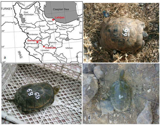 | Figure 1. a)Collection localities. b) Testudo graeca. c) Mauremys caspica. d) Emys orbicularis |
3. Results
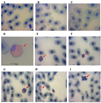
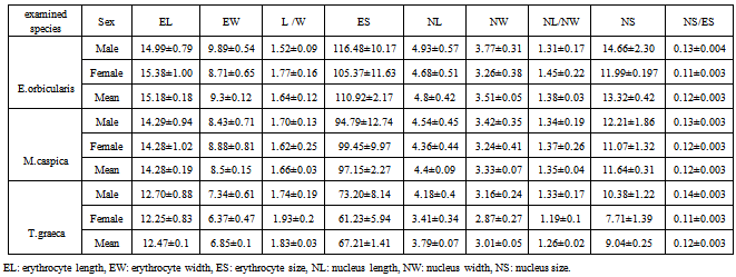
- The erythrocytes or red blood cells of turtles and tortoises are nucleated, oval cells, and their nuclei are also oval and centrally located like those of the other reptile species. The cytoplasm of mature erythrocytes appeared light blue and was homogeneous under Gimsa stain. The nuclei of mature erythrocytes are basophilic. (Figures.1).The largest and widest erythrocytes were found in Emys orbicularis. The largest and widest nuclei were also found in E.orbicularis . Erythrocyte and nuclear sizes and length/width ratios of E.orbicularis are given in table 1.The erythrocyte length of M.caspica was smaller than that of E.orbicularis but larger than that of T. graeca . The nuclear length of M.caspica was smaller than that of E.orbicularis, but larger than that of T.graeca . Erythrocyte and nuclear sizes and length/width ratios of M.caspica are given in Table 1. The smallest erythrocyte length and width were found in T.graeca . The nuclear length of T.graeca was the smallest of the all too . Erythrocyte and nuclear sizes and length/width ratios of T.graeca are given in Table 1. Graph 1 and 2 showes the comparisons between these species.
3.1. White Blood Cells Morphology Granulocytes
- Granulocytes include the leukocytes that contain specific granules that identify the cell lineage. These cells were included eosinophils, and basophils.
3.2. Eosinophil
- The eosinophil (Figures 2D) was distinguished by its round eosinophilic cytoplasmic granules which fill the colorless cytoplasm. The nucleus contains coarse, clumped chromatin and stained purple. It was round to oval, single or bi-lobed and eccentrically placed within the cytoplasm. (Table 2).
3.3. Basophils
- The basophil (Figures 2E) was easily identified by its deeply stained purple, large, round granules that remained tightly adhered to the centrally located nucleus. In some instances the granules completely occluded the nucleus ( Table 2).
3.4. Mononuclear Cells
- The mononuclear cells include lymphocytes and monocytes. These cells generally lack significant lobulation of the nucleus and do not contain an abundance of cytoplasmic granules.
3.5. Lymphocytes
- The lymphocytes (Figures 2F) contained a small amount of blue staining cytoplasm and a round nucleus with a fine reticular pattern. The lymphocyte was more spheroid than the thrombocyte, with a nuclear to cytoplasmic ratio of greater than one (Table 2).
3.6. Monocytes
- The monocyte (Figures 2G) contained a large amount of light blue-gray, finely granular or vacuolated cytoplasm and an oval or indented nucleus with a dense chromatin pattern. (Table 2).
3.7. Heterophils
- It is equivalent to a mammalian neutrophil (Figures 2H). It contained large, eosinophilic and round cytoplasmic granules. The cytoplasm, which can be difficult to visualize, was light blue or clear. The nucleus is segmented and frequently displaced toward the edge of the cell and appeared basophilic with dense chromatin (Table 2).
3.8. Thrombocyte
- The thrombocytes (Figures 2I) were ellipsoidal cells that contained round densely stained nucleus. The cytoplasm was clear and the thrombocytes tended to clump together. These latter two identifying characteristics help in distinguishing the thrombocytes from the lymphocytes (Table 2).
|
|
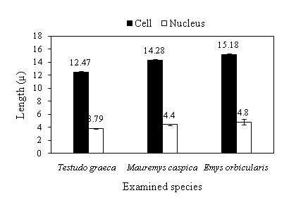 | Graph 1. Erythrocyte and nuclear lengths of examined species |
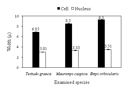 | Graph 2. Erythrocyte and nuclear widths of examined species |
4. Discussion
- One of the most important functions of erythrocytes is to carry oxygen and carbon dioxide, and its surface area to size ratio is also a determining factor in the tissues. Thus, a small erythrocyte offers the possibility of a higher rate of exchange than a larger one. The erythrocyte size reflects the position of a species on the evolutionary scale: in lower vertebrates and those with a not-so successful evolutionary past, in cyclostomes, elasmobranches and urodeles, the erythrocytes are large, but in higher vertebrates (mammals) the same cells are smaller and do not contain nuclei. Investigations carried out by various authors reported that the sizes of erythrocytes vary in members of the 4 orders of reptiles[10-13]. Within the class Reptilia, the largest erythrocytes are seen in Sphenedon punctatus, turtles, and crocodilians[14-15]. The smallest erythrocytes are found in the Lacertidae family [16-18]. The cryptodiran turtles have larger erythrocytes than do other orders of reptiles [19]. Szarski and Czopek (1966) reported that the largest erythrocytes were found Chelydra serpentine (22.5 µ), Trionyx spinifer (22.2 µ), and Emys blandingi (21.0 µ) [20]. Taylor and Kaplan (1961) measured the erythrocytes of Pseudemys scripta elegans (20.5 µ) [21]. Hartman and Lessler (1964) measured the erythrocytes of Gopherus polyphemus (19.1 µ) and Pseudemys ornata (18.6 µ) [22]. Heady and Rogers (1963) measured the erythrocytes of Pseudemys elegans (18.5 µ) [23]. There are smaller numbers of erythrocytes in reptiles than in mammals or birds. Lizards have more erythrocytes than snakes, while turtles have the fewest. Since lizards have the smallest erythrocytes of all reptiles, and turtles the largest, there may be an inverse correlation between the number of erythrocytes and their size; this hypothesis was advanced by Ryerson (1949) (Duguy 1970) [24-25]. Duguy (1970) reported that the number of erythrocytes was 260 000 - 680 000 no./mm in Emys orbicularis and 362 000 - 730 000 no./mm in T. graeca ibera[26]. Among the turtle and tortoise species which were examined in this study, aquatic ones had larger erythrocytes and nuclei than terrestrial T.graeca (Table 1). T.graeca had more ellipsoidal erythrocytes than Aquatic species regarding L/W ratio; however, aquatic species had more ellipsoidal nuclei than T.graeca regarding NL/NW ratio (Table 1). This confirms the findings of Ugurtaş et al. (2003) [27]. Nucleocytoplasmic ratio was found equal in T.graeca and aquatic species (Table 1). Consequently, it can be concluded that T.graeca had more convenient erythrocytes for gas exchange than aquatic ones. However, it is impossible to attribute these differences to the correlation regarding body weight and size, defined by Vernberg (1955) [28]. More probably, these differences were derived from various environmental conditions (e.g. temperature, Air pressure) [29] and/or various activity levels (e.g. healthy, breeding, hibernating, foraging, and daily activity) [30], for erythrocytes were found larger in aquatic species than terrestrials, and smaller in more active species. This view is compatible with the conclusions of Haden (1940) [31], and Gul and Tok (2009) [32].The lymphocytes play several important roles in the reptile, including producing antibodies and attacking foreign materials (natural killer cells). It should be mentioned that the classification criteria of chelonian leukocytes vary among studies. Some cells are not easily identified on the basis of their morphological differences. For example, small lymphocytes may be very similar morphologically to thrombocytes. Most authors agree that reptiles do not have neutrophils, whereas they do have heterophils and eosinophils, which both show acidophilic granules [33]. Some studies classify acidophils (i.e., heterophils and eosinophils) as one type of cell at different stages of maturation [34]. Neutrophils have been reported only in some reports [35-36]. Saint Girons (1970) reported the presence of eosinophils, azurophils, neutrophils and plasma cells in reptiles[37]. While, Sypek and Borysenko (1988) reported eosinophils, heterophils, basophils, monocytes and lymphocytes [38].According to Canfield, the mammalian neutrophil is equivalent to the non mammalian heterophil[39]. Lymphocytes are generally dominant leucocytes in amphibians and reptiles (Campbell, 2004[40]; Allander and Fry, 2008) [41]. Saint Girons (1970) reported that small and large lymphocytes were the dominant cells in blood smears of different reptile species, and the nuclei were not easily be distinguished[42].In this study investigating; the largest eosinophils, hetrophils and lymphocytes were found in T.graeca but the largest monocytes and basophils were in M.caspica and E.orbicularis, respectively (Table 2); small and large lymphocytes were the dominant cells in the blood smears; and the shapes of the nuclei were not distinguished because of the dense granulations in the cytoplasms of both the eosinophils and basophils, which were all compatible with the literature [43]. Though monocytes, heterophils, and eosinophils were present in amphibians and reptiles [44]. Cannon et al. (1996) reported that the heterophils were not observed in Cyrtopodion scabrum[45]. Besides, eosinophils were observed in Crocodilia and Chelonia, but their existence in Squamata is controversial [46]. Even inside a genus of snakes, eosinophils were found in some species and not found in some others [47]. Number and kind of leucocytes could be change environmental and physiological activity [48]. Thrombocytes were defined by some researchers as spindle shaped cells with centrally localized extremely chromophilic nuclei [49-50]. In this study, the largest thrombocytes were observed in T.graeca and the smallest in E.orbicularis (Table 2).The morphological characteristics obtained by light microscopy of erythrocytes, thrombocytes, basophils, eosinophils, monocytes, hetrophils and lymphocytes were similar to those seen in other reptilian species.
 Abstract
 Reference
 Full-Text PDF
 Full-text HTML