International Journal of Textile Science

p-ISSN: 2325-0119    e-ISSN: 2325-0100

2017;  6(6): 153-157

doi:10.5923/j.textile.20170606.03


Impact Analysis of Electro Spun Nano Fiber from Biodegradable Polymer for Tissue Engineering- A Review Article

Md. Rasel 1, Sojib Raihan 1, Israt Zerin 2, Mohammad Tofayel Ahmed 1, Md. Shah Alam 1, Habibur Rahman Abir 1

1Department of Textile Engineering, Southeast University, Dhaka, Bangladesh

2Department of Yarn Engineering, Bangladesh University of Textiles, Dhaka, Bangladesh

Correspondence to: Md. Rasel , Department of Textile Engineering, Southeast University, Dhaka, Bangladesh.

Email:

Copyright © 2017 Scientific & Academic Publishing. All Rights Reserved.

This work is licensed under the Creative Commons Attribution International License (CC BY).
http://creativecommons.org/licenses/by/4.0/

Abstract

Every country in the world is now developing their medical innovation from the problems they are facing. Promising bio-materials as scaffold production from bio polymers is one of great innovation in the field of tissue engineering which has been involved to various application in the field of bio-medical engineering. There are many biopolymer can be used to produce scaffold but we have chosen PCL (POLY CAPRALACTONE) biodegradable Polymer to produce nano fibers or scaffold via electro spinning technique because it’s a easiest way for production nano fibers as scaffold. The target of this study is production of scaffold from biodegradable polymer (PCL) for tissue engineering purpose & this work is includes: - Nano fibers as scaffold production from PCL (POLYCAPROLACTONE) polymers via electrospinnig process. -Observation of the effect of solution viscosity. -Observation of solvents effect on fibers surfaces as scaffold. -Analysis surface or fibers morphology & diameter via microscopic tester.

Keywords: Electro spinning, Scaffold, Biodegradable polymer, Nano fiber

Cite this paper: Md. Rasel , Sojib Raihan , Israt Zerin , Mohammad Tofayel Ahmed , Md. Shah Alam , Habibur Rahman Abir , Impact Analysis of Electro Spun Nano Fiber from Biodegradable Polymer for Tissue Engineering- A Review Article, International Journal of Textile Science, Vol. 6 No. 6, 2017, pp. 153-157. doi: 10.5923/j.textile.20170606.03.

1. Introduction

PCL is a biodegradable polymer with 59-64° temperature, Tg -60° & rubbery state [1-3]. The erosion rate of biodegradable Nano fibers are following the order PGA, PLGA PLLA, PCL [4]. Electros pining is easiest and popular technique and Electro spun Nano fibers are produced from different polymers (natural or synthetic polymers) for different applications [5, 6]. PCL is most promising synthetic/ semicrystaline biodegradable polymer & it degrades very slower [7, 8] and its with a wide range of applications in the field of tissue engineering such as delivery device or commercial sutures, biomedical materials due to physical properties [9-13] & biological properties [14-17] like blood vessel [18-20], bone scaffold [9, 21-24], nonwoven membranes [9, 25] etc. PCL has less mechanical properties but it can be blending with natural polymers [26], synthetic polymers [27, 28] by electro spinning technique for tissue engineering.
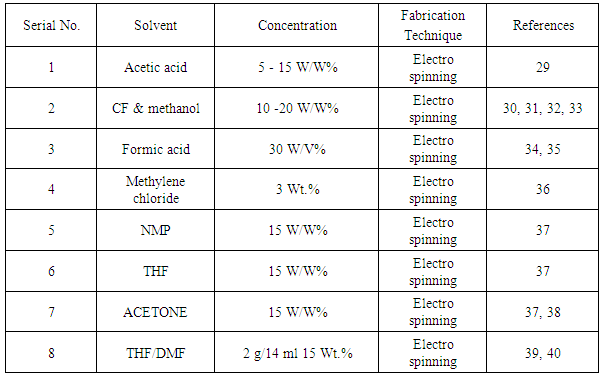
2. Raw Materials

Electro spun PCL Nano fibers which have dissolved into most of solvent but some are given below in a table:-
Table 1. Solvent for Electro spun PCL Nanofibers
     

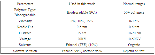
2.1. Parameters of Electro Spinning

There are many parameters can be effected on the electro spinning process such as Viscosity, conductivity, solvent, Surface tension, flow rate, collector take up, Velocity, polymer, temperature, collector, needle design, needle dia etc. Several parameters are used in this work, which are most important for electro spinning these are:-
Table 2. Parameters of Electro spinning
     

3. Methodology

PCL/ethanol & PCL acetone solution viscosity
a) (PCL 3g + METHANOL 27gm) = 10% viscosity
Similarly, acetone
b) 8% & 15% viscosity
These have been used to produce Nano fibers by electrospining technique.
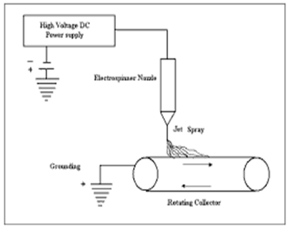
Figure 1. Schematic representation of the electrostatic fiber spinner
The polymer fluid is delivered to the capillary at a constant flow rate .An Electric field is generated by applying a high voltage between the metal capillary and the collector finally output is Nano fibers.
In this work, the Polymer solutions have been dissolved into solvent and then delivered at constant rate to a metal capillary connected to a high voltage supply (20kv) after that the solvent evaporated and charged polymers have been deposited on the collector plate in the form of Nano fibers.

3.1. Flow Rate

Only 10% viscosity has been showed by this figure 2.
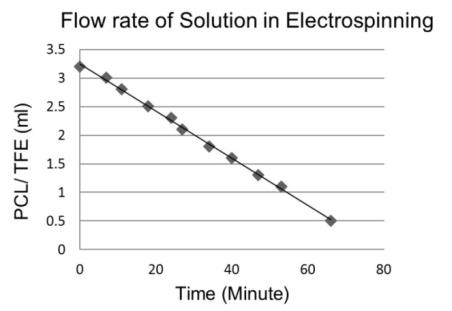
Figure 2. Flow rate of solution
This figure state that time decreases at constant flow rate of 0.0389ml/min & the flow rate is mainly depending on solution viscosity. The solution viscosity depends on solvents & polymer used which can be controlled by Adjusting polymer concentration [41] & the solution temperature. [42]
On the other hand higher polymer concentration increases solution viscosity but higher temperature reduce it. [43]
a) Analysis methods: 1
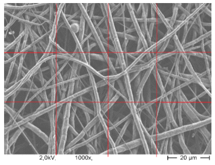
Figure 3. PCL Nano fibers as scaffold has been scanning by electron microscopy & diameter has been evaluated by showing in views a magnifications 200x, 1000x, 2000x times. Diameter has been measured by nm scale with axiovision rel4.8 software for various solution concentrations
b) Analysis methods: 2
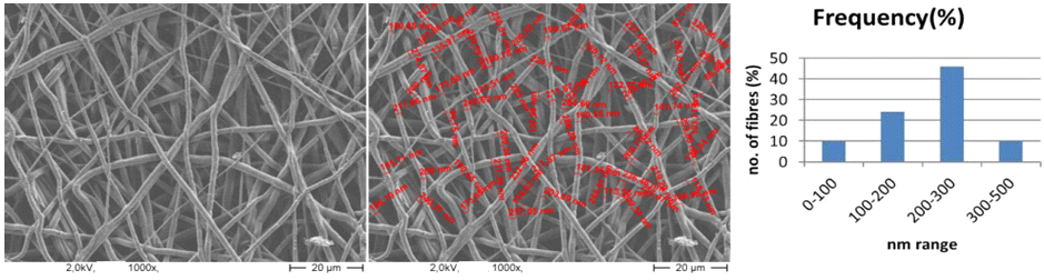
Figure 4. The SEM image has been showed that fiber distribution is most important for cell adhesions in the scaffold as tissue engineering in human body & diameters of Nanofibers 100-500nm
c) Analysis methods: 3
The PCL/ethanol & PCL/Acetone flow rate through the needle has been constant during Nano fibers as scaffold production but an electro spun fiber morphology has been investigated by mixing at various concentrations. Solution viscosity have been observed by Scanning electron micrographs of electro spun fibers at varying solution viscosity (8/10/15%) that increasing solution concentration increased viscosity and the increased solution viscosity which has been resulting in larger fiber diameter. [44-47]
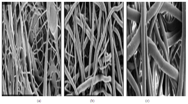
Figure 5. Representative images of fibers spun at (a) low, (b) intermediate, and (c) high viscosity

4. Results & Discussions

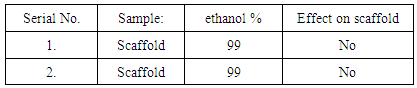
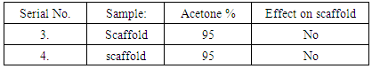
In this work solvents taken ethanol & acetone. Scaffold samples have been produced through acetone 95% & ethanol 99%. These solvent should not modify the fibers.
Table 3. Scaffold samples have been produced using ethanol 99%
     
Table 4. Scaffold samples have been produced using acetone 95%
     
The constant flow rate of solution is most important for properly fiber distributions in the scaffold which have been shown in image. Solvents have not effect on scaffold morphology & almost same. (Image 2) shows that uniform fiber distribution happened in the scaffold and fibers diameter ranges 100-500nm.

5. Conclusions

PCL is versatile polymer for tissue engineering applications. Ethanol/acetone has been used to fabricate Nano fibers as scaffold by electro spinning but they have not effect on scaffold morphology or fiber surfaces but solvents has effect on viscosity of polymer solution which can produces coarser or finer fibers diameter.

References

[1]  N. Mishra, A.K. Goyal, K. Khatri, B. Vaida, R. Paliwal, S. Rai, A. Mehta, S. Tiwari, S. Vyas and S.P. Vyas // Antiinflamm. Antiallergy. Agents. Med. Chem. 7(2008) 240.
[2]  R.S. Benzwada, D.D. Jamiolkowski, I.Y. Lee, V. Agarwal, J. Persivale, S. Trenkabenthgin, M. Erneta, j. Suryadevara, A. Yang and S. Liu // Biomaterials 16 (1995) 1141.
[3]  J.M. Anderson and M.S. Shive //Adv. Drug. Deliv. Rev. 28 (1997) 5.
[4]  Y. You, B.-M, Min, S.J. Lee, T.S. Lee and W.H. Park // J. Appl. Sci. 95(2005).
[5]  L. WJ, L. CT, C.EJ, T. RS and K. FK // J.Biomed. Mater. Res. 60(2002).
[6]  K.H. Rho, L. Jeomg, G. Lee, b. –M. Seo, Y.J. Park, S.-D. Hong, S. Roh, J.J. Cho, W.H. Park and B.-M. Min // Biomaterials 27 (2006) 1452.
[7]  C.G. Pitt, F.I. Chasalow, Y.M. Hibionada, D.M. Klimas and A. Schindler // J. Appl. Polym. Sci. 26 (1981) 3779.
[8]  H. Sun, L. Mei, C. SONG, X. Cui and P. Wang // biomaterials 27 (2006) 1735.
[9]  H. Yoshimoto, Y.M. Shin, H. Terai and J.P. Vacanti // Biomaterials 24 (2003) 2077.
[10]  A.-C. Albertsson and I.K. Varma // Bio macromolecules 4(2003 1466.
[11]  D.W. Hutmacher, T. Schantz, I. Zerein, K.W. Ng, S.H. Teoh and K.C. Tan // J. Biomed. Matures. 55 (2001) 203.
[12]  W.-J. Li, K.G. Danielson, P.G. Alexander and R.S. Tuan // J. Biomed. Mater.Res.A. 67A (2003) 1105.
[13]  N. Lopez-Rodriguez, A. Lopez-Arraiza, E.Meaurio and J. R. Sarasua // Polym. Eng. Sci. 46 (2006) 1299.
[14]  W.-J. Lin, D.R. Flanagan and R.J. Linhardt // Polymer 40 (1999) 1731.
[15]  J. Pena, T. Corrales, I. Lzquierdo-Barba, A.L. Doadrio and M. Valllet-Regi // Pollym. Degrad. Stab. 91 (2006) 1424.
[16]  X. Zong, S. Ran, K.-S. Kim, D. Fang, B.S. Hsiao and B. Chu // J. Bio macromolecules 4 (2003) 414.
[17]  C.H. Kim, M.S. Khil, H.Y. Kim, H.U. Lee and K.Y. Jahng // J. Biomed. Mater.Res. Part B Appl. Biomater. 78B (2006) 283.
[18]  N.E. Zander, J.A. Orlicki, A.M. Rawlett and T.P. Beebe // ACS Appl. Mater. Interfacess 4 (2012) 2074.
[19]  V.Y. Chakrapani, A. Gnanamani, V.R. Giridev, M. Madhusoothanan and G. Sekaran // J. Appl. Polym. Sci. 125 (2012) 3231.
[20]  M.R. Williamson, R. Black and C. Kielty // Biomaterials 27 (2006) 3608.
[21]  N. Hiep and B.-T. Lee // J. Mater. Sci-Mater. Med. 21 (2010) 1969.
[22]  M.-C. Serrano, R. Pagani, M. Vallet-Regi, J. Pena, J.-V. Comas and M.-T. Portoles // Acta. Biomater. 5 (2009) 2045.
[23]  C.Y. Xu, R. Inai, M. Kotaki and S. Ramakrishna // Biomaterials 25 (24) 877.
[24]  S.I. Jeong, S.H. Kim, Y.H. Kim, Y. Jung, J.H. Kwon, B.S. Kim and Y.M. Lee // J. Biomater. Sci. Polym. E.D. 15 (2004) 645.
[25]  J.W. Calvert, K.G. Marra, L. COOK, P.N. Kumta, P.A. DiMilla and L.E. Weiss // J Biomed Mater Res. 52 (2000) 279.
[26]  H. Kweon, M.K. Yoo, I.K. Park, T.H. Kim, H.C. Lee, H.-S. Lee, J.-S. Oh, T. Akaike and C.-S. Cho // Biomaterials 24 (2003) 801.
[27]  A.G.A. Coombes, S.C. Rizzi, M. Williamson, J.E. Barralet, S. Downes and W.A. Wallace // Biomaterials 25(2004) 315.
[28]  H. Chim, D.W. Hutmacher, A.M. Chou, A.L. Oliveira, R.L. Reis, T.C. Lim and J.T. Schantz // Int. J. Oral. Maxillofac. Surg. 35 (2006) 928.
[29]  I. Ahmed, A.J. Parsons, G. Palmer, J. C. Knowles, G. S. Walker and C.D Rudd// J.Biomater.4 (2008) 1307.
[30]  W. Zheng, Z. Wang, L. Song, Q. Zhao, J. Zhang, D.Li, S. Wang, J. han, X.-L. Zheng, Z. Yang and D. Kong // Biomaterials 33 (2011) 2880.
[31]  P. M. Mountziaris, S. N. Tzouanas and A. G. Mikos // Biomaterials 31 (2010) 1666.
[32]  J.K. Hong and S.V. Madihally // Acta Biomater. 6 (2010) 4734.
[33]  E. Pektok, B. Nottelet, J.-.C. Tille, R. Gumy, A. Kalangos, M. Moeller and B. H. Walpoth // Circulation 118 (2008) 2563.
[34]  P. Xiang, M. Li, C. -Y. Zhang, D.-L. Chen and Z.-H Zhou // Int. J. Biol. Macromol.49 (2011) 281.
[35]  J.S. Lim, C.S. Ki, J. W. Kim, K.G. Lee, S.W. Kang, H.Y. Kweon and Y.H. Park // Biopolymers 97 (2012) 265.
[36]  K.S. Tiaw, S.H. Teoh, R. Chen and M.H. HONG // Bio macromolecules 8 (2007) 807.
[37]  X.H. QQin and D.Q. Wu // J. Them. Anal .Clorim. 107 (2012)1007.
[38]  L.A. Bosworth and S. Downes // polym. Degrad. Stab. 95 (2010) 2269.
[39]  J.M. Deitzel, J.D. Klein Meyer, J.K. Hirvoinen and N.C. Beck Tan // Polymer 42(2001) 8163.
[40]  F. Croisier, A.S. Duwez, C.Jeome, A.F. Leonard, K.O. Van der Werf, P.J. Dijkstar and M.L. Bennink //Acta. Biomater. 8 (2012) 218.
[41]  Kulkarni, A.; Bambole, V. A.; Mahanwar, P. A. Polym. Plast. Technol. Eng.2010, 49, 427–441.
[42]  Huang, Z. M.; Zhang, Y. Z.; Kotaki, M.; Ramakrishna, S. Compos. Sci. Technol. 2003, 63, 2223–2253.
[43]  Lurii Sas, Russell E. Gorga, Jeff A. Joines, Kristin A. Thoney Journal of Polymer Science Part B: Polymer Physics, 2012, 50(12), 824–845.
[44]  Tao J. Shivkumar S. Molecular weight dependent structural regimes during the electrospinning of PVA. Mater Lett. 2007; 61:2325.
[45]  Gupta P. Elkins C. Long T.E. Wilkes G.L. Electrospinning of linear homopolymers of poly (methyl methacrylate): exploring relationships between fiber formation, viscosity, molecular weight and concentration in a good solvent. Polymer. 2005; 46:4799.
[46]  Jiang H. Fang D. Hsiao B.S. Chu B. Chen W. Optimization and characterization of dextran membranes prepared by electro spinning. Biomacromolecules. 2004; 5:326.
[47]  Fong H. Chun I. Reneker D. Beaded nanofibers formed during electrospinning. Polymer. 1999; 40:4585.