-
Paper Information
- Paper Submission
-
Journal Information
- About This Journal
- Editorial Board
- Current Issue
- Archive
- Author Guidelines
- Contact Us
International Journal of Surgical Research
2016; 5(2): 15-16
doi:10.5923/j.surgery.20160502.01

Hydatid Cyst in Breast -A Rare Case and Review of Literature
 Abstract
 Reference
 Full-Text PDF
 Full-text HTML
Jyoty Singh, Ashwini Arvind Natekar, Subhasis Basu
Department of Pathology, Chittaranjan National Cancer Institue, India
Correspondence to: Jyoty Singh, Department of Pathology, Chittaranjan National Cancer Institue, India.
| Email: |  |
Copyright © 2016 Scientific & Academic Publishing. All Rights Reserved.
This work is licensed under the Creative Commons Attribution International License (CC BY).
http://creativecommons.org/licenses/by/4.0/

Hydatid disease is a parasitic infestation by a tapeworm of the genus Echinococcus. Most commonly infected organs includes liver and lung. Its occurance at unusual sites can pose a diagnostic challenge. Here we presenting a case with hydatid cyst in right breast. Since its very uncommon to find a hydatid cyst in breast, it is usually not considered as differential diagnosis while evaluating breast lump and can cause serious problems if missed. Our aim of presenting this case is to draw attention to the fact that hydatid cyst should remain in the differential diagnosis while evaluating cystic swellings in unusual sites like breast.
Keywords: Breast lump, Hydatid cyst, Echinococcus
Cite this paper: Jyoty Singh, Ashwini Arvind Natekar, Subhasis Basu, Hydatid Cyst in Breast -A Rare Case and Review of Literature, International Journal of Surgical Research, Vol. 5 No. 2, 2016, pp. 15-16. doi: 10.5923/j.surgery.20160502.01.
1. Introduction
- Echinococcus granulosus, which is the most common cause of hydatid cysts, is a tapeworm from the Cestoidean class. Breast is one of the rare site for hydatid cyst. Clinically it presents as a painless freely mobile lump which can mimic fibroadenoma, phyllodes tumors, chronic abscesses, or even carcinoma. It is not possible to get conclusive diagnosis with only radiological investigations and proper serological and histopathological examination should be done for confirmation. Hydatid cyst is still a public health problem in endemic areas including India. High prevalence in Kashmir, Andhra Pradesh, Tamil Nadu and Central India. [1, 2] Here we reporting a case from eastern India.
2. Case History
- We attended a post operative case of breast lump for histopathological evaluation. Our patient was a 24 years old female who had a right sided painless, palpable cystic breast lump superior to nipple in retroareolar region for 6 months, for which she was operated. On inspection we found a linear incision mark over right breast superior to nipple [Figure 1].
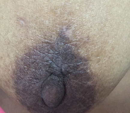 | Figure 1. Linear Incision scar |
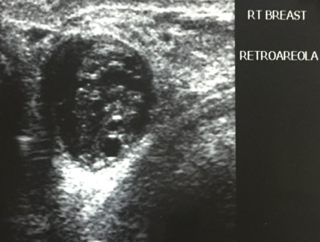 | Figure 2. USG Breast |
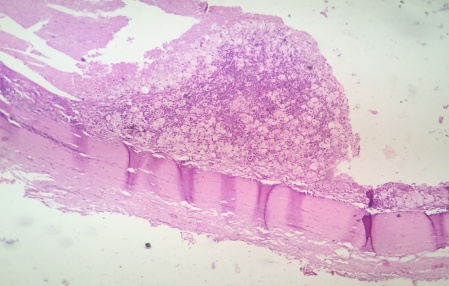 | Figure 3. Laminated Chitinous layer, hydatid cyst and foamy macrophages |
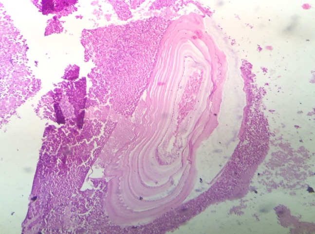 | Figure 4. Hydatid cyst |
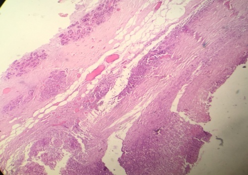 | Figure 5. Hydatid cyst wall and surrounding breast tissue |
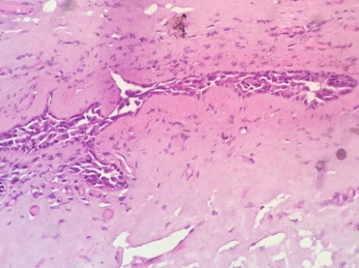 | Figure 6. Fig surrounding Normal breast tissue |
3. Discussion
- In urban India, due to economic development and improved government legislation of abattoirs there is decreasing prevalence of hydatidosis in contrast to rural areas. [3]In 2011, in an article on ‘Continuing challenge of infectious diseases in India’ in the Lancet, Dr T Jacob John observes that Other infectious diseases like zoonoses are not in the process of being systematically controlled and India needs to rethink and revise its health policy to broaden the agenda of disease control with a major focus on public health in addition to the current focus on medical care. [4] This will go a long way in controlling the inadequately controlled and neglected zoonosis like hydatid disease in all parts of India.According to Barret and Tho.mas 60% are found in Liver, 30% in the Lungs, 2.5% in Kidneys, 2.5% in the Heart, 2% in the Bone, L5% in the Spleen, 1% in Muscle and 0.5% in Brain [5]. In 532 cases reported by Bickers one case each was located in the orbit, bladder, heart, chest wall, subcutaneous tissue, tibia, parotid and thyroid. [6] Incidence of unusual site for hydatid cyst is about 8-10% [1]. Reported incidence of breast hydatid cyst is 0.27% in literature [7]. Its Incidence is increasing and it should be considered as a differential diagnosis while evaluating any cystic swelling in unusual sites.It is not possible to reach definitive diagnosis with only radiological investigations and proper serological and histopathological examination should be done for confirmation.
4. Conclusions
- Hydatid cyst in unusual sites can create a diagnostic dilemma. Breast is an unusual site for hydatid cyst to occur. In epidemic areas, it should be consider among differential diagnosis while evaluating breast lumps. Delayed diagnosis and untreated hydatid cyst can cause serious complications related to cyst rupture and hence anaphylactic shock.