-
Paper Information
- Paper Submission
-
Journal Information
- About This Journal
- Editorial Board
- Current Issue
- Archive
- Author Guidelines
- Contact Us
International Journal of Surgical Research
2016; 5(1): 5-7
doi:10.5923/j.surgery.20160501.02

Ameloblastic Fibrosarcoma of Mandible in a Paediatric Patient- A Rare Case Report
 Abstract
 Reference
 Full-Text PDF
 Full-text HTML
Ashwini A. Natekar MD , Jyoty Singh DNB Pgt , Shubasis Basu MD
Department of Pathology, Chittaranjan National Cancer Institute, Kolkata, India
Correspondence to: Ashwini A. Natekar MD , Department of Pathology, Chittaranjan National Cancer Institute, Kolkata, India.
| Email: |  |
Copyright © 2016 Scientific & Academic Publishing. All Rights Reserved.
This work is licensed under the Creative Commons Attribution International License (CC BY).
http://creativecommons.org/licenses/by/4.0/

Background: Ameloblastic fibro sarcoma is a tumour of odontogenic origin. It is frequently seen in the 3rd and 4th decade of life. Case report: We report a case of 17 years old female who presented with left lower jaw swelling. CT scan revealed a large expansile osteolytic soft tissue mass in mandible. The patient underwent left extended hemimandibulectomy. Grossly the large mass measured 16x9x6.5cm, destructing the underlying mandible. Microscopic examination revealed a biphasic tumor composed of islands of benign odontogenic epithelium in abundant mesenchymal component, mild nuclear pleomorphism, and occasional mitosis. Hence the diagnosis of Ameloblastic Fibrosarcoma- Low grade was made. On radiological and clinical follow up patient is free of tumor till date. Conclusions: Ameloblastic fibrosarcoma is rare tumor of paediatric age. Surgical excision is a treatment of choice.
Keywords: Ameloblastic fibrosarcoma, Odontogenic tumor, Paediatric patients
Cite this paper: Ashwini A. Natekar MD , Jyoty Singh DNB Pgt , Shubasis Basu MD , Ameloblastic Fibrosarcoma of Mandible in a Paediatric Patient- A Rare Case Report, International Journal of Surgical Research, Vol. 5 No. 1, 2016, pp. 5-7. doi: 10.5923/j.surgery.20160501.02.
1. Introduction
- Ameloblastic fibrosarcoma (AFS) is a malignant odontogenic tumour characteristically composed of a benign epithelium and a malignant mesenchymal component. [1]Odontogenic tumours and tumour like lesions comprise of a rare group of heterogenous diseases that range from non-neoplastic tissue proliferation to malignant tumor with metastatic potential.They are classified as odontogenic carcinomas and odontogenic sarcomas. [2]The most common presentation is pain and swelling [3] and mandible is the most commonly affected site. [2]Wide local excision is the treatment of choice in AFS and is associated with good prognosis. [4]
2. Case Report
- We report a case of 17 year old female who presented with a painful left mandibular swelling. CT scan revealed a large 12x10x11cm osteolytic expansile heterogeneously enhancing soft tissue mass involving the left side of the body and remus of mandible (Figure 1 & 2). Biopsy from the lesion was inconclusive. Patient underwent a left extended hemimandibulectomy with modified radical neck dissection.
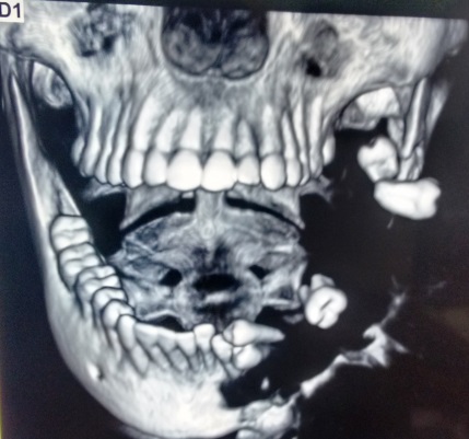 | Figure 1. CT scan image showing bony destruction |
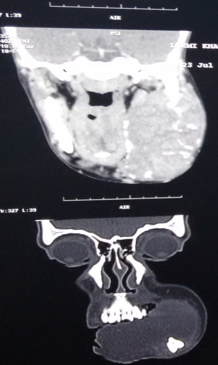 | Figure 2. CT scan image showing expansile soft tissue mass |
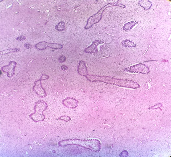 | Figure 3. H & E stain- Biphasic tumor (100X) |
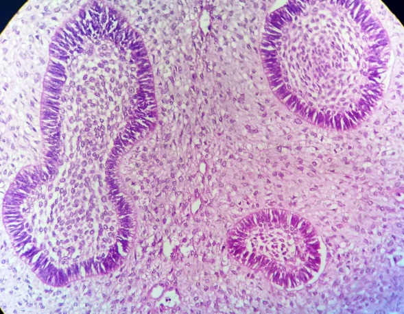 | Figure 4. H & E stain- Biphasic components of tumor (400X) |
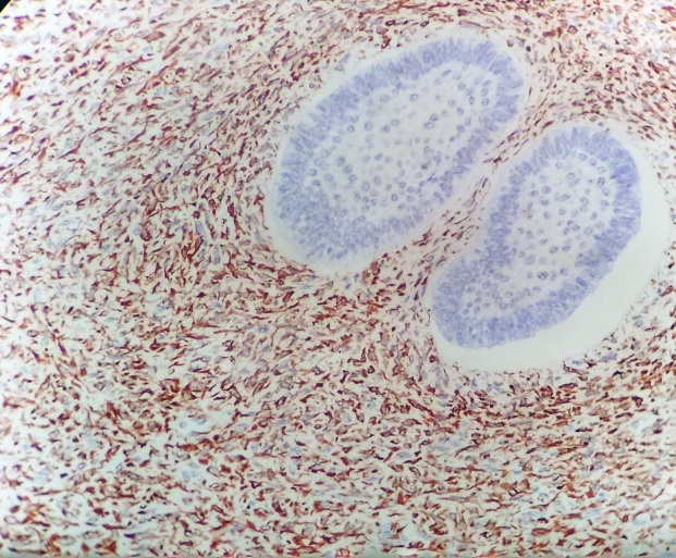 | Figure 5. Vimentin positivity in mesenchyme cells (400X) |
3. Discussion
- AFS was first reported by Heath in 1887 describing it as spindle cell sarcoma that also had epithelial cells resembling the cells of the enamel organ. [5]The mean age of presentation is 28.3 years with wide age range from 3 to 89 years and a male to female ratio of 1.6:1. [6].To best of our knowledge, less than 100 documented cases have been reported in the English literature. [7]The most common clinical presentation is pain and swelling [3] and mandible is the most commonly affected site [2, 6].Radiologically AFS presents as a radiolucent mass with ill-defined borders.AFS is rare in paediatric age group. Till date only three cases of paediatric AFS have been reported in the literature. [8, 9] Our patient was a 17 year female with a posterior mandibular tumor. Her age at presentation was younger than the mean age of presentation.Grossly the tumor may be solid or cystic with fleshy whitish to yellow appearance [8].Microscopically AFS is characterised by benign epithelial cell islands that are composed of peripheral columnar or cuboidal cells arranged in a palisading pattern. At the centre of these islands is polyhedral cell reminiscent of stellate reticulum. The mesenchymal component consists of plump and spindle stromal cells which show mild to moderate cytological atypia. Yamamoto et al. showed the presence of keratin in the columnar and polyhedral cells of the epithelial component and vimentin in the ectomesenchymal component verifying the biphasic nature of this tumor. [10]Surgical excision is the treatment of choice [4]. Other treatment modalities such as adjuvant chemotherapy and radiotherapy were found to be ineffective. [11]Our patient has remained tumor free till date after wide surgical excision which is the recommended treatment for AFS. Some authors have proposed that AFS should be considered as low grade fibrosarcoma [7] in view of its more favourable prognosis in comparison to fibrosarcomas of the orofacial region. The differential diagnosis of AFS include odontogenic sarcoma, ameloblastic carcinosarcoma and spindle cell carcinoma. [8]
4. Summary
- AFS is a rare tumor of paediatric age group with only three cases reported in literature till date with ours being the fourth case. It is a rare malignant odontogenic tumor characterized by benign odontogenic epithelium and a malignant mesenchymal component. Wide surgical excision is the treatment of choice and close follow up is advised.