International Journal of Sports Science

p-ISSN: 2169-8759    e-ISSN: 2169-8791

2016;  6(2): 52-61

doi:10.5923/j.sports.20160602.06


Cardiac Remodeling and Physical Exercise: A Brief Review about Concepts and Adaptations

Paula A. M. Cavalcante1, Mauro S. Perilhão1, Ariana A. da Silva1, Andrey J. Serra2, Aylton F. Júnior1, Danilo S. Bocalini1, 3

1Translational Physiology Laboratory, Postgraduate Program in Physical Education, São Judas Tadeu University (USJT), São Paulo, SP, Brazil

2Postgraduate Program in Biophotonics Applied to Health Sciences, Nove de Julho University (UNINOVE), São Paulo, SP, Brazil

3Translational Physiology Laboratory, Aging Science, São Judas Tadeu University (USJT), São Paulo, SP, Brazil

Correspondence to: Danilo S. Bocalini, Translational Physiology Laboratory, Postgraduate Program in Physical Education, São Judas Tadeu University (USJT), São Paulo, SP, Brazil.

Email:

Copyright © 2016 Scientific & Academic Publishing. All Rights Reserved.

This work is licensed under the Creative Commons Attribution International License (CC BY).
http://creativecommons.org/licenses/by/4.0/

Abstract

Training variable control, is relateed to frequency, duration, and intensity, that respond to the promoting changes in physical fitness. However, adaptations related to different exercise intensities are still controversial, both in regards to cardiovascular disorder prevention and rehabilitation. There are few studies dealing with cardiac adaptations under different exercise intensities, and that a better understanding physiological cardiac remodeling may inspire the design of a strategy to improve ventricular function in cardiac approach. Thus the objective of this review was to assess the influence of chronic aerobic exercise on cardiac adaptations, with a focus on different exercise intensities, as well as presenting a conceptual approach on cardiac remodeling process. Also was included the specific characteristics of cardiac remodeling in each cardiac adequacy step during training. New research trends were also outlined in order to guide studies into developing new therapy strategies for controlling cardiovascular diseases.

Keywords: Cardiac remodeling, Physical exercise, Cardiac adaptations and ventricular function

Cite this paper: Paula A. M. Cavalcante, Mauro S. Perilhão, Ariana A. da Silva, Andrey J. Serra, Aylton F. Júnior, Danilo S. Bocalini, Cardiac Remodeling and Physical Exercise: A Brief Review about Concepts and Adaptations, International Journal of Sports Science, Vol. 6 No. 2, 2016, pp. 52-61. doi: 10.5923/j.sports.20160602.06.

1. Introduction

The chronic effect of aerobic exercise has been studied in order to further and effectively understand the adaptations caused in humans. Based on this perspective, the actions of regular physical exercise and improvements of functional capacity are important factors in improving health. Their beneficial effects on the cardio circulatory system are well described in literature [1-8].
However, in relation to exercise intensity on biological and clinical effects, the guidelines is controversial both in cardiovascular disease prevention and in the rehabilitation field [9,10]. Although increased training intensity may result in further improving physical aptitude and maximum oxygen consumption (VO2max) [2], moderate intensity exercise is usually recommended, as it is equally efficient [9].
This literature review assesses the influence of chronic aerobic exercise on cardiac adaptations considering there are few studies in literature regarding cardiac adaptations in different exercise intensities, and that a better understanding of the physiological cardiac remodeling may lead to a strategy capable of improving ventricular function in cardiac dysfunction [11, 12]. In particular, this review will focus on the discussion regarding the different exercise intensities, presenting a conceptual approach on the cardiac remodeling process and its particular characteristics in each cardiac adaptation step for physical training. New research trends were also outlined in order to guide studies into developing new therapeutic strategies for controlling cardiovascular diseases.

2. Main Body

The most relevant original scientific studies (animals and human) were from 1979 to 2015, analyzed in this review, at the following databases: Science Citation, Index, Scopus, Sport Discus, Scielo, and National Library of Medicine, combining the following: keywords: endurance training, physical exercise, cardiac function, ventricular function, cardiac remodeling, ventricular remodeling and cardiac and ventricular adaptations. Studies investigating the effects of physical exercise and cardiac parameters with and training variables (as well as human and animal studies) were considered inclusion criteria.

3. Cardiac Remodeling

Cardiac remodeling (CR) is a group of genetic, molecular, cellular, and interstitial changes in the myocardium which are anatomically manifested by changes in mass, size, and geometry of the heart chambers and function stemming from hemodynamic overload [74-76].
Recent studies [77-79] suggest epigenetic changes in the DNA, resulting from physical exercise, can also change the functions of the cardiovascular system, although there is insufficient evidence to establish a direct link between epigenetic modulations and changes, caused by exercise, in the heart and blood vessels [78]. This likely occurs due to the cardiovascular field of research not being nearly as advanced as others in investigating epigenetics [77]. Nevertheless, evidence reveal physical exercise is capable of neutralizing the development of pathological epigenetics in diseases such as hypertension, atherosclerosis, and fibrosis [79]. As such, it is possible that epigenetic changes may also be mentioned as part of the concept of cardiac remodeling in the near future.
Following an analysis of new trends, a recent study [80] showed that it is possible to reduce cardiac hypertrophy causing blockage of TLR4 in the brain in mice suffering from arterial hypertension induced by angiotensin II. The actions of receptor toll-like 4 (TLR4) are associated with the development and progression of cardiovascular diseases [81]. Dange et al [80] proved there could be a bidirectional link between the brain suffering from stress and pathological cardiac hypertrophy. Researchers speculated that the toll-like receptors’ activity in the brain, particularly TLR4, is key in structure modulation and cardiac function.
Characterized as end product of several types of hemodynamic stimulus, the CR may be called physiological during normal development, or in athletes [75], or pathological, common to multiple aggressions to the heart. The aggressions can be injury (myocardial infarction), chronic pressure overload (hypertension, aortic stenosis) or volume (aortic or mitral insufficiency), inflammation (myocarditis), or expression of a genetic program leading to cardiomyopathy [82]. These events allow the heart to adapt to new conditions and activate important mediators including wall stress, neuro hormones (sympathetic nervous system, renin-angiotensin, aldosterone and endothelin), cytokines, nitric oxide and oxidative stress [75].
The main adaptation of the heart to physical training is the CR [24, 69, 83, 84], and the cardiac adaptations are related to structural and functional cardiac changes, influenced by the gain of functional ability acquired in training. The major physiological mechanisms attributed to the CR is increased contractility [24, 85-87], improved transient Ca2+ intracellular affecting ventricular function [87-89], increased expansion with significant improvements in myocardial oxygenation [90], and additional endothelium - dependent functions that prevent ischemic events [9]. However, CR’s physiological answers are not uniform and vary according to the type and intensity of the exercise performed, as well as the physical training program [91] and is directly related to maximal aerobic capacity or VO2 max [92, 93].
The beginning of the CR process is driven by a hemodynamic stimulus from a transient overload, such as what occurs in growth and physical training or a persistent overload arising from events such as myocardial infarction or from some diseases such as hypertension, as shown in Figure 1. In this perspective, CR results may have different consequences and outcomes according to the type and the time of the stimulus generator [12, 94]. Thus, the CR, considering one of the most significant results of chronic adaptation to exercise, derives in myocyte hypertrophy in cardiac performance to suit the demands of the body [95, 96].
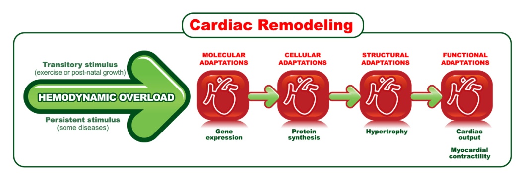
Figure 1. Steps of cardiac remodeling
The growth phase and physical training lead to physiological hypertrophy, is characterized by a lack of fibrosis and altered expression of postnatal hypertrophic genes, and in this case, changes in the expression of fetal genes, classically associated with the development of pathological hypertrophy, do not occur [36, 49, 70, 71, 97, 98]. This suggests physiological hypertrophy induced by exercise depends more on the mRNA transcript for protein synthesis, and the maintenance of synthesized protein [97], as shown in Figure 2.
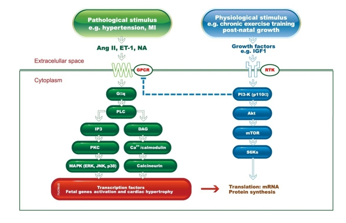
Figure 2. Diagram of the signaling pathways involved in induction of physiological cardiac hypertrophy and pathological. RTK, tyrosine kinase receptor; mTOR, target of rapamycin in mammals; NA, noradrenaline; PLC, phospholipase C; DAG, diacylglycerol; IP3, inositol 1,4,5-triphosphate; MAPK, mitogenactivated protein kinase; JNK, c-Jun amino-terminal kinase; PKC, protein kinase C; PI3-kinase, phosphatidylinositol 3-kinase; ERK, extracellular signal regulated kinase; Ang II, angiotensin II; ET-1, endothelia-1 (endothelin-1); GPCR L-protein coupled receptor; NFAT, nuclear factor of activated T cells; IGF1, similar to insulin growth factor 1; S6Ks, ribosomal S6 kinases. The performance of the physiological stimulus via IGF1-PI3K (p110α) can inhibit the signaling molecules of G protein (GPCRs) activated by pathological stimuli, preventing the progression of heart disease
Although both physiological and pathological cardiac hypertrophy are associated with an increase in the heart mass, the physiological hypertrophy is associated with a structure and a normal or increased cardiac function [12, 25, 26, 49, 97-100] whereas the pathological hypertrophy is associated with a complex series of events, including the upregulation of fetal genes, histopathology and cardiac dysfunction [12, 100].
In physiological CR, the increasing volume of myocytes is done by the synthesis of new components, as the increase in contents of contractile proteins leading to predominant increase of myofibrils. Great changes in the stromal features do not occur, so there is no functional damage of the body [4, 26, 101]. In dynamic exercises, in which athletes perform isotonic exercises such as in swimming, cycling, running and walking, the main hemodynamic patterns are increased heart rate and stroke volume, the two components of cardiac output [101]. In this case, the heart overload is predominantly volumetric, inducing the eccentric left ventricular to hypertrophy, with the addition of sarcomeres in series, which results in an increased cardiac chamber without loss of function [26, 28, 91]. This form of remodeling is homogeneous, and has the effect of increasing the contractile force [102].
Considering that the physiological changes caused by exercises has a favorable effect on the pathological CR, a better understanding of physiological CR can provide a strategy to improve ventricular function after cardiac dysfunction [11, 12, 26, 103]. Therapeutic interventions based on gene stimuli leading to physiological growth may provide an additional strategy for treating or reversing heart failure [11, 12, 26, 103]. In this respect, the role of the physiological stimulus via IGF1-PI3K (p110α) can inhibit the signaling of molecules of G protein (GPCRs) activated by pathological stimuli, preventing the progression of heart disease [103] (Figure 2).
Aerobic endurance training induces chronic enlargement of the myocardium that begins with an enhanced contractile function [87]. From this perspective, the concentration of intracellular free Ca2+ is the critical factor regulating the cardiac myocyte function and primarily responsible for the contractile state of the myocardium [104-107].
At the end of systole, the Ca2+ influx is stopped and the RS is no longer stimulated to release Ca2+. At this time, the RS reuptake cytosolic Ca2+ using a calcium pump, called SERCA2a, which is the main responsible structure for maintaining the homeostasis of the intracellular Ca2+ involved in myocardial relaxation process [105]. The increased expression of SERCA2a protein appears to improve cardiac inotropism due to prolonged ventricular filling time and greater accumulation of Ca2+ in RS [70]. Other studies have also shown high intensity cardio increased the protein expression of SERCA2a [24, 36, 70], in the phospholamban (PLB) phosphorylated at threonine 17 (PLB-Tre17) [36], without changing the protein levels of PLB [36, 70], in the left ventricle in female normotensive rats.
The Frank-Starling Law determined the relationship between stretching and contraction force in the healthy heart, i.e., the greater the amount of blood reaching the heart, the higher the contraction force [105, 109]. A perform better Frank-Starling mechanism may be associated with increased sensitivity of myofilaments to calcium, especially when the muscle fiber was extended, generating more active tension [95, 110].
Several studies [21, 110-114] tested the contractility in rats by analyzing the papillary muscles, and in this respect, the front and rear papillary muscles of the left ventricle are larger and stronger compared to those found in the right ventricle [115]. Thus, in order to analyze the conventional mechanical parameters in the maximum length (Lmax), calculated under isometric contraction [21, 116], the papillary muscles are experimentally used instead of the myocardium muscle fibers themselves, as they are organized in a very complex way [115], preventing the analysis of these parameters.
Studies suggest that individuals well conditioned with aerobic training, have a lower resting HR [117-119], indicating a greater parasympathetic activity [120, 121] or less sympathetic activity [122]. The lower resting HR can still occur due to other factors resulting from a training program, such as increased venous return and systolic volume [123].
The main function of the heart is ventricular ejection, because the stroke volume must be ensured at a minimally compatible level with tissue perfusion. From this perspective, the CR induced by physical exercise is responsible for the improvement of the cardiovascular system capacity, and the main adaptive response is the increase in stroke volume, which contributes to a higher cardiac output observed in trained individuals [105]. Studies in humans [11] and trained animals [20, 19, 24, 9, 21] showed improvement in the ventricular function.

4. Cardiac Adaptation in Chronic Aerobic Exercise

Effects of modality
Within the exercise protocols that were used, positive effects of chronic aerobic training both in humans [2, 11, 13-16] and animals [9, 17, 18] were shown to improve ventricular function, with particular increase in cardiac output and stroke volume. Other studies have also assessed performance in isolated hearts [19, 20], papillary muscles [21, 22], and isolated myocytes [9, 23, 24]. All of these studies highlighted improvements in cardiac function.
Another aspect to consider is the different in magnitude of cardiac hypertrophy, which is directly related to the training protocol that was used [25-28]. In this regard, swimming is frequently used in exercise physiology studies on mice, as swimming is an innate ability [29, 30] with decreased costs compared to using treadmills. Furthermore, studies using this model detected similarities in adapting to exercise, particularly in regards to what was observed in humans [27, 30].
Thus, swimming training in mice is recognized for its efficient introduction of robust cardiac hypertrophy compared to exercising on treadmills [18, 27], promoting significant increase in the end diastolic volume of the left ventricle [18, 31]. While following this line of thinking, Schable & Scheuer (1981) [18] showed that chronic swimming training in mice led to improvements of the left ventricle’s contractile performance, although there was a notable presence of hypertrophy compared to treadmill running, in which they were not observed or hypertrophy or improved function.
Indeed, most studies of treadmill running were unable to observe cardiac hypertrophy in mice [18, 32, 33], but some researchers noticed its presence in trained mice under running protocols [34, 35]. In both of Kemi’s (2002, 2007) [34, 36] studies involving high intensity training on treadmill, there was a significant increase in the size of cardiomyocytes, and both contractility and cardiac function were reinforced. This suggests the high intensity in treadmill or wheel-training programs also appears to be effective in inducing physiological hypertrophy [27].
Effects of intensity
Not much is known from the standpoint of adaptations related to different intensities. In order to reduce the cardiovascular risk factors, the exercise intensity estimate calculated from reserve heart rate usually fluctuates between 60 and 80% [37]. However, professionals and conservative institutions usually prescribe intensity of aerobic exercise for adults and the elderly, regardless of whether or not they are at risk of cardiovascular disease, based on the ventilator threshold offered by ergospirometry. For individuals suffering from cardiac insufficiency, prescription of the upper limit is established to be 10% lower than the value registered on the respiratory compensation point, thus preventing exercise from being carried out with decompensated metabolic acidosis [38].
Although high intensity exercise is generally avoided due to the risks on cardiac disease patients, Rognmo et al. [2], in 2012, revealed that the adverse event rate associated with this kind of training is low. Therefore, it can be considered adequate for patients with coronary arterial disease.
In fact, several studies [1, 2, 11, 14, 39, 40] showed increased effectiveness in obtaining physical capacity, quality of life, and risk factor control when the population was submitted to higher intensity training, showing the importance and safety of increased intensity. These findings may change certain paradigms in the future [38].
It is also known that vigorous exercise may greatly and progressively increase of sudden cardiac death and myocardium failure in susceptible individuals [41]. In this analysis, the ideal dosage of high intensity training is still perceived as a subject of research. It has already been shown that a single weekly session of high intensity exercise can reduce the risks of cardiovascular death in male and female with no records of such diseases [42] and in patients with established coronary disease [43].
The recommendations presented by ACSM and AHA [44] related to chronic diseases prevention in adults and the elderly includes a 5-day routine with 30 minute sessions of moderate-vigorous aerobic activity, or a 3-day routine with 20 minute sessions of vigorous activity or a combination of both intensities. These recommendations also suggest that an increase of aerobic activity dose may be necessary for some groups that attempt to prevent the transition from excessive weight or obesity [45]. Still, there is no prescription of possible exercise prevention excess related to diseases. However, it appears that it is suggested, at least from a public health perspective, to restrict vigorous physical training to 60 minutes a day, with the maximum hours per week not exceeding 5 hours, including 1 or 2 days off from high intensity exercises [45, 46].
In fact, excessive exercise may be cardiotoxic in some responsiveness individuals [47-49] and following this approach, there is a relationship between the accumulated hours of exercise and the risk of atrial fibrillation [47]. Studies on mice, without pathogenic agents [50] and in marathon athletes [51] who underwent excessive training throughout the years revealed an association with myocardium inflammation and fibrosis. This leads to a substrate for arrhythmogenesis and myocardium dysfunction. However, the subjacent mechanisms have yet to be cleared up, although the atrial and structural changes, including dilatation and fibrosis, are present [47].
Additionally, recent studies [52, 53] suggest that long distance runners may show, throughout years of uninterrupted training, increased levels of atherosclerosis and coronary diseases. Excessively high doses of resistance exercise in certain genetically predisposed individuals or with pre-existing diseases [48, 49] may lead to dilatation and cardiac dysfunction [46], particularly on the right side of the heart and the ventricular septum, which conducts the release of cardiac markers, such as troponin and cerebral natriuretic peptide. This results in malignant ventricular arrhythmia and increased risk of sudden cardiac death [46]. Even so, there are still certain segments of the literature dealing with themes capable of establishing the limit of exercise for potential cardiac toxicity, tracking individuals at risk, and designing ideal physical training programs to optimize cardiovascular health [45].
It is for this reason that the evidence that many years of high intensity training may lead to cardiac fibrosis or acceleration of atherosclerosis may appear to be relatively weak, considering there is no definitive argument that excessive endurance training may likely be harmful for the human heart [46]. Finally, evidences may lead to studies [54, 55], signaling that excessively high levels of endurance training may prevent the decrease of complacence and stretching of blood vessels, as seen in the healthy aging.
The importance of exercise intensity was shown by studies on humans [2, 11, 15, 56] and mice undergoing treadmill training [9, 24, 34]. These studies demonstrated that high intensity physical aerobic exercise may be more favorable for adaptations in cardiorespiratory fitness and cardiac function, compared to low and moderate intensity exercise, further suggesting that vascular and peripheral changes may contribute to the effects which depend on intensity, such arterial endothelial function and its regulating mechanisms [17]. From this standpoint, the study conducted by [9] on mice using treadmills, without pathogenic agents, showed that cardiovascular adaptations to training depend on intensity. The close correlation among VO2max, the dimensions of cardiomyocytes, and contractile capability suggested that there is a significant benefit under high intensity compared to the effects on endothelial function caused by regular exercise under moderate intensity [9].
With this aspect in mind, epidemiological and clinical approaches have shown that physical exercise adaptations may depend of both on intensity and total volume of training [15, 16, 57-60]. Such fact may be associated with the metabolic equivalent or maximum consumption of oxygen. In this analysis, it is likely that the oxygen transport system undergoes beneficial adaptation, which becomes more evident through higher VO2max values as a result of function adaptations stemming from the cardiac remodeling process, as caused by aerobic physical training [61]. Thus, peak oxygen consumption (VO2peak) has shown as an important clinical reference, having been identified as one of the greatest independent predicting agents of mortality, both in healthy individuals and those susceptible to cardiac diseases [37, 57, 62, 63, 64].
However, despite the importance of this marker, upon analyzing the general recommendations both for prevention and rehabilitation, literature reveals intensity variations corresponding between 40% to 85% of the VO2max [65, 66], causing controversies regarding the biological and clinical effects between moderate and high intensity exercises [9, 10, 67]. As such, from a physical aptitude standpoint, intensity variation differs between the VO2max gains both in humans [13, 68] and in animals [9, 24, 36, 69, 70, 71], be them healthy [9, 11, 24, 36, 68-73] or at risk of cardiac diseases [13, 15, 16], reiterating the idea that the adaptations are conditioned to exercise intensity.

5. Conclusions

Studies confirm the myocardial adaptations are closely related to the gain of VO2 max. However, much effort is still needed to assess the effects of chronic intensity of aerobic exercise in the cardiovascular system, especially at the molecular level, to find out strategies to prevent and treat cardiovascular diseases. In this context, it is necessary to further studies related to the effects of physical exercise in epigenetic modulation, as this type of intervention may be an interesting strategy in the cardiovascular field.
The integration of studies involving the responses of the immune system in the brain and heart seems to be a trend in a near future. These findings may help develop new therapeutic strategies to control cardiovascular diseases. Future researches will contribute to better understanding if longitudinal follow-up in large groups of endurance athletes (e.g. marathon runners), in order to assess and determine the exercise limit for potential cardiac toxicity, considering the tracking of individuals at risk.

References

[1]  Elliott A., Bentley D., Aromataris E., 2013, Effectiveness of high-intensity interval training in patients with coronary heart disease: a systematic review protocol, JBI Database of Systematic Reviews & Implementation Reports, 11(8) 13- 22.
[2]  Rognmo O., Moholdt T., Bakken H., Hole T., Molstad P., Myhr N.E., Grimsmo J., Wisloff U., 2012, Cardiovascular risk of high- versus moderate-intensity aerobic exercise in coronary heart disease patients, Circulation 126 (12): 1436-1440.
[3]  Freimann S., Scheinowitz M., Yekutieli D., Feinberg M.S., Eldar M., Kessler-Icekson G., 2005, Prior exercise training improves the outcome of acute myocardial infarction in the rat. Heart structure, function, and gene expression, J Am Coll Cardiol 45(6): 931-8.
[4]  Strom C.C., Aplin M., Ploug., T., 2005, Expression profiling reveals differences in metabolic gene expression between exercise-induced cardiac efects and maladaptive cardiac hypertrophy. FEBS J 272(11):2684– 2695.
[5]  Wagner C., Simon A., Evans J., 2002, Physical activity and coronary event incidence in Northern Ireland and France: the prospective epidemiological study of myoardial infarction (PRIME). Circulation 105: 2247-52.
[6]  Shepard R.J. and Balady G.Y., 1999, Exercise as cardiovascular therapy. Circulation 99: 963-72.
[7]  Powers S.K., Demirel H.Á., Vincent K.H., 1998, Exercise training improves myocardial tolerance to in vivo ischemia reperfusion in the rat. Am J Physiol 275: R1468-1477
[8]  Berlin J.Á. and Colditz G.A., 1990, A meta-analysis of physical activity in the prevention of coronary heart disease. Am J Epidem 132: 612-618.
[9]  Kemi O.J., Haram P.M., Loennechen J.P., Osnes J.B., Skomedal T., Wisloff U., Ellingsen O., 2005, Moderate vs. high exercise intensity: differential effects on aerobic fitness, cardiomyocyte contractility, and endothelial function. Cardiovasc Res 67: 161Y72.
[10]  Barinaga M., 1997, How much pain for cardiac gain? Science 276:1324–7.
[11]  Wisloff U., Stoylen A., Loennechen J.P., 2007, Superior cardiovascular effect of aerobic interval training versus moderate continuous training in heart failure patients: a randomized study. Circulation 115: 3086Y94.
[12]  McMullen J.R. and Jennings G.L., 2007, Differences between pathological and physiological cardiac hypertrophy: novel therapeutic strategies to treat heart failure. Clin Exp Pharmacol Physiol 34(4):255-62.
[13]  Amundsen B.H., Rognmo O., Hatlen-Rebhan G., Slordahl S.A., 2008, High intensity aerobic exercise improves diastolic function in coronary artery disease. Scand. Cardiovasc. J 42:110Y7.
[14]  Tjonna A.E., Lee S.J., Rognmo O., Stolen T.O., Bye A., Haram P.M., Loennechen J.P., Al-Share Q.Y., Skogvoll E., Slordahl S.A., Kemi O.J., Najjar S.M., Wisloff U., 2008, Aerobic interval training versus continuous moderate exercise as a treatment for the metabolic syndrome: a pilot study. Circulation 118(4):346-54.
[15]  Rognmo O., Hetland E., Helgerud J., Hoff J., Slordahl S.A., 2004, High intensity aerobic interval exercise is superior to moderate intensity exercise for increasing aerobic capacity in patients with coronary artery disease. Eur J Cardiovasc Prev Rehabil 11: 216-22.
[16]  Adachi H., Koike A., Obayashi T., Umezawa S., Aonuma K., Inada M., 1996, Does appropriate endurance exercise training improve cardiac function in patients with prior myocardial infarction? Eur Heart J 17:1511 – 21.
[17]  Haram P.M., Kemi O.J., Lee S.J., Bendheim M.O., Al-Share Q.Y., Waldum H.L., Gilligan L.J., Koch L.G., Britton S.L., Najjar S.M., Wisløff U., 2009, Aerobic interval training vs. continuous moderate exercise in the metabolic syndrome of rats artificially selected for low aerobic capacity. J Cardiovasc. Dis. Res 81: 723-32.
[18]  Schaible T. and Scheuer J., 1981, Cardiac function in hypertrophied hearts from chronically exercised female rats. J. Appl Physiol 50:1140-5.
[19]  Fuller E.O. and Nutter D.O., 1981, Endurance training in the rat II. Performance of isolated and intact heart. J. App. Physiol 51(4): 941-947.
[20]  Schaible T. and Scheuer J., 1979, Effects of physical training by running or swimming on ventricular performance of rat hearts. J Appl Physiol 46(4): 854-860.
[21]  Bocalini D.S., Veiga E.C.A., Souza A.F.M., Levy R.F., Tucci P.J.F., 2010, Exercise training-induced enhancement in myocardial mechanics is lost after two-weeks of detraining in rats. Eur J Appl Physiol 109: 909-914.
[22]  Chen J., Feller G.M., Barbato J.C., Periyasamy S., Xie Z.J., Koch L.G., Shapiro J.I., Britton S.L., 2001, Cardiac performance in inbred rat genetic models of low and high running capacity. J Physiol 535(Pt 2): 611–617.
[23]  Bozi L.H.M., Maldonado I.R.S.C, Baldo M.P., da Silva M.F., Moreira J.B.N., Novaes R.D., Ramos R.M.S., Mill J.G., Brum P.C., Felix L.B., Gomes T.N.P., Natali A.J., 2013, Exercise training prior to myocardial infarction attenuates cardiac deterioration and cardiomyocyte dysfunction in rats. Clinics (Sao Paulo) 68(4): 549–556.
[24]  Wisloff U., Loennechen J.P., Currie S., Smith G.L., Ellingsen O., 2002, Aerobic exercise reduces cardiomyocyte hypertrophy and increases contractility, Ca2+ sensitivity and SERCA-2 in rat after myocardial infarction. Cardiovasc Res 54: 162-74.
[25]  Hashimoto N.Y., Fernandes T., Soci U.P.R., Oliveira E.M.O., 2011, Molecular Determinants of Cardiac Hypertrophy Induced by Different Amounts of Aerobic Exercise Training. Rev Bras Cardiol 24(3):153-162.
[26]  Fernandes T., Soci U.P., Oliveira E.M., 2011, Eccentric and concentric cardiac hypertrophy induced by exercise training: microRNAs and molecular determinants. Brazilian Journal of Medical and Biological Research 44: 836-847.
[27]  Wang Y., Wisloff U., Kemi O.J., 2010, Animal models in the study of exercise-induced cardiac hypertrophy. Physiol. Res 59: 633-644.
[28]  Barros N.T.L., 1994, Hipertrofia ventricular secundária ao exercício físico. Rev Soc Cardiol Est São Paulo 4: 376-81.
[29]  Kregel K.C., 2006, Resource Book for the design of animal exercise protocols. Available onhttp://www.the-aps.org/mm/SciencePolicy/AnimalResearch/Publications/Animal-Exercise-Protocols/book14824.pdf Accessed 07 October, 2014.
[30]  De Araujo G.G., Papoti M,. Manchado F.B., Mello M.A., Gobatto C.A., 2007, Protocols for hyperlactatemia induction in the lactate minimum test adapted to swimming rats. Com Biochem Physiol A Mol Integr Physiol, 148: 888-892.
[31]  Geenen D., Buttrick P. and SCheuer J., 1988, Cardiovascular and hormonal responses to swimming and running in the rat. Journal of Applied Physiology 65: 116-123.
[32]  Han G.S., 2013, Endurance Exercise Effects on Cardiac Hypertrophy in Mice. J Phys Ther Sci.25(12): 1525–1527.
[33]  Fitzsimons D.P., Bodell P.W., Herrick R.E., Baldwin K.M., 1990, Left ventricular functional capacity in the endurance-trained rodent. Journal of Applied Physiology 69: 305-312.
[34]  Kemi O.J., Loennechen J.P., Wisloff U., Ellingsen O., 2002, Intensity-controlled treadmill running in mice: cardiac and skeletal muscle hypertrophy. J Appl Physiol 93:1301- 9.
[35]  Allen D.L., Harrison B.C., Maass A., Bell M.L., Byrnes W.C., Leinwand L.A., 2001, Cardiac and skeletal muscle adaptations to voluntary wheel running in the mouse. Journal of Applied Physiology 90: 1900-1908, 2001.
[36]  Kemi O.J., Ellingsen O., Ceci M., Grimaldi S., Smith G.L., Condorelli G., Wisloff U., 2007, Aerobic interval training enhances cardiomyocyte contractility and Ca2+ cycling by phosphorylation of CaMKII and Thr-17 of phospholamban. J. Mol. Cell. Cardiol 43: 354Y61.
[37]  Myers J., Prakash M., Froelicher V., Do D., Partington S., Atwood E., 2002, Exercise capacity and mortality among men referred for exercise testing. N Engl J Med 346:793– 801.
[38]  Alves G.B., Roveda F., Camargo E.W., Nunes N., Nery S.S., da Silva C.E.G., de Oliveira P.A., 2010, Reabilitação cardiovascular e condicionamento físico. In: Cardiologia do Exercício Do atleta ao cardiopata, Manole. v.3.
[39]  Gibala M.J., Mcgee S.L., Gamham A.P., Howlett K.F., Snow R.J. & Hargreaves M., 2009, Brief intense interval exercise activates AMPK and p38 MAPK signalling and increases the expression of PGC-1α in human skeletal muscle. J Appl Physiol 106, 929–934.
[40]  Gibala M.J., Little J.P., Maureen J., Macdonald M.J. and Hawley J.Á., 2012, Physiological adaptations to low-volume, high-intensity interval training in health and diseaseJ Physiol 590(Pt 5): 1077–1084, 2012.
[41]  Thompson P.D., Franklin B.A., Balady G.J., Blair S.N., Corrado D., Estes, N.A., Fulton J.E., Gordon, N.F., Haskell W.L., Link M.S., Maron, B.J., Mittleman M.A., Pelliccia A., Wenger N.K., Willich S.N., Costa F., 2007, American Heart Association Council on Nutrition, Physical Activity, and Metabolism; American Heart Association Council on Clinical Cardiology; American College of Sports Medicine. Exercise and acute cardiovascular events placing the risks into perspective: a scientific statement from the American Heart Association Council on Nutrition, Physical Activity, and Metabolism and the Council on Clinical Cardiology. Circulation 1;115(17):2358-68.
[42]  Wisloff U., Nilsen T.I., Droyvold W.B., Morkved S., Slordahl S.A., Vatten L.J., 2006, A single weekly bout of exercise may reduce cardiovascular mortality: How little pain for cardiac gain? The HUNT study, Norway. Eur J Cardiovasc Prev Rehabil. 13:798–804.
[43]  Moholdt T., Wisloff U., Nilsen T.I., Slordahl S.A., 2008, Physical activity and mortality in men and women with coronary heart disease: a prospective population-based cohort study in Norway (the Hunt study). Eur J Cardiovasc Prev Rehabil.15:639–645, 2008.
[44]  Haskell W.L., Lee I.M., Pate R.R., Powell K.E., Blair S.N., Franklin B.A., Macera C.A., Heath G.W., Thompson P.D., Bauman A., 2007, Physical Activity and Public Health: Updated Recommendation for Adults from the American College of Sports Medicine and the American Heart Association. Medicine e Science in Sports e Exercise. Med Sci Sports Exerc 39(8):1423-34.
[45]  O'Keefe J.H., Patil H.R., Lavie C.J., Magalski A., Vogel R.A., McCulloughd P.A., 2012, Potential Adverse Cardiovascular Effects From Excessive Endurance Exercise. Mayo Clin Procv.87(6).
[46]  Lavie C.J., O’Keefe J.H., Sallis R.E., 2015, Exercise and the Heart V the Harm of Too Little and Too Much. Chest and Abdominal Conditions. Volume 14, Number 2.
[47]  Mont L., Elosua R. and Brugada J., 2009, Endurance sport practice as a risk factor for atrial fibrillation and atrial flutter. Europace 11(1): 11–17.
[48]  Sarquella-Brugada G., Campuzano O., Iglesias A., Sánchez-Malagón J., Guerra-Balic M., Brugada J., Brugada R., 2012, Genetics of sudden cardiac death in children and young athletes. Cardiol. Young 24, 1–15.
[49]  Maillet M., van Berlo J.H., Molkentin J.D., 2013, Molecular basis of physiological heart growth: fundamental concepts and new players. Nature Reviews Molecular Cell Biology 14, 38-48.
[50]  Benito B., Gay-Jordi G., Serrano-Mollar A., 2011, Cardiac arrhythmogenic remodeling in a rat model of long-term intensive exercise training. Circulation 123:13-22.
[51]  Sharma S. and Zaidi A., 2012, Exercise-induced arrhythmogenic right ventricular cardiomyopathy: fact or fallacy? European Heart Journal 33, 938–940.
[52]  McCullough P.A. and Lavie C.J., 2014, Coronary artery plaque and cardiotoxicity as a result of extreme endurance exercise. Mo. Med. 111:9Y94.
[53]  Schwartz R.S., Kraus S.M., Schwartz J.G., Wickstrom K.K., Peichel G., Garberich R.F., Lesser J.R., Oesterle S.N., Knickelbine T., Harris K.M., Duval S., Roberts W.O, O’Keefe J.H., 2014, Increased coronary artery plaque volume among male marathon runners. Mo. Med. 111:85Y90.
[54]  Bhella P.S., Hastings J.L., Fujimoto N., Shibata S., Carrick-Ranson G., Palmer D.M., Boyd K.N., Adams-Huet B., Levine B.D., 2014, Impact of Lifelong Exercise “Dose” on Left Ventricular Compliance and Distensibility. J Am Coll Cardiol. 64(12):1257-1266.
[55]  Lew W.Y., 2014, Exercise: commitment to a young heart. J. Am. Coll. Cardiol. 64: 1267–9.
[56]  Swain D.P. and Franklin B.A., 2006, Comparison of cardioprotective benefits of vigorous versus moderate intensity aerobic exercise. Am J Cardiol 97:141–147.
[57]  Belardinelli R., Georgiou D., Cianci G., Purcaro A., 1999, Randomized, controlled trial of long-term Moderate exercise training in chronic heart failure: effects on functional capacity, quality of life, and clinical outcome. Circulation 1173– 82.
[58]  Tanasescu M., Leitzmann M.F., Rimm E.B., Willett W.C., Stampfer M.J., Hu F.B., 2002, Exercise type and intensity in relation to coronary heart disease in men. JAMA 288:1994-2000.
[59]  Gregg E.W., Cauley J.A., Stone K., Thompson T.J., Bauer D.C., Cummings S.R., Ensrud K.E., 2003, Relationship of changes in physical activity and mortality among older women. JAMA 289: 2379-86.
[60]  Lee I.M., Sesso H.D., Oguma Y., Paffenbarger R.S.J.R., 2003, Relative intensity of physical activity and risk of coronary heart disease. Circulation 107:1110– 6.
[61]  S.B.C. (Sociedade Brasileira de Cardiologia), 2005, Diretriz de Reabilitação Cardíaca. Arq. Bras. Cardiol. vol.84 no.5.
[62]  Paffenbarger R.S.Jr., Hyde R.T., Wing A.L., Lee I.M., Jung D.L., Kampert J.B., 1993, The association of changes in physical-activity level and other lifestyle characteristics with mortality among men. N Engl J Med 328:538–45.
[63]  Kavanagh T., Mertens D.J., Hamm L.F., Beyene J., Kennedy J., Corey P., Shephard R.J., 2002, Prediction of long-term prognosis in men referred for cardiac rehabilitation. Circulation 106: 666–671.
[64]  Gulati M., Pandey D.K., Arnsdorf M.F., Lauderdale D.S., Thisted R.A., Wicklund R.H., AL-Hani A.J., Black H.R., 2003, Exercise capacity and the risk of death in women. The St James women take heart project. Circulation 108:1554–9.
[65]  American College of Sports Medicine Position Stand, 1994, Exercise for patients with coronary artery disease. Med Sci Sports Exerc 26: i– v.
[66]  Fletcher G.F., Balady G.J., Amsterdam E.A., Chaitman B., Eckel R., Fleg J., Froelicher V.F., Leon A.S., Piña I.L., Rodney R., Simons-Morton D.A., Williams M.A., Bazzarre T., 2001, Exercise standards for testing and training: a statement for healthcare professionals from the American Heart Association. Circulation 104:1694– 740.
[67]  Manson J.E., Greenland P., LaCroix A.Z., Stefanick M.L., Mouton C.P., Oberman A., Perri M.G., Sheps D.S., Pettinger M.B., Siscovick D.S., 2002, Walking compared with vigorous exercise for the prevention of cardiovascular events in women. N Engl J Med 5;347(10):716-25.
[68]  Gibala M.J. and McGee S.L. 2008, Metabolic adaptations to short-term highintensity interval training: a little pain for a lot of gain? Exerc. Sport Sci. Rev 36:58Y63.
[69]  Kemi O.J., Haram P.M., Wisloff U., Ellingsen O., 2004, Aerobic fitness is associated with cardiomyocyte contractile capacity and endothelial function in exercise training and detraining. Circulation 109:2897Y904.
[70]  Kemi O.J., Ceci M., Condorelli G., Smith G.L., Wisloff U., 2008a, Myocardial sarcoplasmic reticulum Ca2+ ATPase function is increased by aerobic interval training. Eur. J. Cardiovasc Prev Rehabil 15: 145Y8.
[71]  Kemi O.J., Ceci M., Wisloff U., Grimaldi S., Gallo P., Smith G.L. Condorelli G., Ellingsen O., 2008b, Activation or inactivation of cardiac Akt/mTOR signaling diverges physiological from pathological hypertrophy. J. Cell. Physiol 214: 316Y21.
[72]  Wenger H.Á. and Bell G.J., 1986, The interactions of intensity, frequency and duration of exercise training in altering cardiorespiratory fitness. Sports Med 3: 346Y56.
[73]  Warburton D.E., Mckenzie D.C., Haykowsky M.J., 2005, Effectiveness of high-intensity interval training for the rehabilitation of patients with coronary artery disease. Am. J. Cardiol, 95:1080Y4.
[74]  Swynghedauw B., 1999, Molecular mechanisms of myocardial remodeling. Physiol Ver. 79(1): 215-62.
[75]  Cohn J.N., Ferrari R., Sharpe N., 2000, Cardiac remodeling--concepts and clinical implications: a consensus paper from an international forum on cardiac remodeling. Behalf of an International Forum on Cardiac Remodeling. J Am Coll Cardiol 35(3):569-82.
[76]  Zornoff L.A.M., Cicogna A.C., Paiva S.A.R., Spadaro J., 2002, Remodelamento e seu impacto na progressão da disfunção ventricular. Rev Soc Cardiol Estado de São Paulo 12(3):371-8.
[77]  Ordovás J.M. and Smith C.E., 2010, Epigenetics and cardiovascular disease. Nat Rev Cardiol 7(9): 510–519.
[78]  Ntanasis-Stathopoulos J., Tzanninis J.G., Philippou A., Koutsilieris M., 2013, Epigenetic regulation on gene expression induced by physical exercise. J Musculoskelet Neuronal Interact. 13(2):133-146.
[79]  Zimmer P. and Bloch W., 2015, Physical exercise and epigenetic adaptations of the cardiovascular system. Herz. 40 (3): 353-360.
[80]  Dange R.B., Agarwal D., Masson G.S., Vila J, Wilson B., Nair A., Francis J., 2014, Central blockade of TLR4 improves cardiac function and attenuates myocardial inflammation in angiotensin II-induced hypertension. Cardiovascular Research 103, 17-27.
[81]  Mehta J.L., Ding Z., Liu S., Wang X., Khaidakov M., 2014 Hypertension, TLR4 activation in brain and cardiac hypertrophy. Cardiovascular Research 103, Issue 1, 1.
[82]  Pontes M.R.N. and Leães P.E., 2004, Remodelamento Ventricular: dos Mecanismos Moleculares e Celulares ao Tratamento. Revista da Sociedade de Cardiologia do Rio Grande do Sul - Ano XIII nº 03.
[83]  Craig B.W., Martin G., Betts J., Lungren M., Lambret V., Kaiserauer S., 1991, The influence of training-detraining upon the heart, muscle and adipose tissue of female rats. Mech Ageing Dev 57:49-61.
[84]  Wisloff U., Helgerud J., Kemi O.J., Ellingsen O., 2001ª, Intensity-controlled treadmill running in rats: Vo2max and cardiac hypertrophy. Am J Physiol Heart Circ Physiol 280: H1301-10.
[85]  Zhang L.Q., Zhang X.Q., Musch T.I., Moore R.L., Cheung J.Y., 2000a, Sprint training restores normal contractility in postinfarction rat myocytes J Appl Physiol 89: 1099-1105.
[86]  Zhang L.Q., Zhang X.Q., Rothblum L., Musch T.I., Moore R.L., Cheung J.Y., 2000b, Sprint training normalizes Ca2+ transients and RS function in postinfarction rat myocytes. J Appl Physiol 89:38-46.
[87]  Wisloff U., Loennechen J.P., Falck G., Beisva G.V., Currie S., Smith G.L.2001b, Increased contractility and calcium sensitivity in cardiac myocytes isolated from endurance trained rats. Cardiovasc Res 50: 495–508.
[88]  Diffee G.M., Seversen E.A., Titus M.M., 2001, Exercise training increases the Ca2+ sensitivity of tension in rat cardiac myocytes. J Appl Physiol 91:309–15.
[89]  Diffee G.M. and Nagle D.F., 2003, Exercise training alters length dependence of contractile properties in rat myocardium. J Appl Physiol 94:1137 – 44.
[90]  Hambrecht R.M.J., Fiehn E., Weigl C., Gielen S., Hamann C., Kaiser R., Yu J., Adams V., Niebauer J., Schuler G., 1998, Regular physical exercise corrects endothelial dysfunction and improves exercise capacity in patients with chronic heart failure. Circulation 98: 2709-15.
[91]  Morganroth J., Maron B.J., Henry W.L., Epstein S.E., 1975, Comparative left ventricular dimensions in trained athletes. Ann Intern Med 82: 521-4, 1975.
[92]  Montgomery H.E., 1997, Should the contribution of ACE gene polymorphism to left ventricular hypertrophy be reconsidered? Heart 77:89-90.
[93]  Booth F.W., Tseng B.S., Flück M., Carson J.Á., 1998, Molecular and cellular adaptation of muscle in response to physical training. Acta Physiol Scand 162:343-50.
[94]  Dorn G.W., 2007, The fuzzy logic of physiological cardiac hypertrophy. Hypertension 49(5):962- 70, 2007.
[95]  Natali A.J., 2004, Effects of chronic exercise on cardiac myocytes: a review about mechanical adaptations. R. Bras. Ci. e Mov. v. 12 n. 1, p. 91-96.
[96]  Ghorayeb N., Batlouni M., Pinto I.M.F., Giuseppe S.D., 2005, Hipertrofia ventricular esquerda do atleta. Resposta adaptativa fisiológica do coração. Arq. Bras. Cardiol vol.85 no.3.
[97]  Wisloff U., Ellingsen K., Kemi O.J., 2009, High-Intensity Interval Training to Maximize Cardiac Benefits of Exercise Training? Exercise and Sport Sciences Reviews V37, Number 3.
[98]  Kehat I. and Molkentin J.D., 2010, Pathways Underlying Cardiac Remodeling During Pathophysiological Stimulation Circulation 122:2727-2735.
[99]  cMullen J.R., Shioi T., Zhang Li, Tarnavski O., Sherwood M.C., Kang P.M., Izumo S., 2003, Phosphoinositide 3-kinase (p110) plays a critical role for the induction of physiological, but not pathological, cardiac hypertrophy. PNAS 21:12355-12360.
[100]  Bernardo B.C., Weeks K.L., Pretorius L., McMullen J.R., 2010, Molecular distinction between physiological and pathological cardiac hypertrophy: experimental findings and therapeutic strategies. Pharmacol Ther 128(1):191-227.
[101]  De Oliveira E.M., Alves G.B., Brum P.C., Krieger J.E., 2010, Aspectos moleculares da hipertrofia dos músculos cardíaco e esquelético após treinamento físico. In: Cardiologia do Exercício - Do atleta ao cardiopata, Manole. v.3.
[102]  Weber K.T., 2000, Fibrosis and hypertensive heart disease. Curr Opin Cardiol 15(4): 264-72.
[103]  McMullen J. R., Amirahmadi F., Woodcock E.A., Schinke-Braun M., Bouwman R.D., Hewitt K.A., Mollica J.P., Zhang L., Zhang Y., Shioi T., Buerger A., Izumo S., Jay P.Y., Jennings G.L., 2007, Protective effects of exercise and phosphoinositide 3-kinase (p110alpha) signaling in dilated and hypertrophic cardiomyopathy. Proc Natl Acad Sci U S A. 104(2):612-7.
[104]  Fearnley C.J., Roderick H.L., Bootman M.D., 2011, Calcium signaling in cardiac myocytes. Cold Spring Harb Perspect Biol.3(11): a004242.
[105]  Levy M.N., Koeppen B.M., Stanton BA, 2006, Fundamentos de Fisiologia. 4ª ed. São Paulo Elsevier.
[106]  Marks A., 2003, Calcium and the heart: a question of life and death. J. Clin Invest. 111:597–600.
[107]  Bers D. M., 2002, Review article Cardiac excitation– contraction coupling. Nature 415, 198-205.
[108]  Angheben J.M.M. and Ogihara C.A., 2013, Fisiologia cardiovascular. In: Reabilitação cardiovascular e metabólica. 1ª ed. São Paulo. Atheneu.
[109]  Pinnell J., Turner S., Howell S., 2007, Cardiac muscle physiology. Critical Care & Pain Volume 7, Number 3.
[110]  Chen J., Feller G.M., Barbato J.C., Periyasamy S., Xie Z-J., Koch L.G., Shapiro J.I., Britton S.L., 2001, Cardiac performance in inbred rat genetic models of low and high running capacity. J Physiol 535(Pt 2): 611–617.
[111]  Song W., Vikhorev P.G., Kashyap M.N., Rowlands C., Ferenczi M.A., Woledge R.C., Macleod K., Marston S., Curtin N.A., 2013, Mechanical and energetic properties of papillary muscle from ACTC E99K transgenic mouse models of hypertrophic cardiomyopathy. Am J Physiol Heart Circ Physiol 304(11): H1513-24.
[112]  Veiga E.C.A., Portes L.A., Bocalini D.S., Antonio E.L., dos Santos A.A., Santos M.H., Silva F.A, Tucci P.J.F., 2013, Cardiac Implications after Myocardial Infarction in Rats previously Undergoing Physical Exercise. Arq Bras Cardiol 100(1):37-43.
[113]  Bocalini D.S. and Tucci P.J.F., 2009, Developed force of papillary muscle: what index correctly indicates contractile capacity? Int Heart J, 50(5): 643-652.
[114]  Bocalini D.S., Beutel A., Bergamaschi C.T., Tucci P.J.F., Campos R.R., 2014, Treadmill Exercise Training Prevents Myocardial Mechanical Dysfunction Induced by Androgenic-Anabolic Steroid Treatment in Rats PLoS ONE 9(2): e87106.
[115]  Garbelotti Jr. A.S. and Pelozo Jr.O., 2013, Sistema circulatório. In: Reabilitação cardiovascular e metabólica. 1ª ed. São Paulo. Atheneu.
[116]  Serra A.J., Antonio E.L., Bocalini D.S., Santos M.H.H., Higuchi M.L., Levy R.F., Tucci P.J.F., 2010, Exercise training inhibits inflammatory cytokines and more than just prevents myocardial dysfunction in rats with sustained {beta}-adrenergic hyperactivity. J Physiol 588(13): 2431-2442.
[117]  Medeiros A., Gianolla R.M., Kalil L.M.P., Bacurau R.F.P., Rosa L.F.B.C., Negrão C.E., Brum P.C., 2000, Efeito do treinamento físico com natação sobre o sistema cardiovascular de ratos normotensos. Rev. Paul. Educ. Fís 14(1):7-15.
[118]  Almeida M. and Araújo. C.G.S., 2003, Efeitos do treinamento aeróbico sobre a freqüência cardíaca. Rev Bras Med Esporte Vol. 9, Nº 2.
[119]  Wang S., Ma J.Z., Zhu S.S., Xu D.J., Zou J.G. Cao K.J., 2008, Swimming training can affect intrinsic calcium current characteristics in rat myocardium. European Journal of Applied Physiology Volume 104, Issue 3, pp 549-555.
[120]  Aubert A.E., Beckers F., Ramaekers D., 2001, Short-term heart rate variability in young athletes. J Cardiol 37: S85-8.
[121]  Dixon E., Kamath M.V., McCartney N., Fallen E., 1992, Neural regulation of the heart rate in endurance athletes and sedentary controls. Cardiovasc Res. 26:713-9.
[122]  Chacon-Mikahil M.P.T., Forti V.A.M., Catai A.M., Szrajer J.S., Golfetti R., Martins L.E.B., 1998, Cardiorespiratory adaptations induced by aerobic training in middle-age men: the importance of a decrease in sympathetic stimulation for the contribution of dynamic exercise tachycardia. BrazilianJ Med Biol Res.31:705-12.
[123]  Bonaduce D., Petretta M., Cavallaro V., Apicella C., Ianniciello A., Romano M., 1998, Intensive training and cardiac autonomic control in high level athletes. Med Sci Sports Exerc. 30:691-6.