-
Paper Information
- Previous Paper
- Paper Submission
-
Journal Information
- About This Journal
- Editorial Board
- Current Issue
- Archive
- Author Guidelines
- Contact Us
International Journal of Sports Science
p-ISSN: 2169-8759 e-ISSN: 2169-8791
2015; 5(4): 120-127
doi:10.5923/j.sports.20150504.02
The Use of Microsoft Kinect for Human Movement Analysis
 Abstract
 Reference
 Full-Text PDF
 Full-text HTML
Carlos Zerpa1, Chelsey Lees1, Pritesh Patel2, Eryk Pryzsucha1
1School of Kinesiology, Lakehead University, Thunder Bay, Ontario, Canada
2School of Engineering, Lakehead University, Thunder Bay, Ontario, Canada
Correspondence to: Carlos Zerpa, School of Kinesiology, Lakehead University, Thunder Bay, Ontario, Canada.
| Email: |  |
Copyright © 2015 Scientific & Academic Publishing. All Rights Reserved.
The purpose of this study was to provide evidence of reliability and validity for the use of a Microsoft Kinect system to measure displacement in human movement analysis. Three dimensional (3D) video motion systems are commonly used to analyze human movement kinematics of body joints and segments for many diverse applications related to gait analysis, rehabilitation, sports performance, medical robotics, and biofeedback. These systems, however, have certain drawbacks pertaining to the use of markers, calibration time, number of cameras, and high cost. Microsoft Kinect systems create 3D images and are low cost, portable, not markers required, and easy to set up. They lack, however, evidence of reliability and validity for human movement kinematics analysis. Twenty-six participants were recruited for this study. Peak Motus version 9 and Microsoft Kinect system with customized skeleton software were used to collect data from each subject sitting on a platform moving horizontally at the speed of 2.4 meters per minute. The Peak Motus system demonstrated higher degree of reliability for all body joints when compared to the Kinect system. In terms of validity evidence, the Kinect system demonstrated a stronger agreement to the Peak Motus system for the left and right knee joints. The results of this study support the literature and indicate that the Kinect system has potential to be used as a tool to measure and analyze human movement kinematics.
Keywords: Microsoft Kinect, Kinematics, Human movement, Reliability, Validity
Cite this paper: Carlos Zerpa, Chelsey Lees, Pretesh Patel, Eryk Pryzsucha, The Use of Microsoft Kinect for Human Movement Analysis, International Journal of Sports Science, Vol. 5 No. 4, 2015, pp. 120-127. doi: 10.5923/j.sports.20150504.02.
Article Outline
1. Introduction
- When analysing human movement, the musculoskeletal system can be represented as a series of linked body segments to create a spatial human model [23]. The movement of each human body segment in the spatial model can be described in terms of location and orientation in space based on six degrees of freedom (DOF) [23]. These DOF include: moving forward or backward in the sagittal plane, side to side in the frontal plane, or inward or outward in the transverse plane [16, 22].Human movement analyses are usually conducted using three dimensional (3D) video motion systems to measure kinematics (velocity, acceleration, and displacement) of body joints and segments [22]. These 3D video motion analyses systems have many diverse applications related to gait analysis, rehabilitation, sports performance, medical robotics, and biofeedback [23]. Three dimensional video systems can capture movement not just in one plane, but in all three planes and are more reliable than two dimensional (2D) systems. Two dimensional video systems are adequate if movement is only occurring in one plane, perpendicular to the camera [3]. For this reason, researchers would rather use 3D systems over 2D systems. Fenton, Churchill, and Castle [14] completed a preliminary study that examined how useful athletes found 2D analysis compared to 3D analysis. One group of athletes were recorded with a 2D system and another group of athletes were recorded with a 3D system. This study revealed that all the athletes who used the 3D system found the results useful and applicable to their training. However, 62.5% of the athletes who used the 2D system, did not find the results very useful. In addition, athletes who used the 3D system reported the whole experience more positive and confirmed that they would use the system again. These findings revealed that 3D human movement kinematics provided more meaningful information not only for researchers but also for athletes [14].
1.1. Type of 3D Joint Markers
- Three dimensional human motion analysis systems commonly use two types of markers: a) passive markers, which reflect light; or b) active markers, which radiate light [28]. Some 3D human motion analysis systems, however, use bone-pin markers that attach to a pin and insert into the bone. This type of markers is known to provide the most reliable human movement kinematic measures. Although bone-pin markers have stronger evidence of reliability and validity in human movement kinematics measures than passive and active markers, using bone-pin markers can be painful and invasive. There is also a risk associated with the insertion of a pin into the bone [23]. For these reasons, passive and active markers which stick onto the skin or clothing are more commonly used for human movement kinematics measures [23]. Passive and active markers, however, have been found to be less reliable than pin bone markers in human movement kinematic measures because the skin or clothing movement can cause the markers to deviate from their original position causing a measuring error [1, 13].
1.2. Sources of Error in Human Movement Analysis When Using Markers
- It has been recognized that skin movement is the most significant source of error in human movement analysis [17]. Benoit et al. [2] examined the error caused by skin movement when analyzing the kinematics of the tibio-femoral joint. This analysis was done by comparing the kinematics derived from the skin markers with those from the bone-pin markers. Both studies inserted intra-cortical bone-pins into the subject’s tibia and femur and placed reflective markers on the skin of the tibia and thigh, respectively. The markers on the skin provided repeatable results; however, they were not representative of the motion of the underlying bones [2]. Reinschmidt et al. [21] suggested that a standard error measurement should be used when presenting kinematic data from skin markers.Passive and active markers can also make the human movement kinematic task unnatural to perform. For example, active markers are connected to a computer via wires [25] and therefore, the subject needs to be cautious of the wires. This type of setup can cause constraints in the subject movement posing a threat to the reliability and validity of the kinematics measures. Even passive markers can impede the subject’s natural movement as the subject may alter the movement to avoid hitting one of the markers. For example, if a bowling throw was being analyzed, the bowler may move his/her arm away from the body more than usual to avoid hitting the marker on the hip [13]. Another issue that is only associated with active markers was mentioned in an article by Scholz [25], where the reliability and validity of kinematic measures from a motion analysis system using active markers were evaluated. The study reported that there is an error associated with the amount of light reflections. When the camera detects light from a marker and light from a reflection, it creates a “virtual” marker between the two positions. To try and reduce the amount of error associated with light reflections, it was suggested that the walls in the background and the floor should be painted or covered with a dark colour and the subject should wear dark clothes that cover the skin, as the skin is considered another source of reflection. However, light reflections cannot be completely eliminated and therefore will always create some source of error [25]. It is expected that a 3D video analysis system that does not use markers will be much more convenient and will provide more reliable kinematics measures, making it a good topic for further investigation [16, 28].An additional source of error in human movement analysis is caused by manual digitization. Digitizer error is caused by improper manual alignment of the digitizing cursor on the body joints or landmark of interest [30]. A study by Salo and Grimshaw [24] examined the kinematic variability of motion analysis in sprint hurdles. This study found that an unavoidable error is associated with the operator estimating the position of a landmark when it is out of camera view. Another study by Wilson et al. [30] examined the accuracy of digitization. Five operators were used and each operator had a minimum of 16 weeks (an academic seminar) of experience in manual digitization. The data obtained from each operator’s manual digitization were compared to the automatic digitized data. This study concluded that the values were clinically acceptable, in agreement with error ranges reported by other authors; however, it was suggested that improvements in instrumentation or data collection methods should be used as an avenue to reduce error.
1.3. Limitations Pertaining to Traditional 3D Human Motion Analysis Systems
- The main constraint when considering markers for human movement kinematics analysis is the amount of time it takes to attach the markers to the subject. Some systems can have up to 999 markers [11]. Simon [28] found in his study that the set up time for positioning the markers on the subject's body is usually between 30 to 60 minutes. Another limitation is associated with the closeness of the markers when positioned on the subject's body. The closer the markers are to each other, the greater the chance of error occurring. A study comparing commercially available 3D human motion analysis systems by Richards [22], noticed that 5 out of 6 systems confused the identification of two markers when they were 1cm apart. The confusion of marker location would make it difficult to study fine movements. Besides placing markers on the subject’s body to conduct a 3D human movement kinematic analysis, set up of the 3D motion analysis systems also requires calibration of the space where the task will be performed. Calibration is the process used to “ensure that the image coordinates are correctly scaled to size” [23]. This process requires a calibration cube or wand with known coordinates in the X, Y and Z plane, which is filmed so the proper parameters can be formulated. A good system calibration is very critical to produce reliable results when analyzing human movement [16]. The calibrating process, however, is a time consuming process and also requires setting up all the cameras. The number of cameras used usually ranges from 2 to 12 [5, 11, 22]. This calibration process at times can be very impractical for clinicians, ergonomists, and coaches when assessing human movement kinematics.The 3D motion analysis systems are also very expensive and many clinicians, ergonomists, and sport users are not willing to pay the high cost. As a result, clinicians, ergonomists, and sport users are forced to use other techniques for their human movement kinematic assessments, which may produce less accurate results [20]. Research is being conducted to find cheaper and alternative 3D analysis systems compared to the traditional 3D video systems. For example, a study by Carse et al. [5], looked into the marker tracking accuracy of both low-cost (Optitrack) and high-cost (Vicon MX and Vicon 612) 3D human motion analysis systems. This study found that the low-cost system is accurate enough to be used in place of the high-cost system. Although having low-cost systems eliminates the issue of cost, these systems still have the same drawbacks as expensive systems, which relate to the use of markers, multiple cameras, and calibration time.
1.4 Avenues to Minimize Human Movement Analysis Error
- Markerless motion capture is a method that does not use markers, but uses images obtained from multiple cameras placed around the subject to estimate the position of the subject body joints, by using linear transformation algorithms [29]. Robertson et al. [23] and Corazza et al. [10] both agreed that a markerless system would be a major breakthrough in the analysis of human motion and greatly expand the application of human motion capture. By using a markeless motion system, it is possible to overcome some of the limitations of marker-based systems, which relate to makers deviating from their original position when placed on the skin, markers attached to the subject impeding the natural movement, excessive use of time to place the markers on the subject and the need to use a controlled environment to obtain accurate results [10].Some research on markerless motion capture involves the use of grey-level image processing, which consists of recognition and reconstruction of the position of whole parts of the human body [29]. A study by Marzani et al. [18] used this type of grey-level image processing. The study, however, only examined one leg (thigh, calf and the foot) and included three cameras. It was suggested that if more body parts were to be analyzed, more cameras would be needed as each body segment has to be in at least two cameras. A study by Sundaresan and Chellappa [29] also used grey-level image processing to examine the entire body. This study used eight cameras and the researchers stated that the issues with using more cameras were cost increases and the analysis processing time.Another method used for markerless motion capture is the visual hull technique. This method is a 3D reconstruction technique used to build the subject’s 3D representation [27]. The issue with using the visual hull technique is that the quality is based on camera calibration, number of cameras, camera configuration and accurate background segmentation in the image [9]. Two specific studies using this technique were, Corazza et al. [9, 10]. The researchers used a minimum of eight cameras and stated that the processing time is longer as compared to systems that use markers. While there is enough evidence of reliability and validity of human movement kinematic measures obtained from 3D motion analysis systems, these systems have certain drawbacks that pertain to the use of markers, calibration time, number of cameras, background segmentation and cost [10]. Microsoft has released a device called the Kinect. This device uses a pattern of actively emitted infrared light to produce a depth image. When creating the depth image, the value of each pixel depends on the distance of what is being viewed from the device and it is invariant to visible light. This approach allows for a visual representation of human movement in three dimensions using only a single camera [19]. The Microsoft Kinect technology system seems to be promising in providing solutions for markerless motion capture when analyzing human movement kinematic because the system creates 3D images by using an infrared camera to detect heat, which allows for easy identification of body joint landmarks. In addition, the system is easily portable and much cheaper as compared to traditional 3D human motion analysis systems [4]. The set up time is remarkably decreased as the system does not need to be calibrated [19]. Since the Microsoft Kinect system is commonly used as a video gaming system, it is still unknown whether the kinematics measures are reliable and valid as compared to a 3D human motion analysis system. Due to the high demand of using 3D video systems for human movement kinematic motion capture, there is a need to develop or use systems with the least amount of drawbacks, but still able to provide reliable and valid measures. Based on these concerns, the purpose of this study was to provide evidence of reliability and validity for the use of the Microsoft Kinect system as a 3D human movement analysis system. For this research, the question driving the study was: Can the Microsoft Kinect measures of displacement be used to analyze human movement kinematics of body joint and segments? This study was a repeated measures design. The dependent variable for this study was displacement and the independent variable was time (pretest-posttest). Displacement is a vector quantity, which has magnitude and direction. It refers to how far out of place an object or body is from its original location [25].
2. Methods
2.1. Participants
- Twenty-six participants were recruited for this study from the School of Kinesiology at Lakehead University. The participants included seven males and nineteen females. Participants ranged between the ages of 17 and 27 years old. Participants were excluded from the study if they had a condition that caused them to shake or twitch (e.g., Huntington’s disease, Multiple Sclerosis, Parkinson’s disease, Tourette’s syndrome) as they were required to stay very still while the testing took place. Participants were also excluded if they had balance issues or motion sickness as they needed to sit on a moving platform.
2.2. Instruments
- The Vicon Peak Motus human movement analysis system was used as the traditional or standard instrument. This system was composed of two Basler FireWire cameras with a Basler Ricoh lens and fifteen reflective passive markers. The system was calibrated using a three dimensional reference tree, which included 32 points fixed onto 8 rods leveled on a tripod used as base of support. Passive markers, which reflect light and stick onto the skin and clothes were used [22]. The passive markers were configured to create a frontal spatial model view of the subject's body sitting on a movable plate as shown in Figure 2. The Peak Motus version 9 software with automatic digitizing was used to capture, digitize and analyze the data. A Microsoft Kinect camera was also used to capture the 3D human movement. The Microsoft Kinect system was composed of a depth sensor, an accelerometer and RGB cameras (one VGA and one infrared camera) to produce a real time 3D image. Proprietary skeleton software designed for the Kinect camera was used to capture kinematics movement of human joints. Both the Peak Motus and Microsoft Kinect systems were connected to separate computers and synchronized via a LED sensor to start both systems simultaneously as depicted in Figure 1.
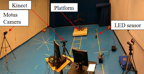 | Figure 1. Peak Motus and Kinect Systems Set up |
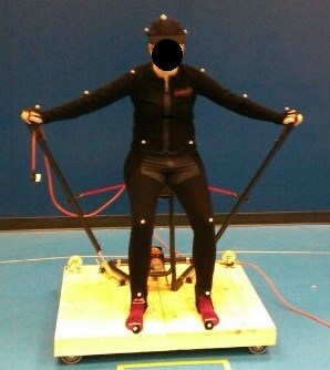 | Figure 2. Sitting Moveable platform |
2.3. Procedures
- Prior to any testing sessions, the equipment was set up as depicted in Figure 1. The Peak Motus set up involved assembling the calibration tree on a tripod as shown in Figure 3. The set up also entailed the use of two basler cameras, with two external lights mounted on the tripod of each camera and connected to the computer, as depicted in
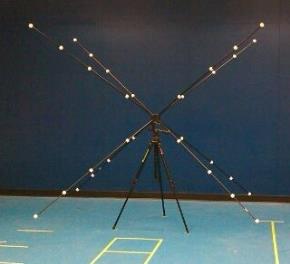 | Figure 3. Calibration Tree for Peak Motus |
2.4. Analysis
- For all trials, the Peak Motus and Microsoft Kinect software were used to collect raw kinematics data. Low pass Butterworth digital filters were used to condition the data and minimize high frequency noise. Excel Microsoft was used to compute displacement measures of each body joint. Evidence of reliability and validity were provided through the data analysis. Reliability is the degree to which the measures are consistent across replication of the test measures [31] and validity is the degree to which theoretical and empirical evidence support the inferences made from test score interpretations [31]. For reliability measures across replications of the test, interclass correlation coefficients were computed separately for the Kinect and Peak Motus systems between trial 1 and trial 2 for each of the fifteen body joints. For concurrent validity measures, interclass correlation coefficients were conducted to compare the Microsoft Kinect displacement measures to the standard Peak Motus displacement measures taken simultaneously for each of the fifteen body joints. These measures were conducted for trial 1 and trial 2 separately.
3. Results
- The results indicate that when comparing trial 1 and trial 2, the Peak Motus traditional system demonstrated a high degree of reliability determined by the ICC values, which can be seen in Figure 4. Fourteen out of the fifteen body joints had a reliability value above r=0.7, p<0.05, n=26; the left ankle exhibited the highest reliability value of r=0.815, p<0.05, n=26 and the head exhibited the lowest reliability value of r=0.534, p<0.05, n=26. The Microsoft Kinect system only demonstrated high reliability displacement measures between trial 1 and trial 2 for only three of the fifteen body joints as depicted by the ICC values charted in Figure 4. That is, the left shoulder, left and right knee had a reliability value above r= 0.7, p<0.05, n=26. Nine out of the fifteen body joints, however, had a reliability value below r=0.4, p<0.05, n=26.
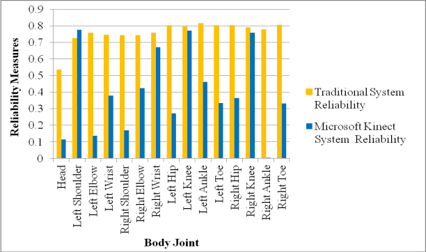 | Figure 4. Reliability of displacement measures from the traditional and Microsoft Kinect system |
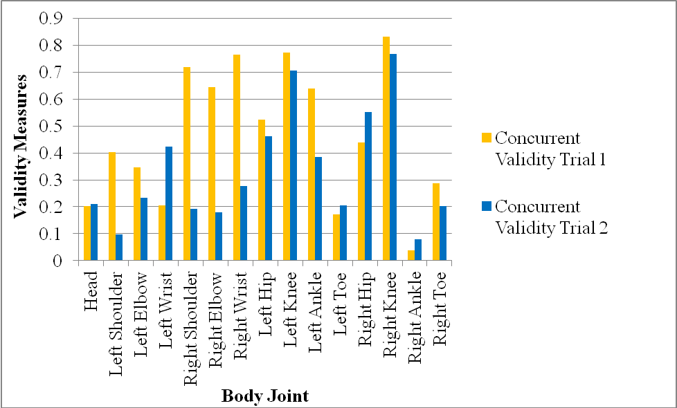 | Figure 5. Concurrent validity of displacement measures between the traditional and Microsoft Kinect system |
4. Discussions
- In the current study, the Peak Motus system produced results with a high degree of reliability across replications of the displacement measures for mostly all body joints as shown in Figure 4. This outcome was expected as the Peak Motus system is considered the gold standard to measure human movement kinematics in three dimensions [5, 20]. The medium correlation value obtained for the head across replications of the test when using the Peak Motus, may be attributed to the instances when it was necessary to switch from automatic to manual digitizing due to marker deviation from the original position. As stated by Richards [22] some automatic digitizing systems (e.g., Peak Motus) lack automatic tracking accuracy due to skin movement or closeness of the markers, which makes it difficult to study fine kinematic movements. The Microsoft Kinect system also provided strong evidence of reliability across replications of the displacement measures, but only for the left shoulder, left and right knee. This outcome, however, supports some of the findings from previous research, which stated that the Kinect system provided a high level of reproducibility of the results across replications of the tests [4]. For example Schmitz et al. [28] conducted a study to assess the reliability of the Kinect system in comparison to a traditional maker-based system. The researcher used a jig and placed it in three static positions: jig flexed, adducted and internally rotated. The researchers assessed the test-retest reliability of the kinect system when measuring angle movement and compared these measures to a 10 camera marker-based system. The researchers found very small differences in the reproducibility of the measures between the two systems across replications of the test. In our current study, however, part of the lack of reproducibility of displacement measures for some of joints across replications of the test when using the Kinect system, may be related to the position of the Kinect camera from the moving object. In our case, the distance from the kinect camera to the subject seating on top of the moving platform was 2 m, which exceeded the recommended distance of 1.8 meters to obtain more reliable results. As stated in the literature, the value of each pixel used to reproduce the image in three dimensions depends on the distance of what is being viewed from the device [19]. Based on this notion, the Kinect system seemed to recognize more consistently the joint locations that were closer to the camera such as the shoulder and knee joints as opposed to the joints locations that were further away. Furthermore, the high degree of reliability measures obtained from the Peak Motus system across replications of the experiment provided an indication that the moving platform was consistent from trial 1 to trial 2. Inconsistent displacement measures obtained from the Kinect system across replications of the experiment for some of the body joints, however, were more likely related to the kinect system not being able to reproduce the proper depth images, due to pixel value variability induced by exceeding the recommended distance from the camera to the human subject, sitting on top of the moving platform. When comparing the Peak Motus joint displacement measures to the Kinect system displacement measures to provide concurrent related evidence of validity, the results indicated that the two systems were highly related for only the left and right knee joints as shown in Figure 5. While this outcome partially supports the work of Clark et al [4], which found high correlation values between the Kinect system and the Peak Motus for upper and lower extremity body joints when performing three postural control tests such as: lateral reach, forward research and single leg standing balance, the results of the current study, however, seem promising and highlight the usefulness of the Kinect system in terms of cost and set up time. Although both systems were measuring displacement of each body joint horizontally and were put under the same scale in order to be compared for either trial 1 or trial 2, the lack of concurrent related evidence of validity for some of the body joints may be attributed to the Kinect system low reliability across replications of the test. Indeed, this outcome supports the notion that for the measurements of a test to be valid, they also need to be reliable [33]. Meaning that evidence of reliability and validity need to go hand in hand in order to provide appropriate and plausible inferences from test data interpretations. As stated by Kane [33], the term validation tends “to have two distinct but closely related usages. In the first usage ‘validation’ involves the development of evidence to support the proposed interpretations and uses... In the second usage ‘validation’ is associated with an evaluation of the extent to which the proposed interpretation and uses are plausible and appropriate” (p.17).Based on the outcome of this study and the literature [4, 19, 29], the Microsoft Kinect system provides desirable advantages over the traditional 3D human motion analysis systems (e.g., Peak Motus, Vicon). For example, the set up time is remarkably decreased as compared to Peak Motus. In addition, the Kinect system does not need to be calibrated; it is easily portable and considerably cheaper. Because it is a markerless system, the Kinect system does not have the potential problems associated with skin movement, set up time, proper marker location over anatomical landmarks, and the possibility of the markers interfering with the movement as compared to the Peak Motus system [4]. Furthermore, marker-based systems require the researcher to digitize the data to produce the results and this process, is time consuming; whereas, the Microsoft Kinect produces real time results when analyzing human movement kinematics.Another factor that may have affected the concurrent-related evidence of validity results was light reflections. Although automatic digitizing was mostly used with the traditional system, it was common to have to manually digitize the left and right foot as the light reflections from the external lights, made it difficult for the computer to recognize where the reflective markers were on the feet. This effect increased the amount of time spent analyzing the data from the traditional system and could have possibly been avoided by painting the platform black. However, Scholz [25] stated that light reflections cannot be completely eliminated and therefore will always create some error. Another issue to consider when providing evidence for the validation of the Kinect system in the current study was the ability of the Microsoft Kinect software to infer the position of a joint when it was not able to detect it. While this procedure is supposed to increase the reliability of the measures, in some instances, the results can also be skewed due to distortions of pixel values when constructing an image [20]. As stated by Galna et al. [15], the inaccuracies of the Microsoft Kinect in properly detecting body joints may be associated with the inability of the system to accurately estimate anatomical landmarks. These inaccuracies may be improved with better spatial resolution, more precise estimation of anatomical landmarks, and using optimal orientation of the Microsoft Kinect system relative to the subject [15].
5. Conclusions
- The purpose of this study was to provide evidence of reliability and validity for the use of a Microsoft Kinect system as a 3D human movement analysis system. The outcome of this study provides some evidence of reliability for the use of the Microsoft Kinect system to measure human movement kinematic of body joints. The outcome of this study also provides some concurrent-related evidence of validity when comparing the Kinect system displacement measures to the Peak Motus marker-based system displacement measures. From completing this research, it has been found that the Microsoft Kinect is much more favourable during the setup, data collection and analysis stages as compared to the Peak Motus. As Robertson et al. [23] and Corazza et al. [10] both stated and agreed that a markerless system would be a major breakthrough in the analysis of human motion and greatly would expand the application of human motion capture. Therefore, it would be worth continuing the research on the Microsoft Kinect as a tool to analyze human movement.There were several limitations associated with this study that should be addressed for future research. To begin with, using a small sample size of only twenty-six participants could have easily skewed the data distribution and affected the correlation coefficient estimates. A larger sample size may be recommended for future studies. In terms of the Peak Motus system, only two cameras were available to collect data, which could cause digitizing error due to passive reflector markers hiding during the motion. To minimize this possibility in the current study, only a subject frontal view was used to allow each passive reflector marker at each body joint to be viewed and captured simultaneously by both Peak Motus cameras and therefore, diminish digitizing error due to markers hiding during the motion. For more complicated human movement, however, it is recommended to use 3 to 6 cameras. The Microsoft Kinect system also has limitations that pertain to the proprietary skeleton software. The Microsoft Kinect software works by detecting body heat, however, it is unable to detect and track objects (e.g. hockey stick or baseball). Future software development should include modules to improve the ability of the Kinect skeleton software to create different spatial models, to assess human interaction with objects or tools for ergonomics, sport, and clinical applications. The system has also a limited viewing range to only 1.8 m. Both of these limitations hinder the system use as a 3D human motion analysis system. Future research studies should also explore different movement situations, as it is still unclear how the Kinect system behaves when measuring human movement kinematics in relation to walking, balance, jumping, jogging, running and hand gripping for clinical, work and sport related situations. Modifications to the Microsoft Kinect skeleton software may also be needed to make it more sensitive when tracking body joints at a further distance for static and dynamic movements. Finally, the outcome of this study supports the work of Schmitz et al. [28] and Clark et al. [4] and highlights the importance of positioning the Kinect camera at a proper distance from the subject when reconstructing an image. The outcome of the study also has implications on the use of the Kinect system as compared to the Peak Motus for work, clinical, and sport applications because it provides an avenue to conduct human movement kinematics analysis by minimizing cost and time when setting up equipment and identifying body joints.
ACKNOWLEDGEMENTS
- The researchers would like to thank the participants. The research would also like to thank Mr. Pretest Patel for designing and constructing the Kinect skeleton software and Mr. Dan Vasilliu for constructing the moving platform.