Resources and Environment

p-ISSN: 2163-2618    e-ISSN: 2163-2634

2017;  7(5): 138-144

doi:10.5923/j.re.20170705.04


Cellular Uptake and Metabolism of High Molecular Weight Polycyclic Aromatic Hydrocarbons by the White-rot Fungus Phanerochaete chrysosporium

Nomathemba Loice Chigu1, 2, Hirofumi Ichinose1

1Faculty of Agriculture, Kyushu University, Fukuoka, Japan

2School of Agricultural Sciences and Technology, Biotechnology Department, Chinhoyi University of Technology, Chinhoyi, Zimbabwe

Correspondence to: Nomathemba Loice Chigu, Faculty of Agriculture, Kyushu University, Fukuoka, Japan.

Email:

Copyright © 2017 Scientific & Academic Publishing. All Rights Reserved.

This work is licensed under the Creative Commons Attribution International License (CC BY).
http://creativecommons.org/licenses/by/4.0/

Abstract

Polycyclic aromatic hydrocarbons (PAHs) are lipophilic molecules that generally partition into lipid rich tissues in living organisms where they can be actively metabolized by cell-associated enzymes. In this study, cellular uptake and accumulation of triphenylene, benzo(a)pyrene, and coronene in lipid vesicles of the fungus Phanerochaete chrysosporium ATCC 34541 was investigated. Numerous bodies that stained with the lipid specific dye Sudan III were observed in fungal hyphae of P. chrysosporium grown with the high molecular weight (HMW) PAHs, implying that they could be taken in by the fungus and stored in lipid vesicles. Furthermore, the subsequent metabolism of the HMW PAHs by the fungus was investigated. This fungus degraded benzo(a)pyrene significantly and showed poor degradation activities for triphenylene and coronene suggesting that fungal intracellular accumulation could not essentially accompany degradation, therefore evaluating transport of compounds in cells may help to discern lack of substrate metabolism due to enzyme specificity or insufficient enzyme contact.

Keywords: High molecular weight polycyclic aromatic hydrocarbons, Cell accumulation, Intracellular enzymes, Lipid vesicles, Ionization potential

Cite this paper: Nomathemba Loice Chigu, Hirofumi Ichinose, Cellular Uptake and Metabolism of High Molecular Weight Polycyclic Aromatic Hydrocarbons by the White-rot Fungus Phanerochaete chrysosporium, Resources and Environment, Vol. 7 No. 5, 2017, pp. 138-144. doi: 10.5923/j.re.20170705.04.

1. Introduction

The white-rot fungus Phanerochaete chrysosporium shown in figure 1 has been shown to be an ubiquitous degrader of an extensive array of xenobiotics compounds ([4, 5, 7, 24, 32, 39, 56, 61]) including polycyclic aromatic hydrocarbons (PAHs).
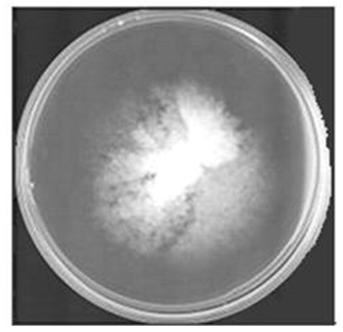
Figure 1. P. chrysosporium maintained on PDA media
A series of PAHs are considered to be organo-pollutants consisting of benzene analogs having two or more aromatic rings in various alignments ([27, 48]). They can be classified into low-molecular-weight (LMW) PAHs with up to three aromatic rings that are known to be very toxic [37], and high-molecular-weight (HMW) PAHs consisting of four and more aromatic rings recognized as highly mutagenic, teratogenic, and carcinogenic for both humans and animals [40]. Over the years, numerous authors have associated the ability of P. chrysosporium and other related fungi to be ubiquitous PAH degraders with the extracellular ligninolytic enzyme system, which includes lignin peroxidase (LiP), manganese peroxidase (MnP), versatile peroxidase (VP), and laccase (LAC) ([4, 8 17, 30, 44, 58]). However, the rates of degradation do not seem to always correlate with the activities of extracellular ligninolytic enzymes ([). An analysis of the catalytic properties of extracellular ligninolytic enzymes suggests that their role in PAH degradation is limited to a narrow range of compounds according to their ionization potentials (IPs) ([20, 26, 52, 53]). Under optimized conditions, the extracellular ligninolytic enzymes are capable of oxidizing PAHs with an IP <7.6. On the other hand, it has been known that cytochrome P450 monoxygenase (P450) activity would play important roles ( 3, 28]) in degradation of PAHs with a higher IP such as triphenylene, benzo(a)pyrene, and coronene. The earlier studies on fungal metabolism by P. chrysosporium ([15, 16, 38, 61]) suggest that the P450-mediated oxidation of PAHs might proceed under ligninolytic conditions.
The P450 enzymes are cell-associated enzymes capable of metabolizing substrates accumulated intracellularly. Many researchers have made an effort to elucidate cytochrome P450-dependent metabolic processes of fungi ([2, 3, 34, 35, 50, 61]). However, these processes are inevitably preceded by cellular uptake of the hydrophobic hydrocarbon in vivo. In the event of cellular uptake in living organisms, PAHs generally partition into lipid-rich tissues where they may be actively metabolized by the organisms’ cell-associated enzymes, or simply accumulated in fatty tissues ([1, 6, 27, 36, 43, 47]). However, the transport of PAHs inside fungal cells can be limited by the size and solubility of these compounds ([11, 27]) and subsequent metabolism impacted by the ionization potentials ([20, 26, 52, 53]). Therefore, it is of interesting to elucidate accumulation and location of PAHs in the fungal cells to better understand fungal metabolism of PAHs. Triphenylene (TRI), benzo(a)pyrene (B(a)P, and Coronene (COR) with properties listed in table 1 are large molecules that may not easily traverse cellular membranes and to become potential substrates for the cell-associated enzymes.
Table 1. Selected Physicochemical Properties of High Molecular Weight Polycyclic Aromatic Hydrocarbons Used in this Study
     
In this study, we examined the uptake of these HMW PAHs by P. chrysosporium cells through direct observation of Sudan III stained cells using fluorescence microscopy. In addition, the metabolism of these HMW PAHs by this fungus was investigated.

2. Materials and Methods

2.1. Chemicals

Anthracene, triphenylene, benzo(a)pyrene and coronene were purchased from Wako pure chemicals (Osaka, Japan). Sudan III was supplied from Kasayama. All other chemicals were reagent grade. Deionized water was obtained with a Milli Q System (Millipore).

2.2. Metabolism of PAHs by the Fungus P. chrysosporium

P. chrysosporium (ATCC 34541) was grown from conidia inocula at 37°C in a stationary culture under air (10 mL of medium in a 100-mL Erlenmeyer flask). The medium used in this study was as previously described [31] with 1% glucose and 1.2 mM ammonium tartrate at pH 4.5.
After a 4-day pre-incubation, substrate was added to the cultures to become final concentration of 25 µM. Coronene in dimethyl formamide (2.5 mM), triphenylene and benzo(a)pyrene in acetonitrile (2.5 mM) were utilized substrates-stock solution. Anthracene (2.5 mM) was used for co-metabolic studies with coronene. NaN3 (1 M) treated cultures were used as controls in the biodegradation studies. To detect abiotic degradation of substrates, blanks without fungi were prepared and processed analogously. Cultures were incubated at 37°C without agitation under 100% oxygen for periods ranging from 20-30 days. Periodically harvested cultures and controls were homogenized in triplicates with 20 ml acetonitrile, centrifuged at 25,000 x g for 10 min at 4°C to separate the mycelium from the aqueous fraction and filtered with a membrane (0.45 µm). The aqueous fraction obtained was analyzed by HPLC.

2.3. High Performance Liquid Chromatography Analysis

HPLC analysis of HMW PAH metabolism was done with a Shimadzu STR ODS-II column (4.6 by 150 mm I.D.) Detection of triphenylene and benzo(a)pyrene metabolites was achieved at 257 and 254 nm respectively with a linear gradient from 20% acetonitrile in water, isocratic for 5 mins, to a 100% acetonitrile (21-31 min) at a flow rate of 1.0 ml/min. Coronene samples were detected at 304 nm with a linear gradient from 50% acetonitrile in water, isocratic for 5 mins, to a 100% acetonitrile (21-31 min) at a flow rate of 1.0 ml/min.

2.4. Fluorescence Observations

Cultures were grown at 37°C without agitation under 100% oxygen for five days after substrate addition. Fungal samples were collected from liquid cultures containing no PAH (control sample) or one of the different PAHs used. Observations were conducted with a fluorescent microscope (Leica). PAHs fluorescence wavelengths range from 210 -380 nm.

2.5. Sudan III Staining Protocol

Stock solution of Sudan III (0.2% w/v) was prepared using 70% (v/v) ethanol and warmed in a water bath. The solution was then cooled down to room temperature, filtered and kept in dark at room temperature. Fungal cells were washed thrice with distilled water, and twice with acetone to remove PAH residues. The cells were immersed in the working solution of Sudan III in the dark for 20 min. Hereafter sections were washed with 3 exchanges of deionized water for 30s each. Cells were mounted for observation in an aqueous medium and or glycerin [21].

3. Results and Discussion

3.1. Uptake of Polycyclic Aromatic Hydrocarbons by P. chrysosporium

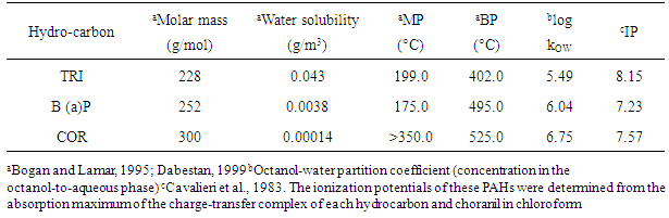
PAHs are lipophilic molecules that are easily incorporated into lipid vesicles for transport and storage ([43, 54]). PAH containing elements in these lipid vesicles can be visualized with Sudan III and other dyes to stain lipid droplets for fluorescence microscopy. In this study, three recalcitrant HMW PAHs (Figure 2) were separately added to cultures of P. chrysosporium after a 4-day pre-incubation. Their uptake by the fungus during its cultivation was investigated using fluorescence microscopy. The results obtained were compared to those analogously done in the absence of PAHs.
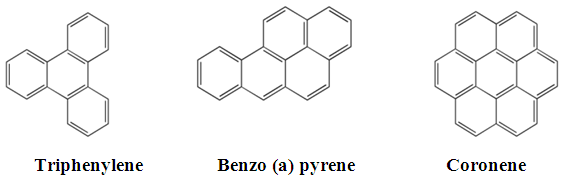
Figure 2. Chemical structures of HMW PAHs used in this study
Figure 3 depicts light microscopic images of fungal cells. Fungal cells incubated with PAH showed small inclusions colored rose-red (Figs. 3A, 3B and 3C) whereas rose-red inclusions were not seen in fungal cell hyphae grown in the absence of PAHs (Figs. 3D). These small inclusions are Sudan III stained lipid vesicles containing PAHs. According to previous literature (), the lipid vesicles in hyphae were also the sites to accumulate PAHs. The absence of these lipid vesicles in fungal cell hyphae grown in the absence of PAHs (Figs. 3D) indicates that these are produced in response to the presence of PAHs in culture. Thus, it is reasonable to speculate that the small inclusions colored rose-red are used as vehicles of transport and storage of polycyclic aromatic hydrocarbons.
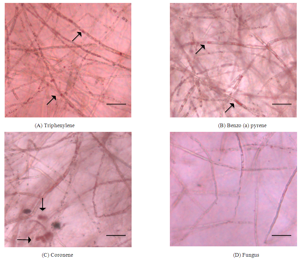
Figure 3. Light microscopic observation of the intracellular elements in P. chrysosporium grown in the presence of (A) - (C) and (D) in the absence of PAHs. The PAH containing lipid vesicles are stained red in the hyphae (20X objective lens): bright field images. Bar = 5 μm
This is in agreement with the report of , who found that the active microorganisms can be viewed both as a biosorbent that retains organic pollutants as well as a bioreactor that degrades them. A similar observation was reported using Fusarium solani that could store PAHs intracellularly in lipid vesicles [54]. It was also shown that the intracellular storage of the PAHs was not restricted to the imperfect fungus F. solani, but could be found in numerous other fungi belonging to different genera including P. chrysosporium P11 [54]. It has been reported that the transport of PAHs inside fungal cells can be limited by the size and solubility of these compounds [11]. However, we revealed that the HMW PAHs used in this study were accumulated in hyphae of P. chrysosporium fungal cells, indicating that PAH transport into fungal cells is not impacted only by the size and solubility of the PAH. This is also in agreement with the results reported by [19] where an isolate of the fungus F. solani incorporated more BaP into cells than phenanthrene (PHE), despite the 400-fold higher aqueous solubility of PHE compared with BaP.

3.2. Metabolism of HMW PAHs by the White-Rot Fungus P. chrysosporium

In , [55] demonstrated that fluorescent PAHs concentrate in the lipid droplets of fungi, which sequester these noxious compounds and perhaps metabolize them to less toxic derivatives. After evaluating the uptake of HMW PAHs, a further study was conducted to evaluate the potential ability of fungal degradation of these high molecular weight PAHs (shown in figure 2). The fungus P. chrysosporium degraded Benzo (a) pyrene significantly the results of which have been published [15]. In contrast, the fungus P. chrysosporium showed poor degradation of both triphenylene and coronene (figure 4). Cometabolism of coronene with anthracene, a relatively simple PAH can have multimechanistic effects on coronene metabolism, such as inhibition or induction [59]. In this study, the presence of anthracene in fungal cultures slightly improved the degradation of coronene.
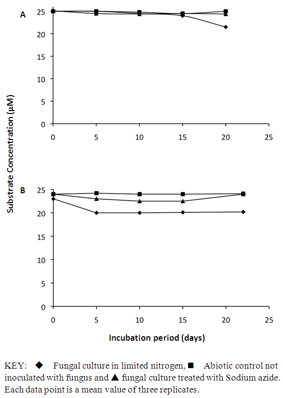
Figure 4. Time course of (A) Triphenylene and (B) Coronene metabolism by the white rot fungus P. chrysosporium ATCC 34541
Intracellular cytochrome P450 monooxygenase is believed to initially metabolize PAH molecules, having an IP of 8.03 eV ([2, 3, 34, 50]) or higher. The IPs of both triphenylene and coronene suggest that these PAHs would be ideal substrates for the intracellular cytochrome P450 monooxygenase and not the ligninolytic enzyme system ([2, 3, 38]). Poor degradation of both coronene and triphenylene could be as a result of their aromaticity that confers electrochemical stability to these compounds. Triphenylene is highly symmetrical and much more resonance stable than its isomers chrysene, benz(a)anthracene, benzo(c)phenanthrene and naphthacene (tetracene). On the other hand, coronene is a highly condensed PAH with seven benzene rings fused together. These attributes hinder the removal of electrons to participate in reactions during a cytochrome P450 monoxygenase activity. This result is in agreement with that reported by [54] that showed that P. chrysosporium was a poor degrader of the compound coronene. Of the PAHs used in this study, B(a)P has an IP of 7.23 making it amenable to biodegradation by both the ligninolytic and cytochrome P450 monoxygenase fungal systems. Benzo (a) pyrene can easily form radical cations by the removal of one electron ([10, 42]), the ease of formation of which depends on the IP of the PAH.
Correlation between the number of aromatic rings of the PAHs, their percentage degradation and solubilities was analyzed. Triphenylene has the least number of aromatic rings, is the most soluble of the HMW PAHs used in this study but showed the least percentage degradation (Figure 5) by the fungus P. chrysosporium. This suggests that no correlation exists between the number of aromatic rings of the PAHs, their solubilities and the extent of degradation. This is in contrast to results described by [12] which showed that degradation was inversely proportional to the number of rings in the PAH molecule.
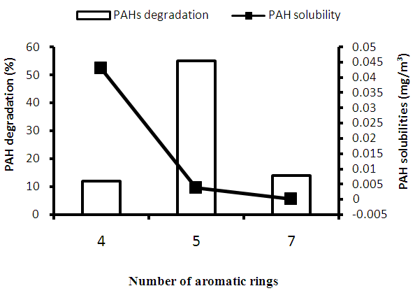
Figure 5. Correlation between the number of aromatic rings of PAHs, their % degradation and solubilities. 4, Triphenylene; 5, Benzo (a) pyrene; 7, Coronene
More attention should be given to studies on the fate of PAH metabolites outside and inside of fungal cell. The result obtained here may be used as a template in future studies to design more improved and useful systems for the study of cytochrome P450s and other intracellular enzymes.

4. Conclusions

This study is one of the few concerned with the correlation between substrate uptake and metabolism. P. chrysosporium cells grown in the presence of triphenylene, benzo (a) pyrene, and coronene accumulated these PAHs and showed small intensely stained rose-red inclusions that were not observed in fungal cell hyphae lacking the PAHs. This fungus degraded Benzo (a) pyrene significantly but showed poor degradation of Triphenylene and Coronene. Therefore, evaluating transport of these recalcitrant HMW PAH compounds in the cells of P. chrysosporium help to discern lack of attack due to enzyme specificity or due to insufficient enzyme contact. This effort is of both a practical and theoretical interest. Effort to engineer modified enzymes or pathways ([14, 29]) for degradation of recalcitrant compounds would be wasted if the recalcitrance were essentially due to a transport problem.

References

[1]  Baussant, T., Sanni, S., Jonsson, G., Skadsheim, A., Borseth, F., 2001, Bioaccumulation of polycyclic aromatic compounds: 1. Bioconcentration in two marine species and in semipermeable membrane devices during chronic exposure to dispersed crude oil. Environmental and Toxicological Chemistry 20, 1175–1184.
[2]  Bezalel, L., Hadar, Y. Fu, P. P. Freeman, J. P. and Cerniglia C. E., 1996a, Initial oxidation products in the metabolism of pyrene, anthracene, fluorine, and dibenzothiophenone by the white-rot fungus Pleurotus ostreatus. Appl. Environ. Microbiol., 62: 2554- 2559.
[3]  Bezalel, L., Hadar, Y. Fu, P. P. Freeman, J. P. and Cerniglia C. E. 1996b. Metabolism of Phenanthrene by the White Rot Fungus Pleurotus ostreatus. Appl. Environ. Microbiol., 62: 2547-2553.
[4]  Bogan, B. W., Lamar, R. T. and Hammel K. E., 1996, Fluorene oxidation in vivo by Phanerochaete chrysosporium and in vitro during manganese peroxidase-dependent lipid peroxidation. Appl. Environ. Microbiol., 62: 1788- 1792.
[5]  Bogan, B. W. and Lamar R. T., 1995, One-electron oxidation in the degradation of creosote polycyclic aromatic hydrocarbons by Phanerochaete chrysosporium. Appl. Environ. Microbiol., 61: 2631-2635.
[6]  Boom M. M., 1987, The determination of polycyclic aromatic hydrocarbons in indigenous and transplanted mussels (Mytilus edulis L.) along the Dutch Coast. Intern. J. Environ. Anal. Chem. 31:251–261.
[7]  Bumpus J.A, and Aust S.D., 1987, Biodegradation of environmental pollutants by the white rot fungus Phanerochaete chrysosporium: involvement of the lignin degrading system. Bioessays 6: 166-170.
[8]  Camacho-Morales, R.L., Gerardo-Gerardo, J.L., Guillén Navarro, K., and Sánchez J.E., 2017, Ligninolytic enzyme production by white rot fungi during paraquat (herbicide) degradation, Rev Argent Microbiol., 49(2):189-196.
[9]  Cavalieri E.L, Rogan E.G, Roth R.W, Saugier K.R and Hakam A., 1983, The relationship between ionization potential and horseradish peroxidase/hydrogen peroxide catalyzed binding of aromatic hydrocarbons to DNA. Chem. Biol. Interactions 47: 87-109.
[10]  Cavalieri E, Rogan E, and Roth R., 1982, Multiple mechanisms of activation in aromatic hydrocarbon carcinogenesis, in Floyd RA (ed). Free radicals and cancer. Marcel Dekker, New York, pp 117-158.
[11]  Cerniglia C. E., 1993, “Biodegradation of polycyclic aromatic hydrocarbons,” Current Opinion in Biotechnology, vol. 4, no. 3, pp. 331–338.
[12]  Cerniglia, C. E., 1992, Biodegradation of polycyclic aromatic hydrocarbons. Biodegradation, 3: 351-368.
[13]  Chen, B. L., Wang, Y. S., and Hu, D. F., 2010, Biosorption and biodegradation of polycyclic aromatic hydrocarbons in aqueous solutions by a consortium of white-rot fungi. J. Hazard. Mater. 179, 845–851.
[14]  Chen W., Zheng L., Jia R., Wang N., 2015, Cloning and expression of a new manganese peroxidase from Irpex lacteus F17 and its application in decolorization of reactive black 5. Process Biochem., 50:1748–59.
[15]  Chigu N.L., Hirosue S., Nakamura C., Teramoto H., Ichinose H., and Wariishi H., 2016, Gene expression profiling and enzymatic function of Phanerochaete chrysosporium cytochrome p450s involved in the metabolism of benzo(a)pyrene. International Journal of Environmental Bioremediation & Biodegradation. 4( 3):85-92.
[16]  Chigu N.L., Hirosue S., Nakamura C., Teramoto H., Ichinose H., Wariishi H., 2010, Cytochrome P450 monooxygenases involved in anthracene metabolism by the white-rot basidiomycete Phanerochaete chrysosporium, Appl. Microbiol. Biotechnol. 87(5): 1907-16.
[17]  Collins, P. J., Kotterman, M. Field, J. A. and Dobson A., 1996, Oxidation of anthracene and benzo[a]pyrene by laccases from Trametes versicolor. Appl. Environ. Microbiol., 62: 4563- 4567.
[18]  Dabestani R, and Ivanov I., 1999, “A compilation of physical, spectroscopic and photo physical properties of poly aromatic hydrocarbons.” Photochem. Photobio. 70: 10- 34.
[19]  Fayeulle, A., Veignie, E., Slomianny, C., Dewailly, E., Munch, J., and Rafin, C., 2014, Energy-dependent uptake of benzo[a]pyrene and its cytoskeleton-dependent intracellular transport by the telluric fungus Fusarium solani. Environ. Sci. Pollut. Res. Int. 21, 3515–3523.
[20]  Field J. A., Vledder R. H., Van Zelst J. G., and Rulkens W. H., 1996, “The tolerance of lignin peroxidase and manganese dependent peroxidase to miscible solvents and the in vitro oxidation of anthracene in solvent: water mixtures,” Enzyme and Microbial Technology, vol. 18, no. 4, 300–308.
[21]  Fukumoto S and Fujimoto T. 2002. Deformation of lipid droplets in fixed samples. Histochem. Cell biol. 118: 423-428.
[22]  Furuno, S., Foss, S., Wild, E., Jones, K. C., Semple, K. T., Harms, H., 2012, Mycelia promote active transport and spatial dispersion of polycyclic aromatic hydrocarbons. Environ. Sci. Technol. 46, 5463–5470.
[23]  Gramss G, Kirsche B, Voight KD, Gunther T & Fritsche W., 1999, Conversion rates of five polycyclic aromatic hydrocarbons in liquid cultures of fifty-eight fungi and the concomitant production of oxidative enzymes. Mycol Res 103: 1009–1018.
[24]  Gong X, Li S, Sun X, Zhang L, Zhang T, and Wei L., 2017, Maturation of green waste compost as affected by inoculation with the white-rot fungi Trametes versicolor and Phanerochaete chrysosporium, Environ Technol. 38(7): 872-879.
[25]  Haemmerli, S. D., Leisola, M. S. A Sanglard, D. and Fiechter A., 1986, Oxidation of benzo(a)pyrene by extracellular lignases of Phanerochaete chrysosporium: veratryl alcohol and stability of lignases. J. Biol. Chem., 261: 6900-6903.
[26]  Hammel K. E., Kalyanaraman B., and Kirk T. K., 1986, “Oxidation of polycyclic aromatic hydrocarbons and dibenzo(p)dioxins by Phanerochaete chrysosporium ligninase,” Journal of Biological Chemistry, vol. 261, no. 36, pp. 16948–16952.
[27]  Hussein, I., Abdel-Shafy, Mona, S.M., and Mansour, 2016, A review on polycyclic aromatic hydrocarbons: Source, environmental impact, effect on human health and remediation, Egyptian Journal of Petroleum, 25, (1), 107-123.
[28]  Kadri T, Rouissi T, Kaur Brar S, Cledon M, Sarma S, and Verma M., 2017, Biodegradation of polycyclic aromatic hydrocarbons (PAHs) by fungal enzymes: A review. J Environ Sci (China). 51:52-74.
[29]  Kellner, D. G., Mavest, S. A. and Sligar S. G., 1997, Engineering cytochrome P450s for bioremediation. Curr. Opin. Biotechnol., 8: 274-278.
[30]  Kim M. I. S., Huh E. J., Kim H. K., and Moon K.W., “Degradation of polycyclic aromatic hydrocarbons by selected white-rot fungi and the influence of lignin peroxidase,” Journal of Microbiology and Biotechnology, vol. 8, no. 2, pp. 129–133, 1998.
[31]  Kirk, T. Schultz, K., E. Connors, W. J. Lorenz, L. F. and Zeikus J. G., 1978, Influence of culture parameters on lignin metabolism by Phanerochaete chrysosporium. Arch. Microbiol., 117: 277-285.
[32]  Kullman, S. W., and Matsumura F., 1996, Metabolic pathways utilized by Phanerochaete chrysosporium for degradation of the cyclodiene pesticide endosulfan. Appl. Environ. Microbiol. 62: 593-600.
[33]  Martinez D, Larrondo LF, Putnam N, Gelpke MD, Huang K, Chapman J, Helfenbein KG, Ramaiya P, Detter JC, Larimer F, Coutinho PM, Henrissat B, Berka R, Cullen D, and Rokhsar D., 2004, Genome sequence of the lignocellulose degrading fungus Phanerochaete chrysosporium strain RP78. Nat. Biotechnol. 22: 695-700.
[34]  Masaphy, S., Levanon, D. Henis, Y. Venkateswarlu K. and Kelly S. L., 1996, Evidence for cytochrome-P450 and P450 mediated benzo(a)pyrene hydroxylation in the white rot fungus Phanerochaete chrysosporium. FEMS Microbiol. Lett., 135: 51-55.
[35]  Masaphy, S., Levanon, D. Henis, Y. Venkateswarlu K. and Kelly S. L., 1995, Microsomal and cytosolic cytochrome P450 mediated benzo(a)pyrene hydroxylation in Pleurotus pulmonarius. Biotechnol. Lett., 17: 969- 974.
[36]  Meador JP, Stein JE, Reichert WL, Varanasi U., 1995, Bioaccumulation of polycyclic aromatic hydrocarbons by marine organisms. Rev Environ Contam Toxicol.; 143: 79-165.
[37]  Moore M., Livingstone D., and Widdows J., 1989, “Hydrocarbons in marine mollusks: biological effects and ecological consequences,” in Metabolism of Polycyclic Aromatic Hydrocarbons in the Aquatic Environment, U. Varanishi, Ed., pp. 291–328, CRC Press, Boca Raton, Fla, USA.
[38]  Ning D., Wang H., Ding, C. and Lu, H., 2010, “Novel evidence of cytochrome P450-catalyzed oxidation of phenanthrene in Phanerochaete chrysosporium under ligninolytic conditions,” Biodegradation, vol. 21, no. 6, pp. 889–901.
[39]  Olicón-Hernández D.R., González-López J., and Aranda E., 2017, Overview on the Biochemical Potential of Filamentous Fungi to Degrade Pharmaceutical Compounds. Front. Microbiol. 8:1792.
[40]  Pahlman R. and Pelkonen O., 1987, “Mutagenicity studies of different polycyclic aromatic hydrocarbons: the significance of enzymatic factors and molecular structure,” Carcinogenesis, vol. 8, no. 6, pp. 773–778.
[41]  Prough, A. Saeki, R., Y. and Capedevilla J., 1981, The metabolism of benzo[a]pyrene phenols by rat liver microsomal fractions. Arch. Biochem. Biophys., 212: 136-146.
[42]  Rogan E, Roth R, Katomski P, Benderson J and Cavalieri E., 1978, Binding of benzo[a]pyrene at the 1,3,6 positions to nucleic acids in vivo on mouse skin and in vitro with rat liver microsomes and nuclei. Chem. Biol. Interact. 22: 35.
[43]  Rufina, C.C., Hoffman, C.L., Opara, L.U., O’Neill, B., Stander, A.M., 2017, Polycyclic Aromatic Hydrocarbons (PAHs) and Organochlorinated Pesticides (OCPs) in Yellowtail (Seriola lalandi) from three Spatially Distinct Locations along the Coast of South Africa: Levels, Sources and Fish Size Effect. J. Food Process Technol, 8: 644.
[44]  Sack U., Hofrichter M., and Fritsche W., 1997, “Degradation of polycyclic aromatic hydrocarbons by manganese peroxidase of Nematoloma frowardii,” FEMS Microbiology Letters, vol. 152, no. 2, pp. 227–234.
[45]  Schützendübel, A., Majcherczyk A., Johannes, C. and Hüttermann A., 1999, Degradation of fluorine, anthracene, phenanthrene, fluoranthene, and pyrene lacks connection to the production of extracellular enzymes by Pleurotus ostreatus and Bjerkandera adusta, Int. biodeterioration and biodegradation, 43: 93-100.
[46]  Shishlyannikov, S.M., Nikonova, A.A., Klimenkov, I.V., 2017, Accumulation of petroleum hydrocarbons in intracellular lipid bodies of the freshwater diatom Synedra acus subsp. radians. Environ Sci Pollut Res 24: 275.
[47]  Simmonich, S.L., Hites, R.A., 1994, Vegetation–atmosphere partitioning of polycyclic aromatic hydrocarbons. Environmental Sciences Technology 28, 939–943.
[48]  Soclo H.H., Budzinski H., Garrigues Ph and Matsuzawa S., 2008, Biota accumulation of polycyclic aromatic hydrocarbons in benin coastal waters Polycyclic Aromatic Compounds, 28: 112–127.
[49]  Sutherland, J. Rafii B., F., Khan A. A., and Cerniglia. C. E., 1995, Mechanisms of polycyclic aromatic hydrocarbon degradation. In L. Y. Young and C. E. Cerniglia (ed.), Microbial transformation and degradation of toxic organic chemicals. Wiley-Liss, New York, N.Y.
[50]  Sutherland, J. B., Selby, A. L. Freeman, J. P. Evans, F. E. and Cerniglia C. E., 1991, Metabolism of phenanthrene by Phanerochaete chrysosporium. Appl. Environ. Microbiol., 57: 3310-3316.
[51]  Thion, C., Cebron, A., Beguiristain, T., and Leyval, C., 2012, PAH biotransformation and sorption by Fusarium solani and Arthrobacter oxydans isolated from a polluted soil in axenic cultures and mixed co-cultures. Int. Biodeterior. Biodegra. 68, 28–35.
[52]  Torres E., Tinoco R., and Vazquez-Duhalt R., 1997, “Biocatalytic oxidation of polycyclic aromatic hydrocarbons in media containing organic solvents,” Water Science and Technology, vol. 36, no. 10, pp. 37–44.
[53]  Vazquez-Duhalt R., Westlake D. W. S., and Fedorak P. M., 1994, “Lignin peroxidase oxidation of aromatic compounds in systems containing organic solvents,” Applied and Environmental Microbiology, vol. 60, no. 2, pp. 459–466.
[54]  Verdin A., Sahraoui, A. L. Newsam, R. Robinson G. and Durand. R., 2005, Polycyclic aromatic hydrocarbon storage by Fusarium solani in intracellular lipid vesicles. Environ. Pollut. 133:283-291.
[55]  Verdin, A., Lounes-Hadj Sahraoui A., and Durant R., 2004, Degradation of benzo (a) pyrene by mitosporic fungi and extracellular oxidative enzymes. International Biodeterioration and Biodegradation. 53, 65-70.
[56]  Vinay Mohan Pathak and Navneet, 2017, Review on the current status of polymer degradation: a microbial approach, Bioresources and Bioprocessing, 4:15.
[57]  Wiseman, A., 1980, Xenobiotic-metabolising cytochromes P-450 from microorganisms. Trends Biochem. Sci., 5: 102–104.
[58]  Xing Qin, Xianhua Sun, Huoqing Huang, Yingguo Bai, Yuan Wang, Huiying Luo, Bin Yao, Xiaoyu Zhang, Xiaoyun Su, 2017, Oxidation of a non-phenolic lignin model compound by two Irpex lacteus manganese peroxidases: evidence for implication of carboxylate and radicals, Biotechnology for Biofuels, 10:103.
[59]  Xuezhu Zhu, Xue Ni, Michael Gatheru Waigi, Juan Liu, Kai Sun and Yanzheng Gao, 2016, Biodegradation of Mixed PAHs by PAH-Degrading Endophytic Bacteria, International Journal of Environmental Research and Public Health, 13, 805.
[60]  Yadav, J. S., and Reddy C. A., 1992, Non-involvement of lignin peroxidases and manganese peroxidases in 2, 4, 5- trichlorophenoxy-acetic acid degradation by Phanerochaete chrysosporium. Biotechnol. Lett., 14: 1089-1092.
[61]  Zahmatkesh, M., Spanjers, H., Toran, M.J., Blánquez, P., and van Lier, J.B., 2016, Bio-removal of humic acid from water by white rot fungi: exploring the removal mechanisms, AMB Express., 6(1): 118.