
-
Paper Information
- Paper Submission
-
Journal Information
- About This Journal
- Editorial Board
- Current Issue
- Archive
- Author Guidelines
- Contact Us
Research In Cancer and Tumor
2026; 13(2): 30-33
doi:10.5923/j.rct.20261302.03
Received: Oct. 19; Accepted: Nov. 8, 2026; Published: Nov. 13, 2026

Key Features of Tumor with an Infiltrative Growth Pattern for Breast Cancer
 Abstract
 Reference
 Full-Text PDF
 Full-text HTML
L. T. Alimkhodjaeva, O. M. Akhmadjanova, S. S. Ziyavidenova
Republican Specialized Scientific-Practical Medical Center of Oncology and Radiology, Tashkent, Uzbekistan
Copyright © 2026 The Author(s). Published by Scientific & Academic Publishing.
This work is licensed under the Creative Commons Attribution International License (CC BY).
http://creativecommons.org/licenses/by/4.0/

The aim of the study was to improve the prognosis and treatment outcomes for patients with infiltrating breast cancer by improving modern methods of molecular genetic diagnostics. Background. Breast cancer remains one of the major challenges in both basic and clinical oncology, as its incidence remains the highest among malignant tumors in women. Despite significant advances in the early diagnosis and treatment of breast cancer, the mortality rate from the disease continues to be high and is showing a persistent upward trend. Currently, the prevalence of infiltrating breast cancer remains unclear, as evidenced by the wide range of incidence rates reported by various authors—from 2 to 62 percent. Material and Methods. This study was conducted at the Republican Specialized Scientific-Practical Medical Center of Oncology and Radiology and all its branches between 2020 and 2024. The research material consisted of 128 tumor tissue biopsy samples, retrieved from the archives, from patients undergoing treatment for breast cancer. Results. Among the primary structures of the tumor’s parenchymal component, we identified large- and small-alveolar, trabecular, cribriform, and tubular structures. The frequency distribution of these structures varied significantly depending on the histological subtype of breast cancer. For instance, trabecular structures were the most common feature in cases of invasive lobular carcinoma, accounting for 89% of all observed structures, whereas they were entirely absent in cases of invasive ductal carcinoma. Conclusion. Thus, overall, it can be concluded that the invasive ductal carcinoma variant of breast cancer, based on its microscopic features, is characterized by a statistically significantly higher degree of malignancy, higher mitotic activity of tumor cells, a higher degree of inflammatory infiltration of the stromal component, and the presence of cribriform structures and necrotic processes in the parenchyma. The invasive lobular carcinoma variant of breast cancer is characterized by pronounced stromal hyalinosis and infiltration of adipose tissue, a predominance of trabecular structures in the parenchyma, and the presence of “dark” cells.
Keywords: Breast cancer, Infiltrating breast cancer, Invasive lobular carcinoma, Invasive ductal carcinoma
Cite this paper: L. T. Alimkhodjaeva, O. M. Akhmadjanova, S. S. Ziyavidenova, Key Features of Tumor with an Infiltrative Growth Pattern for Breast Cancer, Research In Cancer and Tumor, Vol. 13 No. 2, 2026, pp. 30-33. doi: 10.5923/j.rct.20261302.03.
Article Outline
1. Introduction
- Breast cancer (BC) remains one of the major challenges in both basic and clinical oncology, as its incidence remains the highest among malignant tumors in women [1-3]. There has been rapid progress in understanding the fundamental mechanisms underlying the development and growth of these tumors recently. Despite significant advances in the early diagnosis and treatment of breast cancer, the mortality rate from the disease continues to be high and is showing a persistent upward trend [1,4-5]. The study of the morphological mechanisms underlying the origin and development of tumors has been and remains a pressing challenge in medicine and requires the use of modern research and treatment methods to address both theoretical and practical issues in oncology. Currently, the prevalence of infiltrating breast cancer remains unclear, as evidenced by the wide range of incidence rates reported by various authors—from 2 to 62 percent (Semiglazov V.F..., 2016; Vlastos G. et al., 2000). Issues related to the diagnosis, selection of appropriate treatment, and prognosis of infiltrating breast cancer remain particularly challenging. Findings on the prognostic significance of infiltrating breast cancer cells are controversial. Some authors point out that true primary resistance does not affect the prognosis and occurs with equal frequency in patients with both favorable and unfavorable disease outcomes; other researchers consider multifocal tumor growth to be one of the most significant factors determining the risk of disease recurrence [6-8]. There is no consensus regarding the extent of lymphatic metastasis in this form of breast cancer. Infiltrating breast cancer is a significant problem largely due to the characteristics of its clinical course, the lack of clear criteria for preoperative diagnosis, insufficient information regarding its prognostic significance, and the need to select optimal treatment methods, particularly breast-conserving surgeries. [9-10].The aim of the study was to improve the prognosis and treatment outcomes for patients with infiltrating breast cancer by improving modern methods of molecular genetic diagnostics.
2. Material and Methods
- General characteristics of infiltrating breast cancer clinical observationsThis study was conducted at the Republican Specialized Scientific-Practical Medical Center of Oncology and Radiology and all its branches between 2020 and 2024. The research material consisted of 128 tumor tissue biopsy samples, retrieved from the archives, from patients undergoing treatment for breast cancer. In addition to biopsy specimens, the analysis included medical history, clinical and laboratory data, and other materials from the medical records of the patients mentioned above. We analyzed primary documents such as medical records and outpatient charts of the patients. Based on data obtained from medical records and in accordance with the study’s objectives and goals, we analyzed microscopic specimens of infiltrating breast cancer by examining the following data from the patients’ medical records:- age;- clinical stage of the disease;- results of immunohistochemical analysis of tumor tissue for the expression of estrogen, progesterone, and HER-2neu receptors.As part of their combination therapy, all patients underwent 8 courses of preoperative chemotherapy using the CAF regimen (Adriamycin 30 mg/m² IV, on days 1 and 8, 5-fluorouracil 500 mg/m² IV on days 1 and 8, and cyclophosphamide 100 mg/m² IM on days 1 and 8), followed by surgical intervention consisting of organ-preserving and radical mastectomy. During the postoperative period, patients received adjuvant chemotherapy based on medical indications and their clinical condition. antiestrogen therapy with tamoxifen and distant gamma therapy to the regional lymphatic drainage areas were administered based on clinical indications. In cases of HER2/neu-positive status, targeted therapy with Herceptin was additionally administered.The diagnosis of breast cancer was established according to the “Histological Classification of Breast Tumors” (WHO, 2003). The tumor was classified as either infiltrating lobular carcinoma or ductal carcinoma. A diagnosis of infiltrating ductal carcinoma was established in 118 (88.7%) patients, infiltrating lobular carcinoma - in 8 (6.0%) patients, and other forms (mucinous, solid, medullary) - in 7 (5.3%) cases. Infiltrating ductal carcinoma, which occurred in 88.7% of cases, was predominantly of histological grade 2 and was the most common type across all study groups. The patient groups studied were comparable in terms of key clinical and morphological parameters. The surgical specimens were subjected to morphological examination. Sample tissues were placed in a fixative solution—neutral formalin. The specimens were processed according to standard procedures and embedded in paraffin. Sections 5–6 µm thick were stained with hematoxylin and eosin. Morphological examination was performed using a light microscope manufactured by “Carl Zeiss Microimaging,” (Germany). The center and periphery of macroscopically detectable tumor nodules, breast tissue from the four quadrants outside the tumor, and all axillary lymph nodes were evaluated. The center and peripheral regions of the tumor mass were examined, and the ductal structures and infiltrative component were assessed. Within the tumor ducts, solid, cribriform, and papillary structures were identified. In the infiltrative component, trabecular, tubular, microalveolar, solid, and cribriform structures and separate small clusters of tumor cells were identified. Parenchymal structures were assessed using a semiquantitative method based on a 5-point scale (from 1 to 5). In addition, each of the four quadrants of the breast was examined for the presence of possible tumor microfoci. The condition of all axillary lymph nodes was assessed (from 3 to 45 lymph nodes were examined). Metastases were noted.The data obtained were analyzed using the “Statistica 6.0 for Windows” software package. The Student’s t-test for dependent and independent samples, the Mann-Whitney U test, and analysis of variance (ANOVA) were used.
3. Results
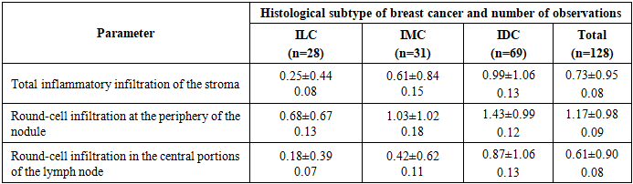
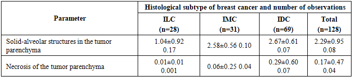
- To identify differences in the morphological characteristics of tumor tissue from different histological subtypes of invasive breast cancer, all parameters used in the histological description were divided into two groups. The first group consisted of qualitative nominal characteristics, which included the frequencies of the main structures of the parenchymal component and the main structures invading the stroma of the tumor nodule at the periphery. All other descriptive histological parameters were classified as semi-quantitative, and their severity was evaluated on a point scale. Among the primary structures of the tumor’s parenchymal component, we identified large- and small-alveolar, trabecular, cribriform, and tubular structures. The frequency distribution of these structures varied significantly depending on the histological subtype of breast cancer. For instance, trabecular structures were the most common feature in cases of invasive lobular carcinoma (ILC), accounting for 89% of all observed structures, whereas they were entirely absent in cases of invasive ductal carcinoma (IDC). Large alveolar structures were detected in the infiltrating mixed carcinoma (IMC) and invasive ductal carcinoma (IDC) subtypes of breast cancer (61% and 36%, respectively). The frequency of small-alveolar structures increased statistical significantly in the order IDC > IMC > ILC. Cribriform structures were observed only in the IDC subtype of breast cancer, while small-acinar structures were observed in specimens of the ILC and IMC subtypes of breast cancer.The mean values of the semi-quantitative evaluation of microscopic parameters characterizing the inflammatory infiltration of the stromal component of a breast cancer tumor node, depending on its histological structure, were as follows (Tab. 1).
|
|
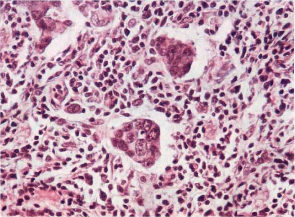 | Figure 1. Microalveolar structures in the infiltrative component of infiltrating ductal breast cancer. The stroma is mildly fibrous, with marked inflammatory infiltration. Stained with hematoxylin and eosin |
4. Conclusions
- Thus, it can be concluded that the IDC subtype of breast cancer is characterized, based on its microscopic features, by a statistically significantly higher grade of malignancy, higher mitotic activity of tumor cells, a higher degree of inflammatory infiltration of the stromal component, and the presence of cribriform structures and necrotic processes in the parenchyma. The ILC subtype of breast cancer, unlike the IDC subtype, is characterized by marked stromal hyalinosis and infiltration of adipose tissue, a predominance of trabecular structures in the parenchyma, and the presence of “dark” cells.
Conflict of Interests’ Statement
- The authors declare no conflict of interest. This study does not include the involvement of any budgetary, grant or other funds. The article is published for the first time and is part of a scientific work.
ACKNOWLEDGEMENTS
- The authors express their gratitude to the management of the multidisciplinary clinic of Republican Specialized Scientific-Practical Medical Center of Oncology and Radiology for the material provided for our study.
Ethical Approval and Consent to Participate
- The Research Ethics Board of our institution does not require review or approval of case reports. Our research was carried out in accordance with the World Medical Association Code of Ethics (Declaration of Helsinki).