Research In Cancer and Tumor

2026;  13(2): 23-29

doi:10.5923/j.rct.20261302.02

Received: Oct. 17; Accepted: Nov. 5, 2026; Published: Nov. 7, 2026

 

Morphological and Molecular Signs of Tissue Hypoxia and VEGF-Dependent Angiogenesis in Uterine Sarcoma and Leiomyoma

B. G. Almukhamedova, D. A. Nishanov, L. T. Alimkhodjaeva

Republican Specialized Scientific-Practical Medical Center of Oncology and Radiology, Tashkent, Uzbekistan

Copyright © 2026 The Author(s). Published by Scientific & Academic Publishing.

This work is licensed under the Creative Commons Attribution International License (CC BY).
http://creativecommons.org/licenses/by/4.0/

Abstract

The aim of the study was to identify the features of malignant mesenchymal uterine tumors course using molecular-morphological methods for the differential diagnosis and prognostic evaluation of uterine sarcoma and leiomyoma. Background. Tissue hypoxia and the vascular endothelial growth factor (VEGF) induced by it, play a key role in the progression of both benign smooth muscle tumors (leiomyomas) and malignant mesenchymal neoplasms (uterine sarcoma). However, the intensity, regulating mechanisms, and morphological consequences of these processes in tumors vary radically. Material and Methods. This retrospective study was conducted at the Department of Reproductive System Tumors of the Republican Specialized Scientific-Practical Medical Center of Oncology and Radiology and its regional branches from 2015 to 2022. The study included 137 patients aged 22 to 60 years. Results. Histological examination evaluated the presence of tumor coagulative necrosis, perinecrotic zones of viable cells, dystrophic changes, interstitial edema, microhemorrhages, vascular heterogeneity, and relatively avascular cellular areas. In uterine sarcoma, the morphological pattern of hypoxia was characterized primarily by a mosaic distribution of viable, hypoxic, and necrotic areas. Conclusion. The results obtained show that uterine sarcoma is characterized not by the isolated activation of individual markers, but by the formation of an interconnected hypoxia-angiogenic phenotype, including tissue hypoxia, HIF-1α-dependent adaptation, activation of VEGF signaling, pathological remodeling of the microvascular bed, vasculogenic mimicry, and tumor necrosis. A comprehensive evaluation of these signs may improve the diagnostic value of the differential diagnosis of uterine sarcoma and leiomyoma.

Keywords: Malignant mesenchymal uterine tumor, Uterine sarcoma, Leiomyoma, Tissue hypoxia

Cite this paper: B. G. Almukhamedova, D. A. Nishanov, L. T. Alimkhodjaeva, Morphological and Molecular Signs of Tissue Hypoxia and VEGF-Dependent Angiogenesis in Uterine Sarcoma and Leiomyoma, Research In Cancer and Tumor, Vol. 13 No. 2, 2026, pp. 23-29. doi: 10.5923/j.rct.20261302.02.

1. Introduction

Tissue hypoxia and the vascular endothelial growth factor (VEGF) induced by it, play a key role in the progression of both benign smooth muscle tumors (leiomyomas) and malignant mesenchymal neoplasms (uterine sarcoma). However, the intensity, regulating mechanisms, and morphological consequences of these processes in tumors vary radically [1].
In leiomyomas, hypoxia is primarily functional and localized in nature. It arises from the mechanical compression of the tumor’s own blood vessels by rapidly proliferating smooth muscle cells and an excess of extracellular matrix [2].
Molecular signs of hypoxia in leiomyomas:
• HIF-1α (Hypoxia-Inducible Factor 1-alpha): It is stable primarily in zones of local ischemic stress (especially following interventional treatment, such as uterine artery embolization). It serves as a central integrator of hormonal signals [1].
• VEGF (predominantly the VEGF-A isoform): It is expressed at moderate levels. Under normal conditions, its production in leiomyocytes is tightly synergistic with estrogen and progesterone levels. VEGFA gene expression is elevated but is regulated by antiangiogenic mechanisms [2].
Morphological signs:
• Vascular architectonics: The blood vessels are properly formed, possessing a mature basement membrane and a complete layer of pericytes/smooth muscle cells. In large or aging nodes, areas of hyalinosis, dystrophy, and fibrosis are often observed due to chronic hypoxia.
• Vascularization: The microvascular density (MVD) increases in an orderly manner, primarily along the periphery of the nodule’s capsule (“pseudocapsule”) [3-6].
In uterine sarcoma (particularly leiomyosarcoma), hypoxia has a critical, widespread nature due to true tissue ischemia: the tumor grows faster than the capillary network can form. Unlike myomas, angiogenesis in this case does not depend on physiological hormones but is triggered by mutations and hypoxic stress [7].
Molecular signs of hypoxia in uterine sarcoma:
• Overexpression of HIF-1α: Consistently high nuclear levels of HIF-1α are observed throughout the tumor, which activates the transcription of hundreds of genes involved in survival and metabolic reprogramming (glycolysis).
• VEGF overproduction: Sharply elevated level of VEGF expression by both the sarcomatous cells themselves and the cells of the microenvironment. The VEGFR-2 receptor signaling pathway is also activated.
• Additional factors: Concurrent increase in cell proliferation markers (Ki-67) and mutations in tumor suppressor genes (e.g., p53, RB) [8].
Morphological signs:
• Pathological neoangiogenesis: The blood vessels are built chaotically, are tortuous, have defective, permeable walls (lacking a smooth muscle layer and pericytes), and often consist simply of endothelial sinuses.
• Tumor Cell Necrosis: This is the main diagnostic criterion for leiomyosarcoma. Necrosis occurs suddenly around “survival zones” (perivascular cuffs) where cells still receive oxygen from damaged blood vessels.
• Hemorrhages and Invasion: Due to the fragility of VEGF-induced blood vessels, extensive areas of hemorrhage are observed. Tumor cells easily penetrate the lumen of such capillaries (vascular invasion) [9].
The aim of the study was to identify the features of malignant mesenchymal uterine tumors course using molecular-morphological methods for the differential diagnosis and prognostic evaluation of uterine sarcoma and leiomyoma.

2. Material and Methods

This retrospective, comparative study was conducted at the Department of Reproductive System Tumors of the Republican Specialized Scientific-Practical Medical Center of Oncology and Radiology and its regional branches for the period from 2015 to 2022. The study included 137 patients aged 22 to 60. The patients were divided into two groups as follows:
The Main Group (n=84) with morphologically confirmed uterine sarcomas (US).
Control Group (n=53) with a histologically confirmed diagnosis of uterine leiomyoma (UL).
Inclusion criteria to the study were as follows:
- Female, age 18 and older;
- Examination and treatment at the institutions serving as clinical research sites during the period specified in the protocol;
- Presence of a morphologically confirmed primary uterine tumor: in the Main group—uterine sarcomas with an established histological subtype; in the Control group—benign uterine leiomyomas, including their morphological variants;
- Availability of medical records to identify key clinical and anamnestic characteristics, examination results, the nature of the procedure performed, and the final diagnosis;
- Availability of representative histological material from the primary tumor—including micro sections and/or paraffin blocks—for diagnostic review and morphological analysis;
- The availability and performance of immunohistochemical and molecular genetic stages of the study.
Exclusion criteria were as follows:
- Lack of morphological confirmation of the diagnosis/inability to reliably distinguish between uterine sarcoma and benign leiomyoma;
- A smooth muscle tumor of uncertain malignant potential – STUMP;
- Other neoplasms not classified under the established diagnostic groups: carcinoma and carcinosarcoma of the uterus, metastatic lesions of the uterus, sarcoma of other primary sites, and other mesenchymal tumors;
- The absence/incomplete nature of medical documentation to confirm inclusion criteria and assessment of key study parameters;
- No material available to perform a reliable morphological verification;
- Conducting antitumor therapy prior to a reliable evaluation of the tumor's baseline morphological and biological characteristics.
Statistical processing of the clinical data was performed using the “EXCEL” and “STATGRAFICS” software packages (StatSoft Inc., USA). The significance of the differences revealed in the analysis of quantitative traits was assessed using the Student-Fisher test. The methods of variational parametric and nonparametric statistics were used, including the calculation of the arithmetic mean (M) of the indicator under study, square mean deviation (σ), the standard error of the mean (m), and relative values (frequency, %). The statistical significance of the obtained measurements in comparing mean values was determined using the Student’s t-test, with the probability of error (p) calculated after testing for normality of distribution (using the kurtosis criterion) and equality of population variances (F-test). A significance level of p < 0.05 was used to determine statistically significant changes.
The “OpenEpi 2009, Version 2.3” software package was used as a computational tool for molecular genetic studies. Data analysis model: Case-control.
Hypoxia and angiogenesis markers
HIF-1α, VEGF-A, CD31, and CD34 were analyzed to evaluate the hypoxic and angiogenic phenotypes.
HIF-1α was evaluated mainly based on nuclear staining of tumor cells, with determination of the percentage of positive nuclei, the intensity of the reaction and the H-score. The location of positive cells relative to necrosis areas was recorded separately, as perineocrotic accumulation of HIF-1α reflects interstitial hypoxia.
VEGF-A was assessed based on the cytoplasmic reaction of tumor cells and, in the presence of membrane reaction it was done with the determination of intensity, extent, and H-score. VEGF-A expression in tumor cells and stromal elements was analyzed separately.
CD31 and CD34 were used to visualize endothelial cells and for evaluating microvascular density. After examining the specimen at low magnification, 3–5 areas of maximal vascularization were selected. Individual positively stained endothelial cells and vascular structures were counted in each section. The result was expressed as the average number of microvessels per 1 mm² of tumor tissue, excluding large vessels with a well-defined muscular wall and vessels in necrosis areas. CD31 was also used together with the PAS reaction for the differentiation of true endothelial vessels and structures of vasculogenic mimicry. PAS-positive, CD31-negative channels formed by tumor cells were interpreted as structures of vasculogenic mimicry.

3. Results

A postoperative histological examination was performed in the majority of patients of both groups: in 86.9% of patients with uterine sarcoma (US) and in 83.0% of patients with leiomyomas (LM). No statistically significant difference was found (p=0.530). The data obtained underscore the key role of surgical specimen examination in the definitive differential diagnosis of benign and malignant mesenchymal tumors of the uterus.
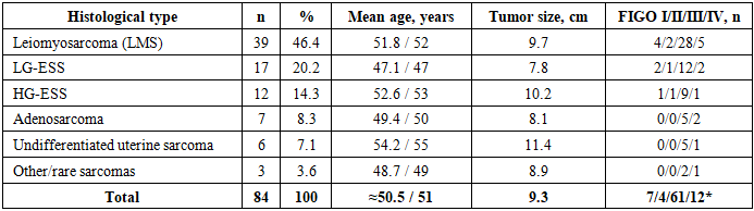
The distribution of US by histological type is presented in the Table 1.
Table 1. Distribution of US by histological type according to morphological revision
     
After morphological revision in the Main group (n = 84), the most common histological variant was LMS, which was identified in 39 (46.4%) of the 84 patients. Low-grade endometrial stromal sarcomas (LG-ESS) ranked second at 20.2%, while high-grade endometrial stromal sarcomas (HG-ESS) accounted for 14.3%; adenosarcoma made up 8.3%, undifferentiated sarcoma - 7.1%, and other rare variants - 3.6%.
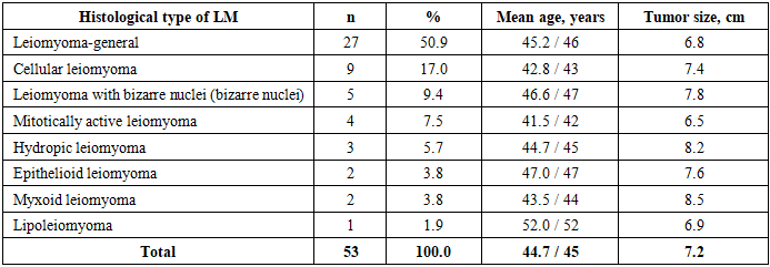
The histological types of LM were examined for a comparative analysis, including an assessment of the patients’ mean age and tumor size (Table 2).
Table 2. Distribution of LM by histological type
     
As it can be seen from the table, the predominant variant in the LM group was leiomyoma-general — 27 (50.9%). The most common types identified among them were: cellular leiomyoma—9 (17.0%), LM with bizarre nuclei—5 (9.4%), and mitotically active LM—4 (7.5%).
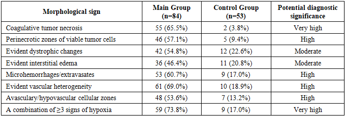
To determine the role of the hypoxic component in the biological behavior of uterine tumors, a comparative assessment of the morphological signs of impaired tissue perfusion was conducted in the Main (n=84) and the Control (n=53) groups of patients.
The presence of tumor coagulative necrosis, perinecrotic zones of viable cells, dystrophic changes, interstitial edema, microhemorrhages, vascular heterogeneity, and relatively avascular cellular areas were evaluated by histological examination (Table 3).
Table 3. Morphological signs of tissue hypoxia in studied patients
     
As it can be seen from the table, the most objective of the listed parameters is tumor coagulative necrosis: 55/84 (65.5%) versus 2/53 (3.8%). Its significant predominance in uterine sarcoma (US) indicates not only the diagnostic significance of necrosis as one of the classic criteria for the malignancy of mesenchymal tumors, but also the formation of significant areas of mismatch between the intensity of proliferation and the blood supply capacity of the tumor tissue.
At uterine sarcoma, the morphological picture of hypoxia was characterized primarily by a mosaic distribution of viable, hypoxic, and necrotic areas. Layers of viable atypical cells persisted in the perinecrotic zones, which was of particular interest for subsequent immunohistochemical analysis of HIF-1α.
Immunohistochemical assessment of HIF-1α
We considered HIF-1α to be a molecular marker of tumor cell adaptation to hypoxia. When oxygen availability decreases, the degradation of HIF-1α is reduced, leading to its stabilization and the activation of the hypoxic response transcriptional program, which includes genes involved in angiogenesis and metabolic adaptation. HIF-1α was detected in a subset of leiomyosarcomas (LMS); however, some comparative studies have found no statistically significant difference in its immunohistochemical (IHC) expression between leiomyomas (LM) and leiomyosarcomas (LMS).
Our proposed method of estimating HIF-1α
It is advisable to evaluate expression semi-quantitatively:
0 - no specific reaction or <5% positive cells;
1+ - weak reaction in 5–25% of cells;
2+ - moderate reaction in 26–50% of cells;
3+ - a significant reaction in more than 50% of tumor cells.
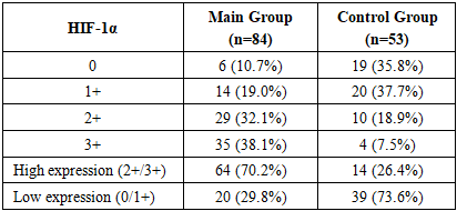
For statistical analysis: low expression – 0–1+; high expression = 2+-3+ (Table 4).
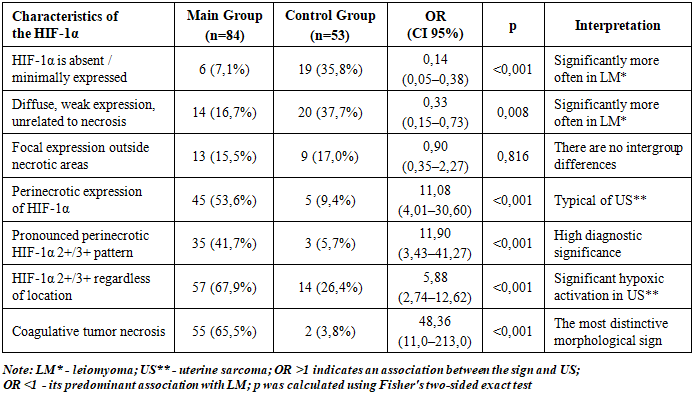
Table 4. Study of the HIF-1α expression
     
According to the study results presented in Table 4, high HIF-1α expression was detected 2.7 times more frequently in uterine sarcoma (US).
The perinecrotic pattern is defined as the predominant accumulation of HIF-1α-positive tumor cells at the periphery of necrotic areas, with the formation areas of increased nuclear or nuclear-cytoplasmic immunoreactivity. Both the intensity of the reaction and its spatial distribution relative to the necrotic focus were taken into account for analysis (Table 5).
Table 5. Characteristics of the HIF-1α expression perinecrotic pattern
     
Absent or minimal expression of HIF-1α was observed in 6 (7.1%) patients with US, compared to 19 (35.8%) patients with LM. The probability of observing this phenotype in US was significantly lower: OR = 0.14; 95% CI 0.05–0.38; p<0.001. Thus, the absence of significant HIF-1α activation is more characteristic of benign LM.
A similar tendency was observed for diffuse, low-level expression of HIF-1α that was not spatially associated with areas of necrosis. It was observed in 16.7% of patients with US and 37.7% of those with LM (OR=0.33; 95% CI 0.15–0.73; p=0.008). Consequently, weak, non-specific HIF-1α expression is not, in itself, an indicator of malignancy; in the material studied, it was actually observed more frequently in cases of LM.
At the same time, focal expression of HIF-1α outside the necrotic areas showed no significant difference between the groups: 15.5% in the Main group versus 17.0% in the Control one (OR=0.90; p=0.816). The absence of a statistically significant difference confirms the low independent differential diagnostic value of this pattern.
Perinecrotic HIF-1α is the most significant spatial pattern. A fundamentally different pattern was observed when evaluating the perinecrotic pattern of HIF-1α expression, which was detected in 45 of 84 (53.6%) patients with uterine sarcoma (US) and in only 5 of 53 (9.4%) with leiomyoma (LM), with a highly statistically significant (p < 0.001) odds ratio of OR = 11.08 (95% CI 4.01–30.60).
Consequently, the presence of perinecrotic HIF-1α expression was associated with an approximately 11-fold increase in the probability of the tumor belonging to the US group compared with the LM group.
Even more telling was the evident perinecrotic pattern (2+/3+), which was observed in 35 (41.7%) US cases but in only 3 (5.7%) LM cases (OR = 11.90; 95% CI 3.43–41.27; p < 0.001). It allows us to consider the expressed HIF-1α positivity of viable tumor cells surrounding necrotic areas as a potentially informative component of the differential diagnosis of US and LM.
High HIF-1α expression (2+/3+)—regardless of its topographic location—was observed in 57 (67.9%) patients with US compared to 14 (26.4%) with LM, with a statistically significant difference (OR = 5.88; 95% CI 2.74–12.62; (p < 0.001).
As it can be seen, high HIF-1α expression increases the probability of a tumor being associated with US by 5.9 times. However, a comparison of two indicators presents an interesting finding: HIF-1α 2+/3+ in general → OR = 5.88 versus perinecrotic HIF-1α 2+/3+ → OR = 11.90.
Taking into account the spatial localization of HIF-1α relative to necrosis doubles the strength of its association with US; not only the number of HIF-1α-positive cells but also the architecture of their distribution within the tumor tissue proves to be diagnostically significant.
The association between HIF-1α and tumor necrosis had the highest OR among the parameters studied; it was observed for coagulative tumor necrosis, which was detected in 55 (65.5%) patients with US and in only 2 (3.8%) with LM (OR = 48.36; 95% CI 10.98–213.03; (p < 0.001).
Thus, coagulative tumor necrosis remains a significantly stronger morphological feature for differential diagnosis than isolated HIF-1α expression.
At the same time, HIF-1α and necrosis characterize different stages of a single hypoxic process: insufficient perfusion → hypoxia → stabilization of HIF-1α in viable cells → cellular adaptation → in the event of a critical oxygen deficiency → irreversible damage → necrosis.
Therefore, the most diagnostic information can be obtained not from a single marker, but from the combination of coagulative tumor necrosis and perinecrotic HIF-1α positivity.
The perinecrotic HIF-1α pattern was identified in 45 of 84 (53.6%) cases of US, compared with 5 of 53 (9.4%) in LM cases of our study, A significant perineocritical reaction (2+/3+) was observed in 46.4% and 5.7% of observations, respectively, indicating a significantly more marked spatially organized hypoxic adaptation of the sarcomas.
It is particularly significant to compare this phenomenon with the established high frequency of coagulative tumor necrosis in US—55/84 (65.5%)—versus 2/53 (3.8%) in LM. However, the presence of necrosis and perinecrotic HIF-1α positivity should be considered as interrelated but not identical signs.
We developed an integrated Hypoxia–Angiogenesis Index (HAI). The development of this index is related to the fact that individual indicators of hypoxia, angiogenic factor expression, and microvascular bed status characterize different stages of a single pathological process, which, when evaluated separately, do not make it possible to distinguish the complex hypoxic-angiogenic phenotype of a tumor.
The HAI model was based on the following components:
• HIF-1α—a marker of the cellular response to hypoxia;
• VEGF-A—a key marker of proangiogenic activation; microvascular density (MVD), which was determined based on CD31 and CD34 levels;
• vasculogenic mimicry (VM)—as an alternative mechanism for the formation of vessel-like channels;
• tumor coagulative necrosis—a morphological sign of disturbances arising from the mismatch between the metabolic demands of tumor tissue and its vascular supply.
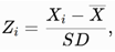
The Z-transformation method was used to standardize the indicators:
where Xi is an individual value of the indicator, X is its mean value in the sample under study, and SD is the standard deviation.
Signs of vasculogenic mimicry and tumor coagulative necrosis were coded as follows: 0 = absence of the sign, 1 = presence, followed by Z-standardization.
Calculation of the integral index
The final HAI was calculated using the following formula:
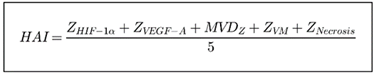
All five components were balanced and had equal weight, which made it possible to evaluate them.
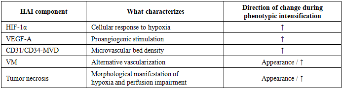
Based on the proposed HAI model, it is possible to assess the sequential and interrelated stages of tumor adaptation to oxygen hypoxia (Table 6).
Table 6. Main signs for the evaluation and the formation of HAI
     
As it can be seen from the table, during the development of tissue hypoxia, HIF-1α is stabilized and HIF-1α-dependent signaling pathways are activated, leading to an enhanced VEGF-dependent angiogenic response, which is manifested by morphological changes in the density of the microvascular network, as determined by the CD31 and CD34 markers.
An additional mechanism of tumor adaptation is vasculogenic mimicry (VM). In cases with a simultaneous marked mismatch between the growth rate of tumor tissue, its metabolic demands, and the capacity of the blood supply, areas of tumor coagulative necrosis are formed. This model of the HAI integrates the molecular, IHC, and morphological levels of evaluation of the underlying pathobiological process:
hypoxia → HIF-1α → VEGF-A → angiogenic response → MVD/VM → morphological consequences of perfusion insufficiency.
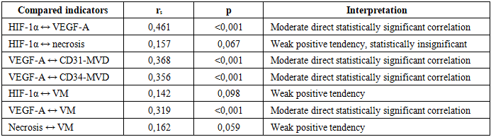
To assess the relationships between indicators of hypoxia, angiogenesis, and vascular phenotype, a correlation analysis was conducted to determine Spearman’s rank correlation coefficient (rₛ) (Table 7).
Table 7. Correlation analysis for evaluation of the relationship between indicators of hypoxia, angiogenesis and vascular phenotype
     
The correlation analysis revealed associations between markers of tissue hypoxia, angiogenic activity, and characteristics of the vascular phenotype in patients with US and LM. A significant association has been established between HIF-1α expression and VEGF-A levels (rₛ=0.461; p<0.001), indicating a coordinated increase in the activity of these markers, which follows the sequence: "hypoxia – stabilization of HIF-1α – activation of a pro-angiogenic response".
At the same time, direct correlations between VEGF-A and microvascular density (MVD) indices, as determined by CD31-MVD (rₛ=0.368; p<0.001) and CD34-MVD (rₛ=0.356; p<0.001), confirming that an increase in VEGF-A was associated with an increase in microvascular density. An analysis of the positive correlation between VEGF-A and vasculogenic mimicry (VM): rₛ = 0.319; p < 0.001, indicates that the formation of VM in the established model is associated with a more pronounced angiogenic phenotype. It allowed us to view the VM not in isolation, but as one of the components of the tumor tissue's complex adaptation to oxygen and vascular insufficiency.
Further evaluation and comparison of HIF-1α–necrosis indicators revealed a weak positive correlation (rₛ=0.157) that was not statistically significant (p=0.067); the same was observed for HIF-1α-VM (rₛ=0.142; p = 0.098) and VM- necrosis (rₛ = 0.162; p = 0.059). The direction of these relationships is consistent with our hypothesis, but they are not statistically significant (α=0.05).

4. Conclusions

The study revealed significant differences in the hypoxic-angiogenic profile between uterine sarcoma and leiomyoma. The morphological pattern of uterine sarcoma was characterized by a combination of tumor coagulative necrosis, perinecrotic areas of viable cells, microhemorrhages, vascular heterogeneity, and hypo-/avascular areas, reflecting impaired tissue perfusion and the formation of localized hypoxic niches. The most significant morphological sign was coagulative tumor necrosis, which was observed in 65.5% of cases of uterine sarcoma versus 3.8% of leiomyoma cases.
A study of VEGF-dependent angiogenesis revealed significant activation of the proangiogenic pathway in uterine sarcoma.
The results obtained show that uterine sarcoma is characterized not by the isolated activation of individual markers, but by the formation of an interconnected hypoxia-angiogenic phenotype, including tissue hypoxia, HIF-1α-dependent adaptation, activation of VEGF signaling, pathological remodeling of the microvascular bed, vasculogenic mimicry and tumor necrosis. A comprehensive evaluation of these signs may improve the diagnostic value of the differential diagnosis between uterine sarcoma and leiomyoma.

Conflict of Interests’ Statement

The authors declare no conflict of interest.
This study does not include the involvement of any budgetary, grant or other funds.
The article is published for the first time and is part of a scientific work.

ACKNOWLEDGEMENTS

The authors express their gratitude to the management of the multidisciplinary clinic of Republican Specialized Scientific-Practical Medical Center of Oncology and Radiology for the material provided for our study.

References

[1]  Porotikova I.E., Demura T.A., Adamyan L.V., Gavrilova T.Yu. Clinical and morphological features of uterine leiomyoma in women of reproductive age following prior uterine artery embolization, MRI-guided focused ultrasound ablation, or myomectomy. Obstetrics and Gynecology. 2016; 6: 94-101. https://dx.doi.org/10.18565/aig.2016.6.94-101.
[2]  Tatavarthi S, Vanos V, Combs AL, Pan A, Saini M, Borahay MA. HIF-1α Signaling in Uterine Fibroids: A Central Integrator of Hypoxic, Hormonal, and Fibrotic Pathways. Oxygen (Basel). 2026 Jun; 6(2): 9. doi: 10.3390/oxygen6020009. Epub 2026 Apr 17. PMID: 42181792; PMCID: PMC13197049.
[3]  Russkikh L.A., Barinov S.V. Specifics of management for pregnant women with large uterine leiomyomas // Siberian Medical Journal. — 2011. — Vol. 100. — No. 1. — pp. 57-59.
[4]  Dobrokhotova Yu.E., Dzhobava E.M., Knysheva I.G., et al. Pregnancy in patients following uterine artery embolization for uterine fibroids: clinical course, characteristics of the fetoplacental complex, and endothelial function // Problems of Reproduction. — 2011. — Vol. 17. — No. 4. — pp. 97-101.
[5]  Grishchenko Ya. V., Konstantinova O. D., Cherkasov S. V. Uterine fibroids in the modern world: current issues in pathogenesis, diagnosis, and treatment // Bulletin of the Orenburg Scientific Center of the Ural Branch of the Russian Academy of Sciences. 2012. No. 3. pp. 2–19.
[6]  Buyanova S.N., Yudina N.V., Gukasyan S.A. Modern aspects of the growth of uterine fibroids // Russian Bulletin of Obstetrician-Gynecologist. 2012. No. 12(4). pp. 42-8.
[7]  Yuldasheva O. S., Negmatshaeva Kh. N., Turaeva G. Yu. Reproductive function status in women with uterine fibroids (literature review) // Molodoy Ucheny [Young Scientist]. 2016. No. 4 (108). pp. 312-315.
[8]  Frantsiyants E. M., Bandovkina V. A., Surikova E. I., Neskubina I. V., Cheryarina N. D., Moiseenko T. I., Menshenina A. P., Rogozin M. A. Characteristics of VEGF-A, VEGF-C, and their receptor levels in tumor tissue and blood of patients with endometrial cancer, in relation to histological structure // Malignant Tumors. 2025. №1.
[9]  Chumak Z. V., Zelinsky A. A., Shapoval N. V., Manasova G. S., Kukhar N. N. // Endometrium: HIF and VEGF as molecular-tissue regulatory factors under conditions of hypoxia. HEALTH OF WOMAN. 2015. 9(105): 78–80.