-
Paper Information
- Paper Submission
-
Journal Information
- About This Journal
- Editorial Board
- Current Issue
- Archive
- Author Guidelines
- Contact Us
Research In Cancer and Tumor
2023; 11(1): 1-5
doi:10.5923/j.rct.20231101.01
Received: Feb. 19, 2023; Accepted: Mar. 5, 2023; Published: Mar. 15, 2023

Immunohistochemical Aspects of the Expression of Markers of Cell Cycle Regulation, Proliferation and Apoptosis (p53, Ki-67, bcl-2, cyclin D1) in Breast Neoplasia
 Abstract
 Reference
 Full-Text PDF
 Full-text HTML
L. T. Alimkhodjaeva, D. A. Nishanov, L. M. Bozarova, M. Kh. Norbekova
Republican Specialized Scientific and Practical Medical Center of Oncology and Radiology, Tashkent, Uzbekistan
Copyright © 2023 The Author(s). Published by Scientific & Academic Publishing.
This work is licensed under the Creative Commons Attribution International License (CC BY).
http://creativecommons.org/licenses/by/4.0/

The aim of study was to develop new criteria for the differential diagnosis of variants of intraepithelial neoplasia of the accessory lobe of the mammary gland based on the use of a comprehensive morphological research method using an optimal panel of markers to improve the quality of diagnosis and estimation the risk of invasive cancer development. Introduction. To date, the pathology of the mammary glands occupies a leading position in the structure of oncological diseases and continues to grow from year to year. Currently, a three-level characteristic of the degree of tumor differentiation, common for oncological pathomorphology, is widely used to determine the malignant potential of preinvasive lesions of the mammary gland and its lobes. Material and methods. The results of a retrospective examination of 182 patients with mammary gland accessory lobe cancer treated at our Center from 2010 to 2020 were included to our research. The patients were divided into 4 groups according to the results of the morphological study. Results. Expression of p53 was least common in patients with dysplasia, lobular and highly differentiated intraductal, making up 1.6%, 3.5% and 14.7%, respectively. The study of cyclin D1 expression showed that among patients with intralobular, intraductal carcinomas and intraductal carcinoma in situ with the onset of invasion, it was observed equally (64.3%, 66.2% and 69%, respectively). Comparison of the apoptosis marker bcl-2 expression values showed that this indicator was most often detected in patients with intralobular carcinoma and highly differentiated intraductal carcinoma (85.5%, 89.3% and 87.5% of cases). Estimation of Ki-67 expression values showed that it was most common in patients with poorly differentiated intraductal carcinoma and patients with microinvasive cancer, the values of the indicators were 24.3% and 50%, respectively. Conclusion. The discovered trend allowed us to propose this set of immunohistochemical markers as additionally characterizing the ability of intraductal carcinoma to further progress into invasive mammary gland accessory lobe cancer. The data obtained on the basis of immunohistochemical studies on the frequency and severity of expression of various immunohistochemical markers provide a basis for determining the individual prognosis of the course of the disease, which, in turn, will contribute to the development of adequate treatment regimens for preinvasive breast neoplasia.
Keywords: Breast cancer, Mammary gland accessory lobe cancer, Immunohistochemistry, Dysplasia, Invasion, Proliferation, Apoptosis
Cite this paper: L. T. Alimkhodjaeva, D. A. Nishanov, L. M. Bozarova, M. Kh. Norbekova, Immunohistochemical Aspects of the Expression of Markers of Cell Cycle Regulation, Proliferation and Apoptosis (p53, Ki-67, bcl-2, cyclin D1) in Breast Neoplasia, Research In Cancer and Tumor, Vol. 11 No. 1, 2023, pp. 1-5. doi: 10.5923/j.rct.20231101.01.
1. Introduction
- To date, the pathology of the mammary glands occupies a leading position in the structure of oncological diseases and continues to grow from year to year. Along with breast cancer (BC), mammary gland accessory lobe cancer (MGALC) has also begun to rise in recent years. The frequency of BC transformation into MGALC is 0.2–0.3% of its total frequency. Currently, a three-level characteristic of the degree of tumor differentiation, common for oncological pathomorphology, is widely used to determine the malignant potential of preinvasive lesions of the mammary gland and its lobes. Large prospective studies have shown, in general, a correlation between the degree of differentiation of preinvasive breast neoplasia, established by morphological criteria, and the risk of developing an invasive tumor after organ-preserving surgeries [1]. However, the morphological characteristics of the tumor are largely subjective, and differences between the conclusions of different specialists often lead to an incorrect assessment of the potential aggressiveness of the process. Besides, the heterogeneity of the structure of preinvasive breast lesions can also lead to significant variations in determining the degree of differentiation [2]. The characteristic of ductal carcinomas accompanied by microinvasion is also of particular interest, the prognosis of which often remains unclear. In this regard, in recent years, studies aimed at selecting the optimal panel of various signs have been conducted, primarily biomarkers, which allow to determine with a high degree of reliability the malignant potential of preinvasive breast lesions, as well as carcinomas with microinvasion foci. In most cases, these studies are based on the study of gene expression profiles and cannot yet be widely used in practical oncology due to the complexity of the method and the high cost of equipment and reagents [3]. The immunohistochemistry (IHC) method has been widely used by researchers in the study of intraepithelial neoplasia of the mammary gland and its proportion for early diagnosis and prognosis in recent years [4]. However, in most published studies using this method, a limited number of markers does not allow to fully clarify the biological profile of the studied diseases, there is no data on the prognostic significance of these types of neoplasia, a comprehensive diagnostic algorithm combining morphological and IHC criteria is not defined [5-9]. The use of techniques based on immunohistochemical detection of protein expression seems more acceptable for the daily work of a pathologist. When studying the latest researches on breast diseases, we found that not so much work has been done that gives clear recommendations for diagnosis and treatment, as well as for the prediction of MGALC, which is why this study seems to be relevant and interesting both in terms of clinical and practical oncology [10]. In most published studies using the IHC method as one of the main methods of diagnosing BC and MGALC, a limited number of markers does not allow to fully clarify the biological profile of the studied diseases, there is no data on the prognostic significance of these types of neoplasia, a comprehensive diagnostic algorithm combining morphological and IHC criteria of the breast is not defined [11-12]. According to most experts, the prognosis of mammary gland accessory lobe cancer (MGALC) is determined by its early diagnosis. This trend requires careful screening among women from the high-risk group, which include, first of all, patients with precancerous lesions of the breast. We identified only four of the most significant and most common types of intraepithelial neoplasia, which were associated with the greatest number of differences of opinion among specialists when establishing a diagnosis and determining the prognosis, and tried to combine morphological and immunohistochemical parameters in order to conduct an adequate diagnosis and identify factors characterizing their malignant potential in our study.The aim of study was to develop new criteria for the differential diagnosis of variants of intraepithelial neoplasia of the accessory lobe of the mammary gland based on the use of a comprehensive morphological research method using an optimal panel of markers to improve the quality of diagnosis and estimation the risk of invasive cancer development.
2. Material and Methods
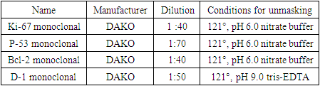
- A retrospective examination of 182 patients with mammary gland accessory lobe cancer treated at the Republican Specialized Scientific and Practical Medical Center of Oncology and Radiology from 2010 to 2020 was included to our study. According to the morphological examination results, the patients were divided into 4 groups. The first group included 62 (34.1%) patients with severe dysplasia, which was defined as "dysplasia". The second group consisted of 71 (39.0%) patients with ductal carcinoma in situ, designated as "intraductal carcinoma". The third group included 28 (15.4%) women with lobular carcinoma in situ (“intralobular carcinoma”). The fourth group included 21 (11.5%) patients with carcinoma in situ with the onset of invasion. We defined this group as “microinvasive carcinoma”. From 3 to 7 pieces of 2x2x0.5 cm in size were cut out for histological examination upon macroscopic detection of the seal. From the tissue surrounding the tumor, from 5 to 8 pieces were cut out. The material was fixed in a 10% solution of buffered neutral formalin for 24 hours, dehydrated in a battery of alcohols in an increasing concentration from 70 to 100%, poured into paraffin wax and sections 5-7 microns thick were prepared on a rotary microtome, which were stained with hematoxylin and eosin according to the generally accepted method. Additional immunohistochemical studies were performed when establishing the diagnosis of dysplasia, carcinoma in situ or carcinoma in situ with the onset of invasion. An assessment to quantify a number of morphological characteristics (necrosis, polarization of cells in relation to the basement membrane) was made according to the principle: 0 - absence of a sign, 1 - presence of a sign. 15 commercial sets of mono- and polyclonal antibodies for immunohistochemical studies, which were distributed according to functional significance were used as follows: markers of proliferative activity (Ki-67); apoptosis markers (bcl-2); tumor growth suppressors (p53); cyclin D1. The material for the study by immunohistochemistry was fixed with 10% neutral formalin for 24 hours, poured into paraffin, prepared sections with a thickness of 4 microns, which were applied to highly adhesive glasses and dried at a temperature of 37°C for 18 hours. Restoration of antigenic activity was carried out in a 2100 Retrieval mini-autoclave (Pick Cell) at 121°C for 20 min followed by cooling for 90 min. The recovery was carried out in a citrate buffer pH 6.0 or in a tris-EDTA buffer pH 9.0. Protease treatment was used for some antibodies. The system "EnVision" ("Dako") was used as a detection system, diaminobenzidine and AES were used as a chromogen. The reactions to obtain results suitable for quantitative processing were carried out using an automatic immunohistostainer "Autostainer Dako". The intensity of reactions localized in the cytoplasm and on cell membranes was evaluated by a semi-quantitative method on a scale from O to 3 using the Leica Q550 image analyzer: 0 - no reaction, 1 - weak reaction, 2 - moderate reaction, 3 - strong reaction. The results of reactions with antigens having nuclear localization (Ki-67, p53, bcl-2, cyclin D-1 were evaluated by counting the number of colored nuclei per 100 nuclei in 3 fields of view. The results were expressed as a percentage (Tab.1).
|
3. Results
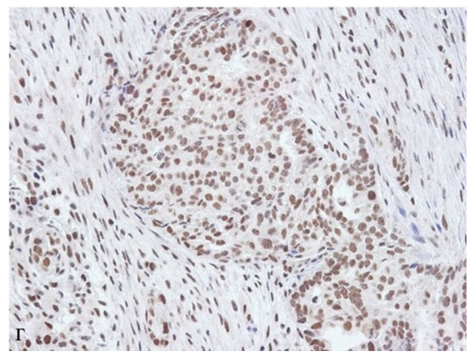
- Expression of p53 was least common in patients with dysplasia, lobular and highly differentiated intraductal carcinoma (1.6%, 3.5% and 14.7%, respectively). The value of this indicator was significantly lower (p<0.05) than the corresponding levels in patients with poorly differentiated intraductal carcinoma (40.5%) and intraductal carcinoma with microinvasion (47.6%). The expression of the p53 marker was most pronounced in patients with microinvasive carcinoma in situ, however, the value of this indicator did not significantly differ (p=0.9024) from that in the group of poorly differentiated ductal carcinoma in situ (Fig.1).
 | Figure 1. Expression of p53 in various types of neoplasia |
 | Figure 2. Expression of cyclin D1 in different types of neoplasia |
 | Figure 3. Expression levels of bcl-2 in various types of neoplasia |
 | Figure 4. Expression levels of Ki-67 in different types of neoplasia |
4. Conclusions
- In view of the above, it can be stated, that a decrease in the degree of differentiation of tumor cells in intraductal carcinoma in situ is associated with an increase in the frequency of necrosis detection, disturbances in cell polarization, a decrease in bcl-2 expression and an increase in p53 and Ki-67 expressions. The discovered trend allowed us to propose this set of immunohistochemical markers as additionally characterizing the ability of intraductal carcinoma to further progress into invasive mammary gland accessory lobe cancer. The data obtained on the basis of the immunohistochemical method of research on the frequency and severity of the expression of various immunohistochemical markers provide a basis for determining the individual prognosis of the disease course, which, in turn, will contribute to the development of adequate treatment regimens for preinvasive breast neoplasia. Determining the expression levels of p53, bcl-2, Ki-67, cyclin D1 is the most important tool for the differential diagnosis of various types of breast neoplasia and an independent factor in estimation of tumor invasiveness. Signs of an increase in the invasiveness of breast neoplasia and its additional lobes are: an increase in the frequency of necrosis detection, disturbances in cell polarization, a decrease in the frequency of estrogen and progesterone receptors expression, a combination of maximum expression of p53 and Ki-67 with minimal expression of bc1-2. Signs of a decrease in the degree of tumor cells differentiation in intraductal carcinoma in situ are also: an increase in the frequency of necrosis detection, disturbances in cell polarization, a decrease in the frequency of estrogen and progesterone receptors expression, a decrease in the expression level of bcl-2, an increase in the frequency of expression of p53, Ki-67. The authors declare no conflict of interest. This study does not include the involvement of any budgetary, grant or other funds. The article is published for the first time and is part of a scientific work.