-
Paper Information
- Paper Submission
-
Journal Information
- About This Journal
- Editorial Board
- Current Issue
- Archive
- Author Guidelines
- Contact Us
International Journal of Plant Research
p-ISSN: 2163-2596 e-ISSN: 2163-260X
2017; 7(2): 36-38
doi:10.5923/j.plant.20170702.02

Comparative Antimicrobial Evaluation of Available Mikania Species in Bangladesh
 Abstract
 Reference
 Full-Text PDF
 Full-text HTML
Rafeza Khatun1, Laila Nasrin2, Sudipta Roy1, Mudasir A. Tantry3, Md Aziz Abdur Rahman1
1Department of Pharmacy, University of Rajshahi, Rajshahi, Bangladesh
2IBSC, Plant Pathology Mycology and Microbiology Lab, University of Rajshahi, Rajshahi, Bangladesh
3Department of Chemistry, S. P. College, Cluster University Srinagar, Kashmir, India
Correspondence to: Md Aziz Abdur Rahman, Department of Pharmacy, University of Rajshahi, Rajshahi, Bangladesh.
| Email: |  |
Copyright © 2017 Scientific & Academic Publishing. All Rights Reserved.
This work is licensed under the Creative Commons Attribution International License (CC BY).
http://creativecommons.org/licenses/by/4.0/

There is considerable interest of using oriental medicinal plants to treat various infections due to multiple and repeated issues with antibiotic efficacy. The vast history of traditional Asian medicine may facilitate the discovery of new antibiotics from herbal products used to treat infections. In present investigation, we selected three available Mikania species (family-Asteraceae) Mikania cordata (MC), Mikania micrantha (MM) and Mikania scandens (MS) for comparative antimicrobial evaluation. The ethanolic extract of these plants were tested by using disc diffusion method. The significant antibacterial activity was shown by M. cordata against Staphylococcus aureus, Bacillus cereus, Escherichia coli, and Shigella sonnii with zone of inhibition in between 30 to 35 mm whereas other two species showed mild to moderate activity with average zone of inhibition 11-30 mm. All the three species showed significant activity against Aspergillus niger with mild to moderate activity against Aspergillus viridis, Fussarium solani and Trichoderma spp. Based on the evaluation, it is concluded that Mikania cordata possesses highest antimicrobial potentials among all the species tested.
Keywords: Asteraceae, Mikania cordata, Mikania micrantha, Mikania scandens, Pathogen, Antimicrobial activity
Cite this paper: Rafeza Khatun, Laila Nasrin, Sudipta Roy, Mudasir A. Tantry, Md Aziz Abdur Rahman, Comparative Antimicrobial Evaluation of Available Mikania Species in Bangladesh, International Journal of Plant Research, Vol. 7 No. 2, 2017, pp. 36-38. doi: 10.5923/j.plant.20170702.02.
Article Outline
1. Introduction
- Nowadays, antibiotics are essential arms for fighting bacterial infections [1]. Antibiotics have become less effective against certain illnesses because many of them produce toxicity as well drug resistance [2]. So, it is emergency to investigate newer drugs of therapeutically and industrially important with lesser resistance to ensure the health-related quality of human life [1]. From the ancient time, plants play an important role to prevent different types of human diseases [3]. Mikania is one of the important plan genus found in the tropics of America and Asia and are widely and combined known as guaco. It comprises about 300 identified species, [4] but only 15-20 of them have been studied. In folkloric medicine, the plants from the genus are used to treat fever, rheumatism, inflammation, oxidative stress, spasmolytic, cancer, respiratory diseases as well as snake bites and scorpion stings [4].Keeping these words, we have selected species from Mikania genus that are native to Bangladesh. We identified four species Mikania cordata, Mikania dioscoreifolia, Mikania micrantha and Mikania scandens. Among these, Mikania cordata has been used in traditional herbal medicine of Bangladesh for a long to treat various ailments including pain, inflammations and some other infectious diseases by folklore people [5-7].A wide variety of biological activities are reported for Mikania species such as antimicrobial, antiviral, anti-inflammatory, antispasmodic, antitumoral, anticoagulant, bronchodilator and antioxidant due to the presence of coumarin, diterpenes, dilactones, and flavonoids [8, 9]. Likewise, terpenes, diterpenes and sesquiterpene lactones are often found, mainly the dilactones type mikanolide and miscandenin derivatives, which have analgesic and antibacterial [10, 11] activities. There were some considerable studies, carried out the evaluation of antibacterial activity of Mikania micrantha, Mikania cordata, Mikania laevigata and Mikania glomerata [12-14]. Though a lot of research have been done on Mikania species but most of them are limited to Mikania micrantha and Mikania cordata, and comparative antibacterial evaluation was not reported elsewhere that might be interesting among biological scientists making the use of active medicinal plants safe.In this study we report comparative antimicrobial study of three available Mikania species: Mikania cordata, Mikania micrantha and Mikania scandens.
2. Materials and Methods
2.1. Collection and Identification of Plant Material
- The whole part of M. cordata was collected from Rajshahi (University of Rajshahi campus) which is northern part of Bangladesh. M. micrantha and M. scandens were collected from Barisal (Southern region) and Kushtia (western region, Kumarkhali area), respectively, during the month of August 2015 and were identified by taxonomist Dr. AHM Mahbubur Rahman, Associate Professor, Department of Botany, University of Rajshahi, Bangladesh. The plants were labeled, air dried for several days and then oven dried at 45°C for 24 hours to assist grinding. The dried plants were crushed separately into course powder.
2.2. Preparation of Extract
- About 170gm dried powdered plant materials of M. cordata (MC), M. micrantha (MM) and M. scandens (MS) taken separately in an amber colored extraction bottle (2.5liter capacity) and the materials were soaked with 70% ethanol (90ml × 3 times). The sealed bottle was kept for 7 days with occasional shaking and stirring. The extracts were filtered through cotton and then Whatman No.1 filter papers and were concentrated with a rotary evaporator under reduced pressure at 45°C and was preserved in refrigerator for further assay. The obtained MC, MM and MS percentage were 11.00%, 9.5% and 7.3%, respectively.
2.3. Antimicrobial Screening
- Antibacterial and antifungal activity of plant crude extract were evaluated by Disc diffusion susceptibility method [15-17]. Bacterial and fungal strains were collected from Department of Botany, University of Rajshahi. Bacterial strains S. aureus, B. cereus, E. coli and S. sonnii were used for antibacterial activity. For antifungal assay A. niger, A. viridis, F. solani and Trichoderma spp. were used. The bacterial and fungal stock cultures were incubated for 24 hours at 37°C on nutrient agar and potato dextrose agar (PDA) medium, respectively, following refrigeration storage at 4°C. The stock solution was prepared by the addition of 20 mg of crude in 1ml MeOH (20µg/ml). Then, sterile discs (made from Whatman filter paper) each about 5mm diameter was impregnated with the specific volume of plant extract to get 300 and 200µg/disc for antibacterial and antifungal assay, respectively. With the help of sterilized forcep, dried discs were then placed on agar plate containing microorganisms. All the plates were incubated at specific temperature (bacteria: 37°C and fungi: 28°C) for 24-48 hours. The zones of growth inhibition around the disc were measured after 48 hours. The sensitivity of the microorganism to the plant extracts was determined by measuring the sizes of inhibition zones (diameter of the zone) on the agar surface around the disc. Standard clindamycin (10 µg/disc) and fluconazole (5 µg/disc) (both purchased from Sigma-Aldrich) were used as positive control for bacteria and fungus, respectively.
3. Results and Discussion
3.1. Antibacterial Activity
- The inhibitory activity of extracts of MC, MM and MS was determined at a concentration 300 µg/disc against two Gram positive and two Gram negative bacteria. The results are shown in Table 1 and Figure 1. Results revealed that all three extracts are sensitive to the organisms tested. Among the extracts, MC was found to be most active other than MM and MS. Extract MC showed strong activity with zone of inhibition 35, 32 and 32 mm against B. cereus, S. aureus and E. coli, respectively (Table 1). Activity of MM and MS was in between 14-30 and 11-21 mm, respectively.
|

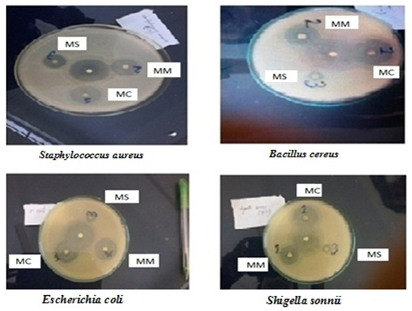 | Figure 1. Zone of inhibition (mm) of MC, MM, MS and standard Clindamycin (Centered disc) against S. aureus, B. cereus, S. sonnii, and E. coli |
3.2. Antifungal Activity
- The plants extract of MC, MM and MS were tested against the four pathogenic fungi at a concentration of 200 µg/disc for each and the result was compared with standard antifungal Fluconazole 5 µg/disc. It was observed that among three plant extract MC showed comparably strong antifungal activity than other two extracts MM and MS (Table 2). All the three extracts showed strong activity against A. niger with zone of inhibition in the range of 25-40 mm. Fungi A. viridis, F. solani and Trichoderma spp. were mild to moderate sensitive to MC, MM and MS.
|

4. Conclusions
- It is concluded that among three plants extract Mikania cordata possesses significant and comparable antibacterial and antifungal activity than Mikania micrantha and Mikania scandens. Considering the broadest range of antimicrobial properties this would suggest that Mikania cordata might possess phytochemicals that can be useful as a broad-spectrum antimicrobial agent. This study revealed the importance of these plant parts as a novel source of antimicrobial agents due to the increasing drug resistance among microorganisms. The mechanism of action will be done in further study on the basis of in vitro experimentation for development of new products. The bioactivity results will provide scientific foundation for rational development and utilization of this plant. This study can easily identify a more potent species which showed a significant pharmacological effect. To utilize the plant extract as a therapeutic agent, further toxicity and pharmacological studies are needed.
ACKNOWLEDGEMENTS
- The authors wish to acknowledge the assistance of taxonomist Dr. A. H. M. Mahbubur Rahman, Associate Professor, Department of Botany, University of Rajshahi, Bangladesh for the identification of the plant samples.