-
Paper Information
- Next Paper
- Previous Paper
- Paper Submission
-
Journal Information
- About This Journal
- Editorial Board
- Current Issue
- Archive
- Author Guidelines
- Contact Us
Research in Otolaryngology
p-ISSN: 2326-1307 e-ISSN: 2326-1323
2017; 6(4): 55-56
doi:10.5923/j.otolaryn.20170604.02

Fungal Ball in the Concha Bullosa: A Rare Entity
 Abstract
 Reference
 Full-Text PDF
 Full-text HTML
Usha SV, Borligegowda Viswanatha
Otorhinolaryngology Department, Bangalore Medical College & Research Institute, Bangalore, India
Correspondence to: Borligegowda Viswanatha, Otorhinolaryngology Department, Bangalore Medical College & Research Institute, Bangalore, India.
| Email: |  |
Copyright © 2017 Scientific & Academic Publishing. All Rights Reserved.
This work is licensed under the Creative Commons Attribution International License (CC BY).
http://creativecommons.org/licenses/by/4.0/

Fungus ball of the paranasal sinuses is a mass of noninvasive matted fungal hyphae within a paranasal sinus. Paranasal sinuses fungal balls are usually seen in maxillary and sphenoid sinuses. A fungus ball in the concha bullosa, without paranasal sinus involvement is rare. A case of fungus ball in the concha bullosa, without paranasal sinus involvement is reported along with review of literature.
Keywords: Fungal ball, Concha bullosa
Cite this paper: Usha SV, Borligegowda Viswanatha, Fungal Ball in the Concha Bullosa: A Rare Entity, Research in Otolaryngology, Vol. 6 No. 4, 2017, pp. 55-56. doi: 10.5923/j.otolaryn.20170604.02.
1. Introduction
- Sinus fungus ball was first described by Plaignaud at the end of the 18th century [1]. Fungal diseases of the paranasal sinuses are categorized as either invasive or non-invasive based on the presence or absence of tissue invasion [2]. Non-invasive fungal rhinosinusitis is subdivided into three types: saprophytic fungal infection, fungal balls, and fungus- related eosinophilic rhinosinusitis [3]. Fungus balls are found in just one paranasal sinus, most frequently in the maxillary sinus and occasionally in the sphenoid sinus [4]. A fungus ball in the concha bullosa, without paranasal sinus involvement is rare. A case of fungus ball in the concha bullosa, without paranasal sinus involvement is reported along with review of literature.
2. Case Report
- A 35 years old female patient presented with history of left side nasal obstruction and recurrent episodes of nasal discharge for the past three years. Patient had taken several courses of antibiotics and did not have any relief. There was no other significant medical history. Nasal endoscopic examination showed prominent middle turbinate on both sides. On computed tomography, paranasal sinuses were clear. There was concha bullosa on both sides and on the left middle turbinate concha bullosa was filled with a high-density material [Figure 1].Through an endoscopic transnasal approach, vertical incision was made in the anterior part of the middle turbinate. Lateral wall of the concha was removed; dark cheesy material was removed and sent for examination. Histopathological examination of the excised specimen revealed Aspergillus. Post operative period was uneventful. No antifungal therapy was given to the patient. Following surgery, patient was relieved from the symptoms and there was no recurrence.
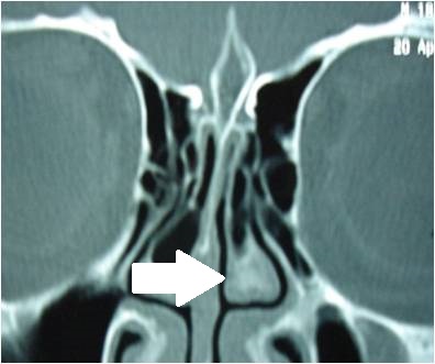 | Figure 1. CT scan image showing the concha bullosa of the left middle turbinate filled with a high-density material (arrow) |
3. Discussion
- Fungus balls are found in just one paranasal sinus, most frequently in the maxillary sinus and occasionally in the sphenoid sinus [4]. Aspergillus species are the most commonly documented pathogens associated with the disease. Most patients are either asymptomatic or present with non-specific complaints similar to those of chronic sinusitis [2, 5, 6]. The host is immunocompetent, but if during the infection the host is immunocompromised, then this noninvasive fungal infection may become invasive and life-threatening [4, 7].Fungal infections of the paranasal sinuses are a well known, but isolated fungal infection of concha bullosa is unusual. Ciger E et al [2] have reported a case of fungal ball in the concha bullosa. Cukurova I et al [8] have reported a case, in a series of 31 cases of concha bullosa pathology. Growth of the fungus balls in the sinuses is slow. Initially fungal hyphae and spores get trapped in a paranasal sinus and in suitable conditions they grow to form the fungus ball. The fungal spores germinate within the sinus cavity and the growth of hyphae further impairs clearance of the fungi and fungi growth proceeds in the involved sinus cavity [4, 9, 10, 11].Computed tomography is essential for diagnosing fungal infections and other types of sinonasal pathology. Fungal ball appears as partial or complete, heterogeneous opacification of the involved sinus, often with calcified material [2, 4]. Cholesteatoma of the concha bullosa should be considered in differential diagnosis [12]. Endoscopic sinus surgery to remove the fungus ball is the treatment of choice. Systemic or local fungal therapy is no required.A fungus ball in the concha bullosa, without paranasal sinus involvement is rare. Only two such cases have been reported in the literature. To best of our knowledge, this is a third case of a fungus ball in the concha bullosa reported in the literature.