
-
Paper Information
- Paper Submission
-
Journal Information
- About This Journal
- Editorial Board
- Current Issue
- Archive
- Author Guidelines
- Contact Us
Research in Otolaryngology
p-ISSN: 2326-1307 e-ISSN: 2326-1323
2015; 4(1): 10-12
doi:10.5923/j.otolaryn.20150401.03
Our Experience in Endoscopic Dacryocystorhinostomy in 203 Patients
 Abstract
 Reference
 Full-Text PDF
 Full-text HTML
Ruchi Rajput, Ravishankar Chandrashekar, Santosh Kumar
Department of ENT, Bangalore Medical College and Research Institute, Bangalore, Karnataka, India
Correspondence to: Ravishankar Chandrashekar, Department of ENT, Bangalore Medical College and Research Institute, Bangalore, Karnataka, India.
| Email: |  |
Copyright © 2015 Scientific & Academic Publishing. All Rights Reserved.
Treatment of Epiphora by endoscopic assistance through endonasal route is a well established procedure. However there are various choices available before the surgeon with respect to methods adopted for the procedure and also postoperative care. We like to share our experience in this regard about the procedure and also postoperative care. In our series Silicone stents and Mitomycin -C were used only in revision cases. Meticulously performed primary surgery (ie.not injuring nasl mucosa and proper exposure of lachrymal sac) saves the patient from discomfort caused by recurrence of symptoms and also cost incurred by using additional gadgets which can be reserved for failures.
Keywords: Epiphora, Endoscopic DCR, Lachrymal stenting, Mitomycin-C
Cite this paper: Ruchi Rajput, Ravishankar Chandrashekar, Santosh Kumar, Our Experience in Endoscopic Dacryocystorhinostomy in 203 Patients, Research in Otolaryngology, Vol. 4 No. 1, 2015, pp. 10-12. doi: 10.5923/j.otolaryn.20150401.03.
1. Introduction
- Epiphora due to obstruction of the lachrymal drainage pathway is a common symptom. Those conditions which require Dacryocystorhinostomy (DCR) to bypass obstructed nasolachrymal duct have to be carefully selected. It is because obstructions at punctum, canalicular/saccular obstructions do not do well with DCR. External DCR was first introduced by Toti in 1904 [1] and a transnasal procedure by West in 1911 [2]. First clinical study of endoscopic endonasal DCR was published in 1989 by Dough and Mevring [3].Endoscopic DCR is now well established procedure. The anatomic location of the sac when the sac is approached from the nasal cavity is well described [4-10]. The relevant intranasal anatomic landmarks, the middle turbinate and frontal process of the maxilla, are vitally important for the identification and exposure of the lachrymal sac and duct while performing endoscopic DCR.
2. Material and Methods
- Between June 2005 and August 2014, 179 cases of primary DCRs and 24 revision DCRs (Fig.1) were performed by the same surgeon. The subjects were chosen after confirmation of obstruction of nasolachrymal duct. We adopted a simple clinical examination to do this; a) lachrymal syringing and b) presence of regurgitation of fluid (mucoid/purulent) on pressing over medial canthus (sac area). All surgeries on adult patients were performed under local anaesthesia with sedation, while paediatric subjects required general anaesthesia.
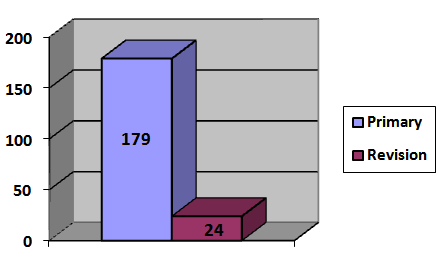 | Figure 1. Distribution of patients in primary and revision DCR |
3. Results
- Out of 179 patients, who underwent endoscopic DCR for the first time, 94 were male and 85 were female. Youngest was 5yrs, and eldest was 78 years. Of this 10 had undergone bilateral DCR. Out of 24 patients, who underwent revision DCRs, 10 were from the above series and the other 14 had undergone surgeries elsewhere. Of these 14, 6 had failed External DCR and 4 had failed Endoscopic DCRs and other 4 had twice failed endoscopic DCR. Among all the patients, 24 were in the age group of 5-14 yrs.Of the 179 DCRs on fresh cases, 147 (82%) improved after the surgery, 24 on whom revision surgery was done 21 (87.5%) were symptom free (table 1) and rest 3 were not symptom free in spite of having good flow of saline during syringing. Later, this was attributed to senile ectropion and they became symptom free 6 months later without any apparent reasons. In revision series, we used silicone stent in 2 patients and Mitomycin -C in 3 patients and it showed 100% results.
|
4. Discussion
- Most studies on DCR, the results range from 85-95% [11-13] and results with external and endoscopic DCRs almost are equal. But it is very well appreciated that endoscopic DCRs have advantage in1. It preserves pump mechanism of orbicularis oculi muscle (as this is dissected in external DCR)2. No external scar3. Revision is easy.It is preferable to do endoscopic DCR after any type of earlier surgery.In our series 82% of patients improved after first surgery and 100% of them improved with revision surgery. It is also noteworthy that we use silicone stent and Mitomycin-c local application only for revision cases. It is seen that meticulously done endoscopic DCR ie. taking care not to damage nasal mucosa and proper opening of the lachrymal sac, results are excellent and are comparable to laser DCRs and also it avoids huge amount of funds needed to be invested in sophisticated equipments and also inherent risks and complications involved with the use of laser. Lastly in view of the complications that occurred in our hands it is recommended that we should be1. Careful in doing lachrymal syringing and probing2. Never pull any tissue from nose side (do always sharp cuts)3. Posterior boundary should be uncinate process, as it is very thin behind and can be easily prone to expose orbital fat.An observation we would like to share on pediatric DCRs. In 14 DCRs which we performed under GA, on children between 5-14 yrs. of age, we found that sac can be exposed with much ease compared to adults with just 2-3 punches and also incision of sac is much easier compared to adults (as in some cases incision on fibrotic sacs is much difficult) But great caution has to be taken as structures are quite small and delicate in them.
5. Conclusions
- The intranasal approach to DCR is very advantageous, as it avoids external scar. Preserves pump action of orbicularis oculi and for revision surgery by this approach is much easier and also revision in previous endoscopic DCR is easier. Use of silicone stenting or application of Mitomycin-c can be done in those cases who don't improve with first surgery. It is important that we use these methods in failures because there is some amount of scarring due to previous surgery. The second reason is that we have to be more careful when doing revision cases as the patient expectations are high and it is necessary to take steps which will prevent the chances of second failure.
References
| [1] | Toti A. Nuovo metodo conservatare di cura radicale delle dacriocistiti croniche (dacriocistorinostomia). Clin Moderna 1904; 10:33-4. |
| [2] | West J.A window resection of the nasal duct in cases of stenosis. Trans Am Ophthalnol sac 1910; 12:659. |
| [3] | McDonogh M. Meining JH. Endoscopic transnasal dacryocystorhinostomy. J Laryngol Otol 1989; 103:585-7. |
| [4] | McDonogh M. Endoscopic transnasaldacryocystorhinostomy. Results in 21 patients. S Afr J Surg 1992; 30:107-10. |
| [5] | Steadman MG. Transnasal dacryocystorhinostomy. Otolarngol Clin North Am 1985; 18;107-11. |
| [6] | Whittet H, Shun-Shin A, Awdry P. Functional endoscopic transnasal dacryocystorhinostomy. Eye 1993; 7:545-9. |
| [7] | Metson R, Woog J, Puliafito C. Endoscpic laser dacryocystorhinostomy. Laryngoscope 1994; 104:269-74. |
| [8] | McDonogh M, Meiring JH. Endoscopc transnasal dacryocystorhinostomy. J Laryngol Otol 1989; 103:585-7. |
| [9] | Weidenbecher M, Hosemann, Buhr W. Endoscopic endonasal dacryocystorhinostomy: results in 56 patients. Ann Otol Rhinol Laryngol 1994; 103:363-7. |
| [10] | Rebeiz E, Shapshay S, Bowlds J, et al. Anatomic guidelines for dacryocystorhinostomy. Laryngoscope 1992; 102: 1181-4. |
| [11] | Dolman PJ. Comparison of external dacryocystorhinostomy with nonlaser endonasal dacryocystorhinostomy. Opthalmology 2003; 1110:78-84. |
| [12] | Moore WMH, Bently CR, Olver JM. Functional and anatomic results after two types of endoscopic endonasal dacryocystorhinostomy: surgical and holium laser. Ophthalmology 2002; 109:628-32. |
| [13] | Woog J, Kennedy RH, Custer PL, et al. Endonasal dacryocystorhinostomy. A report by the American Academy of Ophthalmology. Ophthalmology 2001; 108:2369-77. |