-
Paper Information
- Paper Submission
-
Journal Information
- About This Journal
- Editorial Board
- Current Issue
- Archive
- Author Guidelines
- Contact Us
Research in Otolaryngology
p-ISSN: 2326-1307 e-ISSN: 2326-1323
2015; 4(1): 7-9
doi:10.5923/j.otolaryn.20150401.02
Choanal Atresia: A New Way of Preventing Postoperative Stenosis
Ramabhadraiah Anil Kumar , Borlingegowda Viswanatha , Puneeth Poojar Jayakumar , Suparna Roy
Otorhinolaryngology department, Bangalore medical college & Research institute, Bangalore, India
Correspondence to: Borlingegowda Viswanatha , Otorhinolaryngology department, Bangalore medical college & Research institute, Bangalore, India.
| Email: |  |
Copyright © 2015 Scientific & Academic Publishing. All Rights Reserved.
A 30 year old lady presented to the outpatient department with complaints of watering of left eye and bilateral nasal obstruction, with habituated mouth breathing since 4 years. On diagnostic nasal endoscopy, incidentally she was found to have bilateral incomplete choanal atresia. She underwent transnasal endoscopic surgery for both choanal atresia and dacryocystitis. To maintain the postoperative choanal patency stents were not used. Silver nitrate was applied to the choanal margins. Post operative period was uneventful and at the sixth week of follow-up nasal endoscopic examination showed patent choanae.
Keywords: Choanal atresia, Postoperative stenosis
Cite this paper: Ramabhadraiah Anil Kumar , Borlingegowda Viswanatha , Puneeth Poojar Jayakumar , Suparna Roy , Choanal Atresia: A New Way of Preventing Postoperative Stenosis, Research in Otolaryngology, Vol. 4 No. 1, 2015, pp. 7-9. doi: 10.5923/j.otolaryn.20150401.02.
1. Introduction
- Choanal atresia was first described by Johann Roderer in 1755 [1]. Congenital choanal atresia is unilateral or bilateral occlusion of the posterior nasal orifices. It is a rare congenital abnormality, seen in 1 in 5000-7000 live births [2]; it is associated with other congenital abnormalities in 50% of the cases [3], and is twice as common in females. Seventy percent of the atresia are mixed bony-membranous type and 30% are pure bony type. The atresia may be complete or incomplete [4]. Bilateral choanal atresia is very rare in adults [3, 5-7]. Newborns are obligatory nasal breathers; therefore bilateral choanal atresia is often diagnosed at birth and treated as an emergency with early surgery. This case report is of an adult lady with bilateral incomplete choanal atresia, managed by transnasal endoscopic technique and silver nitrate application (without using nasal stents) and endoscopic dacryocystorhinostomy for epiphora in the left eye.
2. Case Report
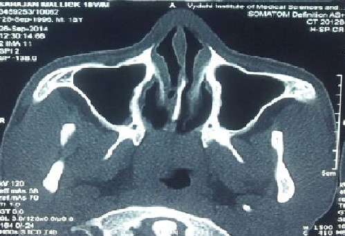
- A 30 year old lady presented to the outpatient department with complaints of watering of left eye and bilateral nasal obstruction, with habituated mouth breathing since 4 years.Obstetric history- she was para 4 and living 2. During her second delivery, which was conducted by trained attendant in her home, she complained of difficulty in breathing during delivery and baby died immediately after birth. The cause of death was not known. During her third delivery, which was conducted by a doctor in nearby health centre, she again had difficulty in breathing and baby died within 1 hour of delivery. The cause of death of the baby was not known. She did not have any other remarkable complaints during early childhood or adolescence. Her parents reported cyanosis in childhood that worsened during feeding and improved during crying; however, they had not visited a physician. Endoscopic examination revealed bilateral partial membranous choanal atresia with left chronic dacryocystitis. Patient was subjected to computerised tomography scan of nose and paranasal sinuses in order to confirm type of choanal atresia and it revealed bilateral membranous atresia [Figure 1] and well developed paranasal sinuses, which confirmed it as an isolated anomaly. She did not have any other congenital abnormalities.
 | Figure 1. Showing membranous atresia on both the sides |
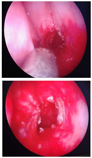
 | Figure 2. Showing patent choanae on the right and left side |
3. Discussion
- Choanal atresia may present as an isolated malformation or in 20% to 50% of cases, as a part of a group of congenital malformations, such as coloboma, heart malformation, mental and growth retardation, genital and ear anomalies or deafness [4]. Thus, a complete physical examination should be done by the pediatrician and otolaryngologist to rule out the presence of these anomalies. Some theories have been proposed to explain the embryological origin of this clinical entity. Among the most accepted, four theories stand out: 1) persistence of buccopharyngeal membrane, 2) failure of rupture of the physiological oronasal membrane of Hochstetter, 3) adherence of abnormal mesodermal tissue located in the nasal choana and 4) medial growth of vertical and horizontal processes of palatine bone [5, 6].Panda et al [3] reported a 22-year-old patient with bilateral choanal atresia. The passage in this patient was established via a transnasal endoscopic approach using a 2.5 mm diamond burr. Subsequently, a number 6 portex cannula was inserted for 6 weeks. Computerised tomography of this patient one year after the operation showed that adequate opening was preserved. There was no other congenital abnormality in this patient. Yasar and Ozkul [6] reported a 51-year-old patient with bilateral choanal atresia. Using a transnasal endoscopic approach, they raised mucosal flaps and achieved an opening that allowed passage of a number 7.5 portex cannula. The stent was removed 7 days later, and follow-up examination at 18th month showed that patency was preserved. El-Sawy et al [5] reported a 24-year-old patient with bilateral nasal obstruction and total loss of sense of smell and hypogammaglobulinemia. Computerised tomography of this patient revealed aplasia of frontal and sphenoid sinuses and hypoplasia of the inferior and middle turbinates. Adequate opening was achieved with transnasal endoscopic approach; however, restenosis occurred requiring a second operation. In this case report, a 30-year-old patient with bilateral partial membranous choanal atresia is presented. In our case, we preferred the transnasal endoscopic approach as reported in the previous literatures [3, 5-7]. In order to prevent stenosis, silver nitrate was applied to the neo-opening. Intranasal stent was avoided due to the requirement of intense antimicrobial therapy, foreign body reaction and fear of skin necrosis in the columella. On endoscopic examination at the 6th week following surgery, choanae were patent on both the sides.
4. Conclusions
- Bilateral choanal atresia is very rare in adults and is often diagnosed at birth. Rarely, it can be found during the evaluation of adults with bilateral nasal obstruction, as reported in this case. In the present case, stent was avoided due to the requirement of intense antimicrobial therapy, foreign body reaction and fear of skin necrosis in the columella. Intra operative silver nitrate application to the neo-orifice in choana was found to be useful in preventing postoperative stenosis.
 Abstract
 Reference
 Full-Text PDF
 Full-text HTML