International Journal of Nursing Science

p-ISSN: 2167-7441    e-ISSN: 2167-745X

2019;  9(2): 25-29

doi:10.5923/j.nursing.20190902.01


Effect of Apus Bamboo Shoot Extract on Total Interleukin 17 and Leukocyte at Atherosclerosis Process

Edy Soesanto1, Edi Dharmana2, Soeharyo Hadisaputro2, S. Fatimah Muis2

1Department of Nursing, University of Muhammadiyah Semarang, Indonesia

2The Great Teachers Faculty of Medicine Diponegoro University of Semarang, Indonesia

Correspondence to: Edy Soesanto, Department of Nursing, University of Muhammadiyah Semarang, Indonesia.

Email:

Copyright © 2019 The Author(s). Published by Scientific & Academic Publishing.

This work is licensed under the Creative Commons Attribution International License (CC BY).
http://creativecommons.org/licenses/by/4.0/

Abstract

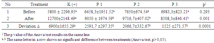
Atherosclerosis is a consequence of inflammation and excess of oxidative stress. High level of IL-17 and leucocytes are considered as the risk of rupture of plaque, thrombus, and embolism due to the acceleration of blood vessels lession and blockage Bamboo shoots apus possesses antioxidant content and it possibly inhibits the atheroslerosis progress. The purpose of this study was to prove the influence of bamboo shoot extract to decrease interleukin 17 and leucocyte count in New Zealand White rabbit with atherogenic diet. This research used 1-2 weeks freeze dried apus bamboo shoot extract and administered in New Zealand White rabbit. The atherogenic diet consisted of standard feed added by 0.5% egg yolk and 5% pork oil. This research used Randomized pre and post-test with control group design by dividing (jumlah kelinci) rabbits into 4 groups. The results showed that there was a significant difference of IL-17 levels between treatments and all groups had decreased levels of IL-17 except in the positive control group that increased by 57.1% and the highest decrease occurred in treatment group 3 which was 65.4%. There was a significant difference in the number of absolute leukocytes between treatments (p = 0.001). The increase of absolute leukocyte count of positive control group with treatment group 1, 2 and 3 was significantly different (p = 0.0001). In conclusion, bamboo shoot extract is able to decrease interleukin 17 level and absolute leukocyte count.

Keywords: Bamboo shoot, Bamboo shoot extract, IL-17, Leukocytes

Cite this paper: Edy Soesanto, Edi Dharmana, Soeharyo Hadisaputro, S. Fatimah Muis, Effect of Apus Bamboo Shoot Extract on Total Interleukin 17 and Leukocyte at Atherosclerosis Process, International Journal of Nursing Science, Vol. 9 No. 2, 2019, pp. 25-29. doi: 10.5923/j.nursing.20190902.01.

1. Preliminary

Atherosclerosis is the most common cause of coronary artery disease, carotid artery disease, peripheral artery disease and is the global major cause of death. Atherosclerosis is a progressive disease, progressing slowly but surely and increasing by 3 percent annually in the age of 20 (Nurahmi, et.al 2016). Heart disease and blood vessels, which lead to atherosclerosis as the cause of death in Indonesia, raised from 5.1% to 8.7% in the age range 45-54 years and the increased incidence of atherosclerosis in patients aged more than 40 years is 17-40% (Indonesia DKR, 2010).
Atherosclerosis is the consequence from multiple factors, including inflammatory processes and oxidative stress. Endothelial dysfunction, caused by ox-LDL, and inflammatory processes, resulting in activation of monocyte migration into the intima, leading to atherosclerosis (Li D, 2005). The occurrence of inflammatory processes in atherosclerosis is related to the role of cytokines and leukocytes. High levels of IL-17 in the vascular worsen the stability of atherosclerotic plaques bylesions formation. These lessions in turn promote plaque rupture (Hashmi, 2006). One of the indicators in initiation and the onset of integrated atherosclerosis from acute and chronic inflammatory responses is an increase of leukocytes number (Lee D.C et.al. 2001). An elevated circulating leukocytes may increase blood vessel blockage and increase risk of plaque rupture which result in thrombus and emboli ending with myocardial necrosis (Homenta et al. 2009).
In the world of health is currently being intensified the concept of "food as medicine" and well known as functional food which overcome the disease. Functional food ingredients are expected to inhibit the progression of atherosclerosis due to its antioxidant content. Several ingredients which contribute as an inflammatory process in atherosclerosis, which is a vitamin E, pholipenol, plavonoid, vitexin and orientin, palmitic acid, curcumene, limonene, toluene, naphthalene, 1,3,5-trimethyl benzene, (Lu B. Et.al. 2010). Other antioxidant compounds present in bamboo are vitamin A, thiamin, riboflavin, vitamin C, curcumin (Choudhury D, 2010). The purpose of this study was to prove the influence of bamboo shoot extract to decrease interleukin 17 and leucocyte count in New Zealand White rabbit with atherogenic diet.

2. Materials and Methods

The bamboo shoot aged 1-2 weeks was gained from Banyumeneng village, Mranggen district. The bamboo shoot was extracted by freeze dried method using ethanol 90% (Soesanto E, 2016). The standard feed for rabbits used was Feed Premium rabbit produced byPT Cargill Indonesiaand has nutritional content per 100 gr: 5% fat, 18% protein, crude fiber 14%, 1% calcium, 0.8% phosphorus, ash 12% and water 12%. The diet was given maximum 5% of the rabbit's weight and beverages weregiven maximum 10% of the rabbit's weight.
The atherogenic diet used was standard diet added by 0.5% egg yolk and 5% pork oil (Soesanto, E. 2017). Cholesterol flour is made from egg yolks dried oven at 60 °C for 24 hours to dry and blend until smooth. The experimental animal used was 4-month-old New Zealand White rabbit weighed between 2000-2500 grams, and male sex. Standard diet as well as atherogenic dietwere provided as much as 100 grams / day / tail and ad libitum water. Absolute leukocyte examinedfrom whole blood plus EDTA and analyzed by Hematology Analyzer while IL-17 was assessed using quantitative sandwich enzyme immunoassay technique which was conducted at Integrated Research and Testing Laboratory (LPPT) Gajah Mada University Yogyakarta. This research used pure experimental, Randomized pre and post-test design with control group design which have got approval from Health Research Ethics Commission FK UNDIP and Dr. Kariadi Semarang no. 23 / EC / H / FK-RSDK / 2017 dated May 22, 2017. This study used 24 rabbits which were divided into 4 groups ie control group:the group given Atherogenik diet, treatment group I, II and III respectively were given Atherogenik diet plus bamboo shoot extract with concentration 130 mg / kg Weight, 260 mg / kg Weight, and 520 mg / kg Body Weight.

3. Result and Discussion

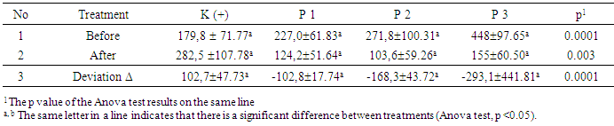
The mean of IL-17 level of all samples before treatment was 281.7 ± 130.14 pg / mL with a range of 124.8-570.5 pg / mL, while the mean of IL-17 concentration of all samples after treatment was 166.3 ± 98.53 pg / mL with a hose between 51.2-496,7 pg / mL.
Table 1. Mean of IL-17 Level (pg/mL)
     
In the initial sample (pre-test), the average of IL-17 level was significantly different between groups (p=0.0001). The delta showed that there was a significant difference between the groups (p=0.0001).
Post-hoc LSD test showed that the mean change of rabbit IL-17 level (adjusted) in the positive control group was significantly different from the treatment group I, II, and III (p=0.001; p=0.0001; p=0.007 respectively). The mean change of the largest IL-17 level was 177.24 pg / mL and the smallest was -441.51 pg / mL. Changes in IL-17 levels between the positive control group and the treatment group were significantly different (p = 0.0001), and between treatment groups and other treatment groups there was also a significant difference (p <0.05 Table 1 shows that all groups experienced a decrease in IL-17 levels with the highest decrease by 65.4% in group treatment III, while the positive control group had an increase of IL-17 by 57.1%. Thus this research can prove that there is a decrease of IL-17 level after bamboo shoot extract tretament in various doses, the higher dosage of bamboo shoot extractwas related to decrease level IL-17 in the blood (dose effect relationship). Interleukin-17 is produced by T cells, macrophages, CD8 + T cells, mast cells, neutrophils, dendritic cells and natural killer cells and is a proinflammatory cytokine. IL-17 as an inflammatory mediator is also produced by Th 17 Cells with IL-23, IL-1, and IL-21 activations that plays a role in the process of atherosclerosis (Ciric B. et.al. 2009; Liuzzo G. 2013). Regular T cells are an important cell in providing protection against atherosclerosis through the production of IL-10 as well as controlling pro-atherogenic responses by Th1 cells (Taleb, 2009). LSD post-hoc assay in this study showed that the mean change of IL 17 rabbit level (adjusted) in the positive control group was significantly different from the treatment group (p <0.05), all groups experienced a decrease level of IL 17 except in the positive control group. This study is consistent with that of Smith and Erbel that by blocking IL-17 in rat apoE / mouse successfully decreased the incidence of atherosclerosis (Erbel C. Et.al, 2009; Smith E. Et.al, 2010).
Atheroschlerosis, IL-17 plays a role in plaque formation through extravasation adhesion, cell activation, T cell proliferation, triggering the formation of MMP 9 (Matrix Metalloproteinase -9). IL-17 has a proatherogenic effect by inducing the production of cytokines, chemokines, and matrix metalloproteinases (Chen S, 2010). In addition, Th17's response is against vascular inflammation and the development of atherosclerosis. Similarly, the deficiency of SOCS3 in T cells leads to IL-17 reduction in the development of lesions and vascular inflammation by increasing the production of IL-17 and IL-10, as well as by inducing the anti-inflammatory macrophage phenotype.
IL-17 is generally a proinflammatory cytokine, IL-17 and IL-17F are primarily bound to IL-17RA and IL-17 RC in epithelial, endothelial vascular, and fibroblast cells. Then include nuclear factor (NF) kB and activated Mitogen activated protein kinese (MAPKs). Finally, the induced receptor cell expresses a variety of proinflammatory mediator types such as TNF, IL-1, IL-6, chemokines (CXCl1, IL-8, CCL-2), MMP (MMP-1, MMP-3, MMP-9) and C-reactive protein (CRP), causing inflammatory cell adhesion and retrieval of neutrophils, T cells, macrophages, and other types of cells at local inflammatory sites (Gaffen SL, 2009). IL-17 plays a role in the progression of inflammation and considers as a host defense against bacterial infections and also maintains the stability of atherosclerotic plaques and their complications (Iwakura Y. et.al, 2011). Interleukin-17 has the potential to cause the incidence of acute coronary syndromes through 3 pathways, ie 1) in macrophages, produced cytokines / chemokines, and foam cell formation; 2) in smooth muscle cells also produced chemokines and proliferation; and 3) in endothelial cells also occurring chemokine secretion, adhesion molecule up-regulation, and leukocyte adhesion (Chen S. et.al, 2010). IL-17 stimulates proinflammatory mediators from various cells including IL-6 and IL-8 production of stromal cells. The attachment of IL-17 to receptors increases Ca2 + influx, decreases cAMP levels, mitogen activation by protein kinase, and stimulation of NF-kB activation. The IL-17 family has no implications for atherogenesis, but the proinflammatory effects on macrophages, endothelial IL-2 stimulation, also IL-17F, IL-17 production by T cells and the extent of IL-17 receptor expression make this interleukin family potential as proatherogenic causing the syndrome coronary acute.
IL-17 acts as a proinflammatory mediator through some mechanisms including: stimulation of the production of other proinflammatory mediators (cytokines), such as: TNF-α, IL-1, and IL-6 and chemokines CXCL1 and CXCL2; stimulating the production of ICAM-1 adhesion molecules; stimulation and production of C-reactive protein and nitric oxide. IL-17 also stimulates chemotactic neutrophils and monocytes to inflammation sites through the induction of chemotactic mediators such as IL-8, MCP-1, and growth protein. IL-17 is currently found in human plaques, but the presence of these cells is often concomitant IFN γ. IL-17 depicts the presence of macrophage activation in atherosclerotic lesions inducing systemic IL-17 with local effects on blood vessel walls. Systemic activation of macrophages by IL-17 contributes to the progression of lesions by releasing proteolytic factors. The production of vascular IL-17 appears to worsen pro-atherogenic conditions. High levels of vascular IL-17 produce a proinflammatory environment that promotes the growth of vascular atherosclerotic disease. Inhibition of IL-17A reduces the area of atherosclerotic lesions and induced plaque stability. Production of IL-17 by activated T cells and its expression on IL-17 receptors, makes the IL-17 family potential as pro-atherogenic.
Antioxidants in bamboo shoots by cutting the oxidation reaction chain from free radicals or by capturing free radical (free radical scavenger). As a result, free radicals will not react with cellular components (Hashmi S, 2000). Flavonoids in bamboo shoots contributes to decline the amount of monocyte as a part of foam cell formation by alleviating the expression of Vascular Cell Adhesion Molecule-1 (VCAM-1) and Monocyte ChemotacticProtein-1 (MCP-1). Flavonoid is also capable of reducing the inflammatory process by inhibiting the release of pro-inflammatory cytokines as well as reducing platelet aggregation. Thus, the application of bamboo shoot bamboo shoot extract will decrease the levels of IL-17 in the blood, the higher the dose of bamboo shoot bamboo shoot extract will be the decrease of blood-dose effect of IL-17.
The average number of absolute leukocytes throughout the sample before treatment was 6722.9 ± 1644.22 g / dL with a range between 3000-8750 gr / dL. The average number of absolute leukocytes throughout the sample after treatment was 9993.8 ± 2186.63 gr / dL with a hose between 6400-14800 gr / dL. Based on the data in table 2, all groups experienced an increase of absolute leukocytes and the highest increase of absolute leukocyte was found in positive control group that was 119% and the lowest was treatment group 3 of 21.8%.
Table 2. Average Number of Absolute Leukocytes (gr / dL)
     
There was no significant difference (p=0.293) of absolute leukocytes in the preliminary sample. The result (present in table 2) shows there was a significant difference of absoulte leukocytes in the end of study between the groups (p=0.001) The post-hoc LSD test showed that the mean increase of absolute leukocyte count (adjusted) in the positive control group was significantly different with the treatment group I, II, II (p=0,001; p=0.004; p=0.0001 respectively).
The largest increase in absolute leukocyte count was 6900 g / dL and the smallest was 1525 g / dL. The increase of absolute leukocyte count of positive control group with treatment group 1, 2 and 3 was significantly different (p = 0.0001), and between treatment group 1 and treatment group 3 there was significant difference (p = 0,028), trend of increasing absolute leukocyte the highest occurred in the control group. Thus this study can prove that there is a decrease in absolute leukocyte content after administration of bamboo shoot extract in various doses. Bamboo shoot extract have an effect on the decrease of absolute leucocyte content, the higher the dose of bamboo bamboo shoot extract will decrease the absolute leukocyte level in blood (dose effect relationship).
Leukocytes are a unit that always moves actively to protect the immune system, moving into an inflamed area, thus providing a fast and strong defense against foreign body attack. Leucocytes tend to aggregate and colonize into small blood vessels under low blood flow conditions (Guyton, 2015). In this research, the result of one way anova test followed by post-hoc LSD test showed that the average increase of absolute leukocyte count of rabbit (adjusted) in the positive control group was significantly different with treatment group (p <0.05). Increase in absolute leukocyte group positive control with treatment group 1, 2 and 3 were significantly different (p = 0.0001), and between treatment group 1 and treatment group 3 there was a significant difference (p = 0.028). Difference highest increase of absolute leukocyte count in positive control group that is equal to 6900 ± 1655.29 gr / dL or by 118.97% and the lowest is treatment group 3 equal to 1525 ± 271.57 gr / dL or equal to 21,84%. According to a study conducted by Milas in 2009, the number of normal absolute leucocytes in New Zealand White male rabbits was in the distribution of 4.2-12.3x10⁹ / L (Poljicak Milas N. et.al, 2009). In this study, the average increase of absolute leukocyte above normal only occurs in the control group that is 12.7 x 10⁹ / L whereas in all treatment groups the absolute leukocyte count is still in the normal range. An acute increase in absolute leukocytes may be triggered by infection and other stressor states while chronic ones may be triggered by persistent exposure by toxic substances such as hyperlipid foods. An increase in the number of absolute leukocytes in the body indicates an inflammatory process, the occurrence of a vascular injury that affects the process of atherogenesis which is the development of an atherosclerotic plaque rupture, and thrombosis (Grzybowski M. et.al, 2004; Wu TH et.al, 2013).
The high leukocyte count corresponds to the extent of infarction occurring in the process of atherosclerosis (Rohani A. et.al, 2011). Plaque stabilization is influenced by leukocytes by secreting myeloperoxidase (MPO). The higher the activation of leukocytes due to inflammation, the higher the MPO will be released, thus affecting the plaque stabilization (Homenta RS et.al, 2009). An increase in the number of leukocytes results in increased thrombus formation in ruptured plaque areas resulting in reduced epicardium blood flow and myocardial perfusion, resulting in an increase in the number of leukocytes to be a marker of coagulation hyper or thromboresistance (Barron HV et.al, 2000). In patients with acute myocardial infarction with leukocytosis has a 10.71 times mortality risk compared to patients with normal leukocytes (Kurniawan LB, 2015). Research conducted by Atherosclerosis Risk in communities study found an association between changes in leukocyte count and increased incidence of ischemic stroke and death due to cardiovascular disease (Lee DC. et.al, 2001).
Leukocytes, especially monocytes and neutrophils, trigger coagulation by releasing granular content and forming aggregates with platelets to form leucocyte platelet aggregates. Aggregates or clumps that form can clog the blood vessel. This is in accordance with the opinion expressed by Oliver that one of the factors that cause atherosclerosis is the buildup of blood clots that form thrombus in the blood vessels. High leukocyte counts lead to increased production of free radicals and toxic substances triggering the release of proinflammatory cytokines in inflammatory reactions (Lakhan SE, 2009). Leucocytes can also cause occlusion in the brain circulation. Decreased perfusion pressure can trap leucocytes in the circulation and form contact with the capillary endothelium. Leucocyte aggregation causes the capillary occlusion resulting in tissue hypoperfusion and ischemic brain (Rudijanto A, 2007). The deployment of leukocytes (especially monocytes and T lymphocytes) into the blood vessel wall (intima layer) is considered as the key stage of atherogenesis which then turns into macrophages and secretes the enzyme metalloproteinase The elevating deployement of leukocytes result in atherosclerosis progress acceleration. Leukocytes also play a role in the destabilization of atherosclerotic plaques, the inflammatory process can lead to decreased synthesis and increased degradation of fibrous capsule structural components by degradation and cytokine enzymes produced by macrophages at the site of rupture of plaque.
Flavonoids in bamboo shoots are one type of antioxidant components that can inhibit adhesion, aggregation, and platelet secretion. These may due to inhibition of flavonoids in the arachidonic acid metabolism by cyclooxygenase. Decrease in platelet count by the activity of antioxidants will also lead to a decrease in the number of leukocytes (Zhang Z. Et.al, 2006). Therefore, the higher intake of bamboo bamboo shoot extract will affect the decrease in the number of absolute leukocytes in the blood (dose effect relationship).

4. Conclusions

The bamboo shoot extract is found to have a beneficial effects on the decrease of IL-17 and absolute leukocyte count in New Zealand White rabbit given atherogenic diet. The dose 520 mg / kg / day is an effective dose to decrease IL-17 and leukocytes level.

Suggestion

Further research is needed to examine the toxicity before continuing the clinical trial to human. Furthermore, apus bamboo shoot extract can be one of the standardized ingredient alternatives of traditional medicine.

References

[1]  Barron HV, Cannon CP, Murphy SA, Braunwald E, Gibson CM. Association between white blood cell count, epicardial blood flow, myocardial perfusion, and clinical outcomes in the setting of acute myocardial infarction: a thrombolysis in myocardial infarction 10 substudy. Circulation. 2000; 102(19): 2329-34.
[2]  Chen S, Crother TR, Arditi M. Emerging role of IL-17 in atherosclerosis. Journal of innate immunity. 2010; 2(4): 325-33.
[3]  Chen S, Shimada K, Zhang W, Huang G, Crother TR, Arditi M. IL-17A is proatherogenic in high-fat diet-induced and Chlamydia pneumoniae infection-accelerated atherosclerosis in mice. The Journal of Immunology. 2010; 185(9): 5619-27.
[4]  Choudhury D, Sahu JK, Sharma G. Biochemistry of bitterness in bamboo shoots. Assam University Journal of Science and Technology. 2010; 6(2): 105-11.
[5]  Do Lee C, Folsom AR, Nieto FJ, Chambless LE, Shahar E, Wolfe DA. White blood cell count and incidence of coronary heart disease and ischemic stroke and mortality from cardiovascular disease in African-American and White men and women: atherosclerosis risk in communities study. American Journal of Epidemiology. 2001;154(8): 758-64.
[6]  Gaffen SL. Structure and signalling in the IL-17 receptor family. Nature Reviews Immunology. 2009;9(8):556-67.
[7]  Grzybowski M, Welch RD, Parsons L, Ndumele CE, Chen E, Zalenski R, et al. The Association between White Blood Cell Count and Acute Myocardial Infarction In‐hospital Mortality: Findings from the National Registry of Myocardial Infarction. Academic Emergency Medicine. 2004; 11(10): 1049-60.
[8]  Erbel C, Chen L, Bea F, Wangler S, Celik S, Lasitschka F, et al. Inhibition of IL-17A attenuates atherosclerotic lesion development in apoE-deficient mice. The Journal of Immunology. 2009; 183(12): 8167-75.
[9]  Hall JE. Guyton and Hall textbook of medical physiology e-Book: Elsevier Health Sciences; 2015.
[10]  Hashmi S, Zeng QT. Role of interleukin-17 and interleukin-17-induced cytokines interleukin-6 and interleukin-8 in unstable coronary artery disease. Coronary artery disease. 2006; 17(8): 699-706.
[11]  Homenta R.S KH, Karo-karo S, Soerianata S, Harimurti G.M, Rahayoe A.U. Mieloperoksidase pada penderita infark miokard akut. Majalah Kedokteran Indonesia. 2009:69.
[12]  Indonesia DKR. Pedoman pengemdalian penyakit jantung dan pembuluh darah: Departemen Kesehatan Republik Indonesia; 2010 [cited 2012 April 24th]. Available from: www.hukor.dep.kes.go.id.
[13]  Iwakura Y, Ishigame H, Saijo S, Nakae S. Functional specialization of interleukin-17 family members. Immunity. 2011; 34(2): 149-62.
[14]  Kurniawan LB, Bahrun U, Darmawaty E, Arif M. Pengaruh jumlah leukosit terhadap mortalitas pasien infark miokard akut selama perawatan. CDK-233. 2015; 10: 729.
[15]  Li D, Mehta JL. Oxidized LDL, a critical factor in atherogenesis. The Oxford University Press; 2005.
[16]  Lu B, Xia D, Huang W, Wu X, Zhang Y, Yao Y. Hypolipidemic effect of bamboo shoot oil (P. pubescens) in Sprague–Dawley rats. Journal of food science. 2010;75(6).
[17]  Nurahmi N, Aprianti S, Arif M, Hardjoeno H. Nilai small dense LDL remaja dan kaitannya. Indonesian Journal of Clinical Pathology and Medical Laboratory. 2016;13(1):17-9.
[18]  Poljičak-Milas N, Kardum-Skelin I, Vuđan M, Marenjak TS, Ballarin-Perharić A, Milas Z. Blood cell count analyses and erythrocyte morphometry in New Zealand white rabbits. Vet Arhiv. 2009;79:561-71.
[19]  Rohani A, Akbari V, Moradian K, Malekzade J. Combining white blood cell count and thrombosis for predicting in-hospital outcomes after acute myocardial infraction. Journal of Emergencies, Trauma and Shock. 2011;4(3):351.
[20]  Rudijanto A. The role of vascular smooth muscle cells on the pathogenesis of atherosclerosis. Acta Med Indones. 2007; 39(2): 86-93.
[21]  Smith E, Prasad K-MR, Butcher M, Dobrian A, Kolls JK, Ley K, et al. Blockade of interleukin-17A results in reduced atherosclerosis in apolipoprotein E–deficient mice. Circulation. 2010; 121(15): 1746-55.
[22]  Soesanto E. Antioxidant Activity of Extracts from Bambusa vulgaris and Gigantochloa apus Kurz Bamboo Shoots. Pakistan Journal of Nutrition. 2016;15(6):580.
[23]  Soesanto E, Khoiriyah, editors. Efektifitas Ekstrak Rebung Bambu Apus terhadap Penurunan Kadar MDA pada Kelinci New Zealand White Hiperkolesterolemia. Prosiding Seminar Nasional dan Internasional; Universitas Muhammadiyah Semarang. 2017; 131-135.
[24]  Taleb S, Romain M, Ramkhelawon B, Uyttenhove C, Pasterkamp G, Herbin O, et al. Loss of SOCS3 expression in T cells reveals a regulatory role for interleukin-17 in atherosclerosis. Journal of Experimental Medicine. 2009; 206(10): 2067-77.
[25]  Tedgui A, Mallat Z. Cytokines in atherosclerosis: pathogenic and regulatory pathways. Physiological reviews. 2006; 86(2): 515-81.
[26]  Wu T-H, Chien K-L, Lin H-J, Hsu H-C, Su T-C, Chen M-F, et al. Total white blood cell count or neutrophil count predict ischemic stroke events among adult Taiwanese: report from a community-based cohort study. BMC neurology. 2013; 13(1): 7.
[27]  Zhang Z, Machac J, Helft G, Worthley SG, Tang C, Zaman AG, et al. Non-invasive imaging of atherosclerotic plaque macrophage in a rabbit model with F-18 FDG PET: a histopathological correlation. BMC nuclear medicine. 2006; 6(1): 3.