Nanoscience and Nanotechnology

p-ISSN: 2163-257X    e-ISSN: 2163-2588

2018;  8(1): 11-24

doi:10.5923/j.nn.20180801.03


Recent Progress in Biomedical Applications of Nanodiamonds

Santos-Adriana Martel-Estrada

Instituto de Arquitectura, Diseño y Arte, Universidad Autónoma de Ciudad Juárez, Ciudad Juárez, México

Correspondence to: Santos-Adriana Martel-Estrada, Instituto de Arquitectura, Diseño y Arte, Universidad Autónoma de Ciudad Juárez, Ciudad Juárez, México.

Email:

This work is licensed under the Creative Commons Attribution International License (CC BY).
http://creativecommons.org/licenses/by/4.0/

Abstract

Carbon based nanomaterials have unique advantages in several areas due to its electronic, optical, thermal, and mechanical properties. In these terms, nanodiamonds have attracted considerable attention in recent years in different research areas such as biological sensing, medical therapy, fluorescent markers, enzyme immobilization and so on. Nanodiamond (ND) is a new member of carbon nanoparticle family with a truncated octahedral architecture that showed superior characteristics of diamond. This review is focus on synthesis methods, dispersion, functionalization, medical applications and toxicity of nanodiamonds.

Keywords: Nanodiamonds, Polymers, Composites, Biomaterials

Cite this paper: Santos-Adriana Martel-Estrada, Recent Progress in Biomedical Applications of Nanodiamonds, Nanoscience and Nanotechnology, Vol. 8 No. 1, 2018, pp. 11-24. doi: 10.5923/j.nn.20180801.03.

1. Introduction

Carbon based nanomaterials have unique advantages in several areas due to its electronic, optical, thermal, and mechanical properties [1]. Fullerenes, carbon natubes (CNT), carbon nanohorns, carbon nanodots, nanodiamonds (ND), and graphene (Figure 1) have attracted considerable attention in recent years [1]. Nevertheless, it is important to mention that while graphite is the stable carbon form at ambient conditions, diamond is metastable [2].
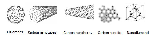
Figure 1. Carbon based nanomaterials [3-6]
Nanodiamond (ND) is a new member of carbon nanoparticle family with a truncated octahedral architecture that showed superior characteristics of diamond [7-9]. NDs are characterized by a size of crystal grains less than 100 nm [10], a surface area per unit of particle larger than CNTs, carbon nanofibers and graphene oxide ones [11]. Their diameter is around 2-10 nm [12]. They have superior physical and chemical properties of versatile functionalization, large surface area, high adsorption capacity and good biocompatility. They can form spontaneously nanometer sized clusters in aqueous solution, which could facilitate the loading of therapeutic agents on the surface or in the internal nano-scaled pores of ND clusters by non-covalent interactions [13]. NDs are widely used as nanomaterials for biomedical applications, especially in drug delivery and imaging systems [9, 14, 15]. They exhibit remarkable luminescence properties with emission of great stability and of high quantum yield which originate from color centers, as Nitrogen-Vacancy centers emitting in far-red/near infrared, perfectly adapted for biological labeling [16].
The main applications of NDs include its use as a carrier for biologically active substances, biomarkers, biosensors, high efficiency adsorbents, coatings for surgical instruments, cosmetic compositions, UV screening creams [17] and additives for dental materials (Figure 2) [18].
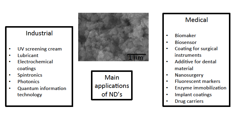
Figure 2. Main applications of NDs and SEM micrograph of commercial nanodiamonds purchased to Sigma-Aldrich
Synthetic diamond can be obtained by high pressure high temperature method since 1950 [2]. They exist in interstellar dust [19] and were discovered in 1980s by Lewis in meteorites [20, 21].
These materials combine nano-dimensionality, chemical inert core and reactive peripheral shell [22]. They are synthesized by detonation of explosives in an oxygen-deficient atmosphere, characterized by a narrow size distribution [23]. They are particles around 4-5 nm and during its manufacturing process forms oxygen containing functional groups at the stage of the removal of non-diamond sp2-component from the detonation soot [24]. The physical properties of ND surface are strongly affected and determined by impurities, morphology and bonding configuration, so the extent of H and O surface termination has a profound effect on the wettability, charge density and surface electronic properties [25]. Also, NDs are excellent precursors of carbon onion structures during annealing-induced graphitization [26].
NDs are the most abundant presolar grain types, occupying 1440 ppm and with the smallest size, approximately 3 nm of diameter [27] and large surface (300-500 m2/g) [28]. They originate as dust formed in the winds of low-mass stars or in the ejects of supernova events [29]. The theoretical surface area of NDs is about 250-450 m2/g, which makes it potential superior adsorbents to use in biopharmaceutical industry [30, 31]. The size of diamonds nanoparticles is related to their structure. The smaller the particle size is, the bigger part of their volume is defect, so only particles with size greater than 100 nm, can be considered monocrystallic [32]. The shape, size and structure of NDs depend of the process in which they were produced [33]. Nevertheless, the critical size of the stability of ND is 4 nm. Below this size the particle is unstable and rapidly oxidizes in air or transforms into other with a graphite-like structure [34]. The properties of nanoparticles are associated to large surface area to mass ratio, as well as an increase in surface reactivity, altered physico-chemical properties, such as changes in melting point, solubility, electrical conductivity and resistivity, crystalline structure, biocompatibility, etc. [35, 36].
As it was mention before, properties of NDs depend on how they are produced: high-pressure high-temperature (HPHT), chemical vapor deposition (CVD) and detonation synthesis (DND) [37]. HPHT reproduces pressures of 70-80 kbar and temperatures of 1400-1600ºC. In CVD synthesis, hydrocarbon gas mixtures are ionized and through a series of reactions, the carbon atoms are deposited on a substrate to grow diamond films. DND forms a process where carbon-containing explosives are detonated in the absence of oxygen [37]. Nevertheless, diamond grown under non-optimum conditions, and gives films with small grain size [28]. Luminescence is produced from all of these different synthesis techniques and it is due to the incorporation of defects in the crystal lattice [37]. ND can locate lattice point defects and nitrogen-vacancy center, which has an electronic spin resonance between the triplet ground state sublevels that can be detected optically and could be used as a nanoscale magnetic field sensor due to these sublevels are sensitive to external magnetic fields [38]. Nanovacancy centres are atomic impurities in the diamond lattice that include a substitutional nitrogen atom adjacent to a hole in the lattice. So, this fact enables a negative or neutral charge in diamonds [37]. In order to get fluorescence, particles with diameters around 7 nm are favored to the appropriate amount of NV centers in them [39].
As it was previously mention, NDs can be produced by detonation synthesis, chemical vapor deposition high pressure or high temperature synthesis [40]. These processes give to NDs different purity, surface composition and reactivity [41]. If the diamond cores exhibit the chemical and mechanical inertness of the bulk material, their surface can be widely functionalized through the carbon-related groups such as carboxylic acids, alcohols or alkenes [16]. During the preparation, it could be produced some impurities like metal-oxide microparticles, carbides, silicon dioxide, mercury or insoluble salts [42]. Even the presence of nitrogen is expected as it is the most common impurity in diamond [43].
Due to during the detonation nanodiamonds (DND) production exist the possibility of DNSs being contaminated, them medical uses requires strict inspection of their impurity composition in accordance with medicinal standards [31]. Usually the removal process is carried out by a treatment of the detonation soot with nitric acid in autoclave at 200-230oC or by the treatment with other oxidizing agents [24]. In spite of this inconvenience, NDs have excellent biocompatibility, chemical stability, scalability, fluorescent properties and easy functionalization [44], due mainly to its high concentration of surface functional groups [18]. The biocompatibility and not toxicity of NDs to cells has been proved [7, 8, 45]. They have high strength and modulus, superior hardness, attractive optical properties and highest coefficient of thermal conductivity [19, 46-49]. They are composed of aggregates of primary particles of average size of 5 nm, with a diamond core partially covered by layers of graphitic and/or amorphous carbon with carboxyl, hydroxyl, carbonyl, amida and sulfur groups on its surface [32, 46]. They are predominantly spherical in shape and is a supramolecule with a single-crystal core surrounded by a shell with functional groups [50]. These particles are currently used in many areas, such as biological sensing and medical therapy [51, 52], nanosurgery [31], composite materials and lubricants [51], fluorescent markers [33], electrochemical coatings, enzyme immobilization, implant coatings and drug carriers [9]. Some applications of NDs include spintronics, photonics, quantum information technology [53], cosmetology and cryptography [54]. In these applications, nuclear spin relaxation process plays a significant role [54]. Other studies includes the use of thermoplastic polymers such as poly(vinyl alcohol), polyamide, polyisoprene and poly(L-lactic acid) [28].
NDs are used in electrodeposited metal, diamond coatings, various lubricants, lubricating-cooling fluids for physical modification of ferrolacquer coatings of magnetic tapes and disks, as composite materials in polymeric films and membranes [55], mirror polishing, antifriction liquids and as biomarkers and drug delivery vehicles [10, 56].
NDs are used as a nanoreinforcement in a variety of polymeric materials to overcome their limitations and to develop a composites with improved mechanical and functional properties [47]. It has been reported nanocrystals with a Young’s modulus of about 950 GPa [57]. The main advantages of ND includes: a) diamond structure that provides superior mechanical properties, such as Young’s modules, hardness, high thermal conductivity, electrical resistivity and biocompatibility, b) small and uniform size, c) spherical shape, d) large external surface, e) rich surface chemistry with good flexibility for rational design [11].
It has been investigated that NDs can be modified by the presence of copper producing multiple carboxyl and anhydride groups, suggesting that DND are suitable for targeted surface modification by double-charged metal ions [24].
So, due to the importance of ND in research, the author of this review only pretend to analyze some publications to demonstrate basic medical applications of these important carbon particles. Specially, this review is focus on synthesis methods, dispersion, functionalization, medical applications and toxicity of nanodiamonds.

2. Synthesis Methods

A large variety of methods have been developed for synthesis of NDs, such as direct synthesis at ultrahigh pressures and temperatures, electron and ion beam techniques, chemical deposition of a carbon containing vapor at high temperatures and pressures, electrochemical anodic deposition, and detonation synthesis (Figure 3) [58].
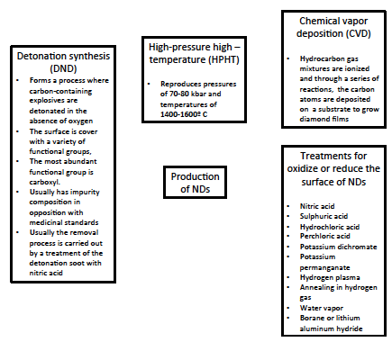
Figure 3. Production methods of nanodiamonds
Detonation process in one of the most popular techniques commercially applied for the synthesis of NDs. DNDs are also called ultra dispersed diamond and have a narrow size distribution around 4-5 nm [19, 52]. ND was synthesized for industrial and research purposes by the detonation technique or mechanical milling of high pressure and high temperature diamond microcrystals (HPHT) [52].
DND are produced by detonation of carbonaceous explosives [59] On the other hand, NDs produced by HPHT has median size of 18 nm and contain minimum lattice defects and uniform structure [52]. DNDs are synthesized from excess carbon contained in the explosive detonation products inside an explosion chamber. ND are formed under high pressure, temperature and fast cooling conditions [60].
Another way of the NDs production implies the use of laser irradiation of water suspension of graphite powders at room temperature and normal pressure. Through this process it can be obtained particles of 3-5 nm size [61].
Different techniques have been used to modify DND particles. One of them is gas discharge plasma treatment that has been demonstrated that can deagglomerate DND particles and improve their dispersability in water, change wettability and electric conductivity and introduce new surface functionality [62]. On the other hand, N2 plasma is more efficient than O2 plasma in deagglomeratting DND particles [62].
ND surface can be oxidize or reduce it. Several approaches have been used to reach these properties. In order to oxidate or carboxylate, it can be used acid treatments with nitric acid, sulphuric acid, hydrochloric acid, perchloric acid, potassium dichromate and potassium permanganate. On the other hand, hydrogen plasma, annealing in hydrogen gas, water vapour, borane or lithium aluminum hydride have been used to reduce it [37].

3. Detonation Nanodiamonds

Detonation nanodiamonds (DND) were discovered in the 1960s. The interest in them rapidly grew over time due to their high potential for different applications [32, 63]. Through this preparation method nanoparticles maintain most of the physical properties of bulk diamond and exhibit a rich surface chemistry [62].
The main disadvantage of DNDs is the tendency to aggregate. At elevated temperature, starting from the outer layers, DNDs transform into concentric shells of graphitic sp2C and eventually resembles and onion. These onion ND particles exhibit layers of graphitic shells based on the degree of onionization [64].
DNDs are relatively low cost material of around $5-10 per gram [65]. They contain 80-88% of carbon in diamond phase, oxygen 10%, hydrogen 0.5-1.5%, nitrogen 2-3%, and incombustible residue of oxides, carbides or salts (0.5-8.0%) [50]. It has been reported that NDs are formed by a structure of mainly three layers [22, 66]:
a) A diamond core 4-6nm (70-90% of all ND carbon atoms)
b) A surface containing carbon atoms and different functional groups.
c) A translational carbon shell around the core made from X-ray amorphous structures with a width of 0.4-1-0 nm.
NDs produced by this method have a negative oxygen balance in a nonoxidative medium. Then, the diamond fraction is contained in a carbon stock [55].
DND are considered suitable candidate for drug delivery. They contains primarily –OH, -CH3, -CH2 and –C=O groups. They are produced by the detonation of explosives such as trinitrotolueno (TNT) and hexogen (RDX) [67]. NDs are optically transparent and capable of fluorescing from point defects. The carbon surface of NDs is covered by a variety of oxygen functional groups which serves as the foundation for covalently attaching of bioactive molecules or other functional groups [9]. So, the surface can be functionalized with radioactive labels and compounds with biological activity [68]. Also NDs are photoluminescent, an important property in most applications [53], due that fluorescent labeling capacities can be used for both diagnostic and therapeutic purposes [45].

4. Fluorescent NDs

NDs have excellent chemical stability, good biocompatibility and can be bonded due to the abundant surface functional groups [69]. They are biocompatible and have extraordinary optical properties include photostability, absence of photoblinking, fluorescent -vacancy (NV) centers within NDs that give a broad emission peak (maximum around 680 nm) [41], emitting in far-red/near infrared, perfectly adapted for biological labeling [16]. An NV center comprises a pair of substitutional nitrogen atom and lattice vacancy on the nanoparticle. These centers has two charge states: NV0, a neutral species and NV, a negatively charged species [43]. Fluorescent nanodiamond (FND) has a center that can emit bright far-red fluorescence at 700 nm wavelenght when excited by Green-yellow light and nearly 70 % of the emission lies in the near infrared window of biological tissue [70]. It has been reported that the first NV FND was prepared in laboratory by Beveratos using HPHT [71, 72]. Using the same method, fluorescent diamond nanocrystals (10nm diameter) were prepared for life science applications with fluorescent intensity and spin decoherence time depending on the nature of the post-milling chemical treatment [72].
FNDs have been produced in mass based on direct irradiation of NDs in aqueous colloidal solution with an homogenous distribution [38]. When the FNDs are carboxylated with oxiding acid, it can be produced carboxylated FND-COOH that can react with biomolecules to form covalent amide bonds [73].
Frequent elements introduced in diamond are nitrogen, silicon, nickel, phosphorus, boron, and cobalt [74]. Silicon diamond is an ideal photon source fluorescence [49, 74]. Chromium-based and niquel-based single photon emitters are attractive due to their room temperature narrow bandwidth emission [49]. FNDs uses in biomedical imaging include diamond with nitrogen vacancy sites, extrinsic dyes and blue fluorescence [56, 75].
FNDs are considered biocompatible luminescent probes with exceptional optical properties for bioimaging and a center photostable showing no photobleaching or photoblinking [76, 77]. The fluorescence lifetime of NV center is 10 ns, so it is longer than lifetime of auto fluorescence, that it is 4 ns [76]. Also, they have been studied as targeted probes for long-term imaging and single particle tracking in vivo. They have photostability with photobleaching and bright-red fluorescence [2, 78]. Furthermore, it was shown that hydrogenated NDs are candidates as radiosensitizing agent associated with antisense molecular therapy [8].

5. Functionalization of NDs

Applications of most of nanocarbons, including NDs have been limited because of inherent no compatibility with solvents, polymers and other matrices. The surface of NDs contains primarily -OH, -CH3, -CH2, CO2 and –C=O groups [68]. So, several methods to develop surface modification for NDs have been studied. The methods include mechanical disruption of the bundles, non-covalent and covalent methods, such as conventional chemical techniques (milling, sonication and refluxing). Many of these reactions required to be carried out over long periods of time. For example, for carboxylation, the reaction mixture was typically fluxed with strong acids of oxidizing agent for 10-50 h. Also, it has been proposed functionalization by microwave assisted process [79].
The ND particle surface can be functionalized with a large number of surface ionogenic groups (ether — C – O – C, peroxide — C – O – O –, carbonyl — C_O, and hydroxyl-type C – O – H bonding, etc.) as well as hydrocarbon fragments. The surface can also be modified with biologically active molecules by adsorption, covalent or non-covalent chemical immobilization [80]. If it is used a treatment with strong acids, it could be produced a formation of COOH groups on the surface which can react with alcohols and amines derivatives [18]. Another approach consist in reducing all oxygen-containing surface groups to OH functions with borane, then allowing the grafting of variety of silanes of long alkyl chains. Others procedures are halogenations and cold plasma functionalization and microwave plasma chemical vapor deposition [81]. Oxidation or hydrogenation of NDs produces carbon-hydrogen bond on the particle surface and the formation of positive zeta potential in colloidal solution. On the other hand, when NDs have groups containing oxygen on the surface it is produced a negative zeta potential of oxidized particles in colloidal solution [67].
The functional groups that it has been produced in NDs include: amines, silanes, butyl, hexyl, aminoacid, carboxyl, aryl, azid, benzoquinone, thymidine, cyanide, fluorine, alkyl, methyl, perfluorootyl ester, aliphatic, chlorocarbonyl, chlorine, amino, glycine, ether, phenyl, halides, and thiols. On the other hand, some biological moieties have been covalently bonded to nanodiamonds using amide bonds such as transferrin, green fluourescent protein, bovine serum albumin, porcin trypsin, growth hormone, mitochondria, actin, paclitaxel, DNA, biotin, folic acid, amino acid, etc. (Figure 4)
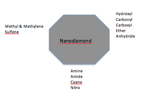
Figure 4. Common functional groups on the nanodiamond surface [56]
Some biological moieties have been physically absorbed by nanodiamonds such as bone morphogenetic protein-2, catalase, histone, lysozyme, obelin, bovine serum albumin, ovalbumin, fetuin, ribonuclease B, doxorubicin, paclitaxel, bovine insulin, DNA, Cellobiose, Poly-L-lysine, sodium dodecyl sulfate, etc. [82]. Bondar, et al. [83], used detonation ND to separate apoobelin and recombinant luciferase from bacterial cells of Escherichia Coli.
Other methods to functionalize NDs includes gas phase functionalization and, non-convalent functionalization with surfactans, peptides, proteins and polymer, microwave radiation [79] and cathepsin B-sensitive peptides like Phe-Lys with anticancer drugs [84]. Hydrogen termination of NDs is achieved by a hydrogen plasma treatment or by annealing in hydrogen gas at elevated temperature [67]. Usually amorphous silicon can be used as a primary coating on any oxidized ND to normalize its surface. The silanols are created in situ by catalyzed hydrolysis of a silyl ether, often tetraethylorthosilicate (TEOS) [41].
Because of its high specific surface area, high adsorption capability, functional groups with oxygen and sp3 diamond cores, NDS are considered an excellent candidate for drug delivery applications [7]. These functional groups constitute an integral part of its structure, providing chemical properties, stability and thermal properties [11]. Depending on the production method it is possible to find NDs with sizes ranging from 100 nm to 5 nm [8], with polyhedral structure with almost 20 % of the total atom number exposed on the surface [85]. This makes them compatible with in vivo applications, and one can expect their elimination by kidney [45].
It has been studied, NDs functionalized with carboxilation of oxidation to facilitate chemical or physical conjugation with biomolecules [86]. These particles are typically covered in a variety of oxygen functional groups including esters, hydroxyls and carboxylic acids. The process to functionalize these particles includes a treatment with nitric or sulphuric acid, or reducing agents to produce a surface with hydrogen or hydrophobic [37]. Also, it has been shown that carboxylated NDs exhibit strong negative zeta potential when dispersed in water at a pH> 5 [8]. Furthermore, NDs have been functionalized with CTAC to obtain Langmuir-Blodgett films with regular deposit and a monoparticle height [87].

6. ND Dispersion

NDs increase the initial elasticity modulus and breaking strength of composite materials [22]. Although, NDs tend to form aggregates, this can be used for a more effective treatment in vivo, in order to provide a better and a constant release of the drug from within the aggregates or means limitations in its application in biomedical area [46]. This strategy could decrease damage to remote healthy cells and reduce side-effects [37]. Also, NDs have high affinity in its surface with proteins [41]. Although, ND composites have poor dispersion and show agglomeration [88], it is a nanoparticle that can be made monodispersed with functionalizable surfaces [64]. Nevertheless, it could be found only one research that reports a stable water suspension with particles less than 100 nm in size [10]. Other researchers report that 75-80 % wt of primary aggregated NDs particles can be converted into nanoparticles, 10-20 nm in size by chemical modification [18]. For these reasons, usually strong aggregates are dispersed using grinding, milling with salts, sonication with the addition of salt and the addition of surfactant [56]. Then, in order to use NDs in biomedical area, it is needed a stable dispersion of particles in aqueous solution [56].
NDs are highly stable in water when are coated by polymers produced from living polymerization, and in acidic and basic conditions, even in 1M NaCl and ethanol [41]. Nevertheless, DNDs have shown instability in hydrosolutions forming aggregates even when they are thawing of ice. So, some efforts have been made to solve it modifying its surface [89]. Moreover, salt and sugar with pH adjustment can produce stable aqueous ND colloidal solution [90]. Amine functional groups could be successfully grafted on DNDs by ball milling technique using ammonium bicarbonate or salt as a milling medium [19].
It is possible to control the zeta potential depending of the kind of ND used. For example, carboxylated NDs exhibit a negative zeta potential when dispersed in water at a pH>5. On the other hand, surface graphitization lead a positive zeta potential in water [8]. ND surface charge allowed the electrostatic loading of biomolecules such as siRNA [8, 75] and plant metabolites [8, 75].
In addition, other research observed and characterized the influence of salts and proteins in the aggregation process and suggested strategies to improve the dispersion of particles for biological applications. The authors first suspended the particles in fetal bovine serum and then diluted in DMEM [91]. Furthermore, other studies purposed modified NDs by explosion synthesis that showed high colloidal stability in water and glucose solutions when used to binding lysozyme [92].

7. Medical applications

Advances in nanomedicine such as imaging and diagnosis, drug delivery and gene therapy have demonstrated the benefits of nanoparticles therapy [93]. Nanoparticle-based nanoscience have great promise in medical applications, such as drug and gene delivery [94]. NDs were used in biomedical applications such as drug delivery, biomedical imaging and biosensors, due to their small size (2–10 nm), surface structure, and inertness (Figure 5) [95]. Nevertheless, until today, it is necessary to improve methods of drug delivery to maximize therapeutic effects while decreasing associated complications due to nonspecific or over-elution.
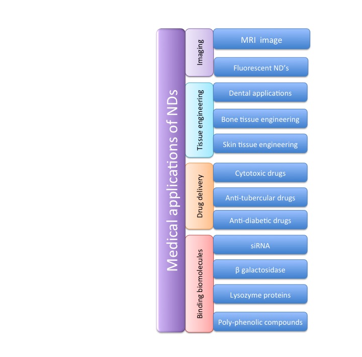
Figure 5. Medical applications of nanodiamonds
The advantages of NDs include stability, biocompatibility [17, 96] and the ability to resist bacterial adhesion [59]. NDs have low toxicity and good optical properties for biomedical applications. For instance, they are biocompatible with cells of different types [86] and show low toxicity for neurons, stem cells, pulmonary epithelium, blood cells, fibroblast, ovary tissues, etc. [42]. Nevertheless, at relatively high doses were found to cause cardiovascular system impairment, liver fibrosis and granuloma formation in liver and spleen of rats [96]. Also, NDs are made of thousands of atoms and their enhanced hardness is used to create specific coatings. This property permits that they could be used as biological markers for analysis in confocal microscopy [97].

7.1. Use of NDs for Imaging

The potential applications of NDs as nanoreinforcements in polymeric matrices have been limited due to the difficulties associated with their dispersion and aggregation during processing and the poor interfacial interaction with polymers [47]. For biomedical imaging, NDs should remain separate from one another to facilitate the attachment to relevant molecules [37].
The use of coordinated water molecules on the surface of nanodiamonds make these particles a promising platform for MRI contrast agent, due that strong electrostatic potential on the nanodiamond cause a surface-mediated attraction toward water molecules creating a nanophase of water on the solvent interface. This method was used to monitoring solid hepatic tumors [98].
Furthermore, as it was discussed before, NDs present nitrogen-vacancy centers that have intrinsic fluorescence properties so they are interesting tools for imaging and diagnostics [1]. Moreover, NDs have a refractive index higher than cytoplasm, so they show a strong light scattering signal that permit clearly distinguish them in cell by optical microscopy [75].
In other research, FNDs for biological imaging were produced by ultrafast photo-initiated RAFT polymerization. The nanoparticles were functionalized with poly(2-methacryloyloxyethyl phosphorylcholine). During the experimental procedure, the cytotoxicity test showed that the composites were nontoxic [99]. Also, NDs have attracted attention in particle tracking, such as HeLa cells and gonads of C. elegans [100]. Fu, et al. conjugated fluorescence nanodiamonds with BnK CT, a chlorotoxin like peptide isolated from the venom of Buthus mantensii Karsch. This conjugated was used to visualize and confirm glioma cells by confocal flurescence assay [101].

7.2. Tissue Engineering Applications of NDs

Because of NDs are inert, non-toxic and biocompatible [56], they could be used to reinforce polymers for bone surgery and tissue engineering [11]. Some researchers have suggested that these nanoparticles have bioactive and antibacterial properties useful for dental implants [102]. Lee, et al., developed a composite of ND with gutta percha, a typical dental filler material for endodontic therapies. Their results showed an improvement in mechanical properties and bacterial growth inhibition when the composite was functionalized with amoxicillin [103].
On the other hand, it has been shown that particles uptaken by cells are minimally cytotoxic and biocompatible, which means that they do not affect mitochondrial function or ATP production at the cellular level [56]. In this sense, it was studied that NDs are able to enter directly in cell nucleous and they could be thrown out the cytoplasm by exocytosis [75]. Nevertheless, other researches showed that ND did not influence cell viability, cell membrane or intracellular oxidative stress and their cellular influences are less than other nanocarbons [75, 104].
Because of their size one can expect the elimination of the particles by kidney [45]. Nevertheless, although some publications consider NDs biocompatible and no toxic, other works suggest that biocompatibility should not be overgeneralized [76]. Some studies showed that NDs accumulate in the lung, liver, spleen, and kidney in a day and its clearance lasted over 10 days [56].
In spite of these results, NDs have been used as fillers of biocomposite scaffolds that promote cellular attachment, differentiation and proliferation with bioactive glass (BG) particles produced by the method of electrophoretic deposition. In that study, it was used alginate as a matrix and the in vitro bioactivity assay showed the formation of hydroxyapatite on the surface of the composite films [105].
On the other hand, in bone tissue engineering were obtained nanocomposites with properties close to the human cortical bone using fluorescent nanodiamonds with poly(l-lactic acid) (PLLA) [106]. Poly(vinylidene fluoride) with NDs was evaluated morphological, structural, optical, thermal and electrically. Therefore, it was evaluated the cell culture viability with pre-osteoblast cells. During this study, it was proven that porous samples were more appropriate than films for proliferation [107]. Also, NDs were used conjugated with alendronate for bone target delivery. It was evaluated the ALP activity and specific uptake for MC3T3-E1 of the drug system. The results showed high affinity of alendronate/NDs with HAp, so it could be potentially used for osteoporosis treatment [108].
Other research showed that poly(L-lactic acid)-FNDs composite for bone engineering, reached uniform dispersion and good affinity between the matrix and the nanoparticle. Also, the composite was nontoxic to murine osteoblasts [106].
In skin tissue engineering, it was used irradiated NDs and polycaprolactone, creating a superior cell-interfacing material with a less hydrophobicity. This substrate was capable of in vivo detection [109].
Cai, et al., evaluated the effect to add NDs to Poly(Lactic Acid) in nanofiber membranes. The fiber diameter decreased as ND content increased. Also, the nanoparticles improved the thermal and mechanical properties of the composite, so they have potential applications in biomedical engineering [110]. Furthermore, poly(lactic-co-glycolic acid) loaded with NDs phospholipid showed high mechanical properties, good in vitro and in vivo biocompatibility NDs. The composite was evaluated for in vitro proliferation and differentiation of human fetal osteoblastic cells. It was found that the material could promote osseointegration [111].

7.3. Use of NDs for Drug Delivery

Nanoparticles are immensely used in cancer management due to their versatile attractive features such as small size, stability, inertness, increased surface-to-volume ratio, highly tunable optical properties, and suitability for surface modification [6]. Several researches show the successful conjugation of nanoparticles to therapeutic drugs, imaging agents, polymers and cancer targeting ligands [46-48]. Nanoparticles could be customized for delivering the chemotherapeutic or cancer diagnostic agents selectively to the cancer cells [112]. For example, chemotherapeutic agent could be conjugated to ligand target nanoparticles allowing the discrimination of cancer cells from the fast growing healthy cells of the body. Then the cytotoxic drugs could be delivered only to the cancer cells [112]. Also, they could be functionalized with doxorubicin to use as an inhibitor of lung metastasis in mice [75] or with ciproten and quercetin for cancer treatment [75].
Anticancer agents can functionalize the surface of nanomaterials via chemical activation of surface groups or by non-covalent binding [68]. So, previously a research group developed a polysaccharide sodium alginate functionalized NDs for chemotherapeutic drug delivery of cis-diamminedichloro platinum (II) [12].
ND drug delivery systems could be obtained by adsorption of a drug on the surface of NDs and by covalent binding of a drug with surface functional groups [18]. So, NDs have been used too as a adsorbent of antidiabetic and antitubercular drugs [18]. Also, the particle could be used for separation and purification of proteins, such as immunoglobulins of human blood [113] or other components of human blood serum [114]. Furthermore, NDs were functionalized with N-O-carboxymethyl chitosan as an ideal candidate for drug delivery [7]. Modification of the NDs surface with biologically active compounds and drugs offers a potential application of it with anticancer therapeutic agent such as doxorubicin, daunorubicin or cisplatin [68]. They have been tested as a potential carries of myramistin showing easy drug adsorption and drug release under physiological conditions only [68]. Moreover, it has been shown that ND at 25 μg/ml concentration did not promote apoptosis, an inflammatory response, or inhibited proliferation at the level of the transcriptional response [56].
Wang, et al., [115] used a transferrin-doxorubicin complex with carboxylated ND. Their results showed that the complex could deliver the drug inside living cells via a clathrin-dependent and transferrin receptor-mediate endocytosis pathway. Also, they found that the complex reduced the volume of a tumor more than DOX-treated.
On the other hand, in order to used NDs as a drug delivery systems it is required colloidal stability in physiological medium [68]. The stability could be increased using surfactants [68]. Previously, NDs were functionalized with a beta cyclodextrin and hyperbranched polyglycerol polymers to explore their biomedical applications, improving dispersion in water and biocompatibility. Also, they showed that DOX could be absorbed by the composite with high efficiency [116]. Other research group showed that it could be produced NDs that includes amino acid, peptides, oligonucleotides, sugars, etc., for drug delivery [117].
Furthermore, inhibition of angiogenesis with VEGD inhibitors could be an important and effective strategy for the treatment of cancer metastasis. Sorafenib, is an efficient anti-proliferative and antiangiogenic drug that block Raf signaling and VEGF. Nevertheless, sorafenib is almost insoluble in water or buffered solution at certain pH values (from 1.2 to pH 7.4) and the oral bioavailability is extremely low (about 8.43 %) which greatly restricts its therapeutic efficacy on cancer metastasis. So, the particles were used in a lipid-coated ND as a drug delivery platform, improving the oral bioavailability of lipophilic drugs [13].
NDs are particles among 5 and 100 nm that could be functionalized and conjugated with biomolecules [75, 80], so they have been used as a vehicle for the delivery of insuline [93] and for easy biomolecule immobilization [118]. Furthermore, NDs have antibacterial activity that it is influenced by their surface chemistry and size [119]. Nevertheless, biomedical applications of NDs commonly require that NDs be delivered and used in aqueous solutions [120].
For medical applications such as biomolecule immobilizations, it is highly desirable to have diamond in a thin fiber or film form [40]. Mainly the chemical vapor deposition is the technique that is used for preparing biocompatible diamonds films [40].

7.4. NDs for Binding Proteins and Lipids

It is possible to modify NDs surface to improve the interaction between the particle and its environment [51]. Hydrogenated NDs are stable in aqueous solutions, so these cationic particles were used to efficiently deliver siRNA to human cells [16]. In addition, cellular toxicity of ND-pani nanoparticles and films was evaluated using embryonic kidney cells. The study showed that the toxicity was dependent of the NDs concentration [121].
It was shown that NDs support neural differentiation over glial differentiation with potential results to improve tissue integration and prevent the glial scar formation in implants [59]. Also, the particles were evaluated for enzyme immobilization, such as β galactosidase with broad applications in lactose-free dairy products [9].
Other medical applications includes NDs functionalized with lysozyme proteins showing antibacterial activity [86]. Also, it was shown that NDs inhibit EWS/FLI-1 cells expression in culture [45]. NDs were used to inhibit the lung metastasis of breast cancer, inducing tumor cell apoptosis with reduced systemic toxicity [48]. Furthermore, nanocomposites elaborated with carboxylated NDs and cellulose have been evaluated as a wound dressing showing potential use in skin engineering [69].
Recently, Cheng, et al., [122] functionalized NDs with vitamin E TPGS to improve oral absorption of curcumin, a lipophilic poly-phenolic compound that exhibits biological activities such as anti-oxidant, anti-inflammatory and anti-cancer properties. Their results showed that the absorption of the nanocomplexes was significantly improved in the GI tract biodistribution.
NDs have high affinity in its surface with proteins. Then, proteins can remain attached with them, even after washing. Mainly, protein adsorption includes electrostatic, hydrophilic and hydrophobic interactions, hydrogen bonding, and van der Waals forces [41]. Due to this high affinity for proteins, they were used to find biomarkers of the secreted proteins from carbapenemase-producing A. baummannii. The spread of carbapenem-resistant Acinetobacter baumannii is a challenge for optimization of antibiotic therapies and outbreak prevention, so this method could be used for the detection of the bacteria [123].
Li, et al., evaluated the receptor-mediated endocytosis of fluorescent NDs linked with transferrin. Their results showed that the crosslinking of the nanoparticles with proteins promote better stability and efficiency for cellular uptake [124]. Furthermore, NDs tend to precipitate at physiological ionic strength, and proteins are readily adsorbed on the exposed surface [41]. So, the apoobelin protein was separated and purifying using NDs in a chromatography [83]. On the other hand, it has been proved that ND can adsorb peptides [125] and amplify signals and detection of miRNA-21 [126].
Other research used as a suspension for relieves the state of oncological patients, decreasing probably the level of intoxication. The particle was used to correct protein and lipid peroxide oxidation processes associated with malignant tumor growth [65].
The particle has been used in advanced composites for biomedical purposes. For instance, nanofibers with NDs and polycaprolactone were produced by electrospinning using acetone as a solvent exhibiting bright flourescence [39]. It has been reported polymer nanoparticles from poly(vinylpyrrolidone) (PVP) using ultrasonication with a uniform dispersion of the NDs[88] and NDs with iodine-125 labeled immunoglobulin and bovine serum albumin and Rabbit Anti-Mouse Antibody by covalent immobilization [127].
Also, it is possible to modify NDs with a positively charged polymers but those kind of materials do not ensure shielding from protein and cell interactions. Nevertheless, they can serve as a platform for nucleic acid complexation and as gene deliver vectors [41].

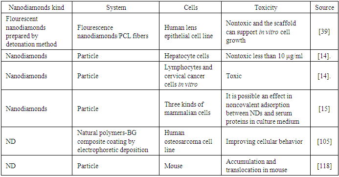
7.5. Some Considerations about Toxicity of Nanodiamonds

NDs, like several particles of nanometric dimensions, have been subject to continuous evaluation of their toxicity (Table 1). Depending on the type of application, a certain degree will be desirable, or maybe a totally undesirable characteristic. Although, MTT (3-(4,5-dumethylthiazol-2-yl)-2,5-diphenyltetrazolium bromide) and adenosine triphosphate (ATP) assays have shown that NDs are non-toxic to a variety of cell types [105], it was found significant lung and systemic toxicity of NDs after intratracheal instillation suggesting further toxicological studies [128]. So, inevitably, the biological behavior and potential toxicity of nanodiamonds are topics of interest [118].
Table 1. Different efforts to evaluate toxicity of nanodiamonds
     
Some cells, such as HeLa cells, human lung adenocarcinoma, keratinocytes, lung fibroblasts human kidney cells, etc., have shown good biocompatibility with NDs in vitro. Cells can tolerate concentrations up to about 400 μg/mL of functionalized NDs. Also, the protein expression, cellular proliferation and apoptosis are evidence of good biocompatibility [37]. Nevertheless, unattached nanoparticles can have toxic effects to cells at concentration >50 μg/ml due to their easy penetration into cells [59].

8. Conclusions

During the last decades, nanodiamonds have been studied due to their superior physical and chemical properties of versatile functionalization, such as large surface area, high adsorption capacity and good biocompatibility. These particles have been used in many research areas such as biological sensing, medical therapy, composite materials, lubricants, fluorescent markers, electrochemical coatings, enzyme immobilization and implant coatings. NDs have shown biocompatibility for many biomedical applications. Nevertheless, due that it not exist an agreement about its toxicity, more experiments could be developed in order to elucidate the potential risks in different tissues.
In spite of its unique characteristics, it has not been explored enough the possibility to improve its compatibility with different solvent and polymers. Furthermore, until today, it is a challenge to disperse NDs in a solution for an effective application in biomedical area, so future studies should explore particle systems for this purpose.

References

[1]  K. Bhattacharya, S.P. Mukherjee, A. Gallud, S.C. Burkert, S. Bistarelli, S. Bellucci, M. Bottini, A. Star, B. Fadeel, Biological interactions of carbon-based nanomaterials: From coronation to degradation, Nanomedicine: nanotechnology, biology, and medicine, 12 (2016) 333-351.
[2]  J.C. Arnault, Surface Modifications of Nanodiamonds and Current Issues for Their Biomedical Application, in: N. Yang (Ed.) Novel aspects of diamonds, Springer International, Switzerland, 2015.
[3]  C. Murphy, Two ways to make nanoparticles, in, Sustainable nano, http://sustainable-nano.com/2014/06/10/two-ways-to-make-nanoparticles/, 2014.
[4]  Sympatec, Application field Science Et Reseach, in, SympaTEC, https://www.sympatec.com/en/applications/fields-of-application/science-research/, 2018.
[5]  R. Bhisey, Carbon Nanotube Manufacturers Can Expect Significant Increase in Demand from Polymer Producers, reports TMR, in, The edition truth, https://0x9.me/ajfqv, 2017.
[6]  P. Suvarnaphaet, S. Pechprasarn, Graphene-Based Materials for Biosensors: A Review, Sensors, 17 (2017).
[7]  H.-D. Wang, Q. Yang, C.H. Niu, Functionalization of nanodiamond particles with N,O-carboxymethyl chitosan, Diamond and Related Materials, 19 (2010) 441-444.
[8]  R. Grall, H. Girard, L. Saad, T. Petit, C. Gesset, M. Combis-Schlumberger, V. Paget, J. Delic, J.C. Arnault, S. Chevillard, Impairing the radioresistance of cancer cells by hydrogenated nanodiamonds, Biomaterials, 61 (2015) 290-298.
[9]  S.A. Ansari, R. Satar, S.K. Zaidi, M.I. Naseer, S. Karim, M.H. Alqahtani, M. Rasool, Nanodiamonds as an effective and novel matrix for immobilizing β galactosidase, Food and Bioproducts Processing, 95 (2015) 298-303.
[10]  M.V. Baidakova, Y.A. Kukushkina, A.A. Sitnikova, M.A. Yagovkina, D.A. Kirilenko, V.V. Sokolov, M.S. Shestakov, A.Y. Vul’, B. Zousman, O. Levinson, Structure of nanodiamonds prepared by laser synthesis, Physics of the Solid State, 55 (2013) 1747-1753.
[11]  V.N. Mochalin, Y. Gogotsi, Nanodiamond–polymer composites, Diamond and Related Materials, 58 (2015) 161-171.
[12]  Z. Cui, Y. Zhang, J. Zhang, H. Kong, X. Tang, L. Pan, K. Xia, A. Aldalbahi, A. Li, R. Tai, C. Fan, Y. Zhu, Sodium alginate-functionalized nanodiamonds as sustained chemotherapeutic drug-release vectors, Carbon, 97 (2016) 78-86.
[13]  Z. Zhang, B. Niu, J. Chen, X. He, X. Bao, J. Zhu, H. Yu, Y. Li, The use of lipid-coated nanodiamond to improve bioavailability and efficacy of sorafenib in resisting metastasis of gastric cancer, Biomaterials, 35 (2014) 4565-4572.
[14]  J. Mytych, A. Lewinska, J. Zebrowski, M. Wnuk, Nanodiamond-induced increase in ROS and RNS levels activates NF-κB and augments thiol pools in human hepatocytes, Diamond and Related Materials, 55 (2015) 95-101.
[15]  J. Li, Y. Zhu, W. Li, X. Zhang, Y. Peng, Q. Huang, Nanodiamonds as intracellular transporters of chemotherapeutic drug, Biomaterials, 31 (2010) 8410-8418.
[16]  H.A. Girard, P. Benayoun, C. Blin, A. Trouvé, C. Gesset, J.-C. Arnault, P. Bergonzo, Encapsulated nanodiamonds in smart microgels toward self-assembled diamond nanoarrays, Diamond and Related Materials, 33 (2013) 32-37.
[17]  M.S. Wu, D.S. Sun, Y.C. Lin, C.L. Cheng, S.C. Hung, P.K. Chen, J.H. Yang, H.H. Chang, Nanodiamonds protect skin from ultraviolet B-induced damage in mice, J Nanobiotechnology, 13 (2015) 35.
[18]  R.Y. Yakovlev, A.S. Solomatin, N.B. Leonidov, I.I. Kulakova, G.V. Lisichkin, Detonation diamond—A perspective carrier for drug delivery systems, Russian Journal of General Chemistry, 84 (2014) 379-390.
[19]  M. Khan, N. Shahzad, C. Xiong, T.K. Zhao, T. Li, F. Siddique, N. Ali, M. Shahzad, H. Ullah, S.A. Rakha, Dispersion behavior and the influences of ball milling technique on functionalization of detonated nano-diamonds, Diamond and Related Materials, 61 (2016) 32-40.
[20]  R.S. Lewis, Anders, E., Draine, B.T., Properties, detectability and origin of interstellar diamonds in meteorites, Nature, 339 (1989) 117-121.
[21]  I. Shugalei, Voznyakovskii, A., Garabadzhiu, A., Tselinskiii, I., Sudarikov, A., Ilyushin, M., Biological Activity of Detonation Nanodiamond and Prospects in Its Medical and Biological Applications, Russian Journal of General Chemistry (2013) 5.
[22]  V.Y. Dolmatov, Composition Materials Based on Elastomer and Polymer Matrices Filled with Nanodiamonds of Detonation Synthesis, Nanotechnologies in Russia, 4 (2009) 556-575.
[23]  A. Speltini, D. Merli, A. Profumo, Analytical application of carbon nanotubes, fullerenes and nanodiamonds in nanomaterials-based chromatographic stationary phases: a review, Anal Chim Acta, 783 (2013) 1-16.
[24]  V.Y. Osipov, A.E. Aleksenskiy, A.I. Shames, A.M. Panich, M.S. Shestakov, A.Y. Vul’, Infrared absorption study of surface functional groups providing chemical modification of nanodiamonds by divalent copper ion complexes, Diamond and Related Materials, 20 (2011) 1234-1238.
[25]  X. Liu, Z. Yu, Q. Wei, G. Wen, X. Duan, X. You, Modification of polycrystalline nanodiamonds by using periodic magnetic field enhanced hydrogen plasma and the application on nanogrinding of thin film magnetic head, Colloids and Surfaces A: Physicochemical and Engineering Aspects, 416 (2013) 9-15.
[26]  A.V. Ukhina, D.V. Dudina, A.G. Anisimov, V.I. Mali, N.V. Bulina, I.A. Bataev, I.N. Skovorodin, B.B. Bokhonov, Porous electrically conductive materials produced by Spark Plasma Sintering and hot pressing of nanodiamonds, Ceramics International, 41 (2015) 12459-12463.
[27]  J.B. Lewis, D. Isheim, C. Floss, D.N. Seidman, C12/C13-ratio determination in nanodiamonds by atom-probe tomography, Ultramicroscopy, 159 Pt 2 (2015) 248-254.
[28]  E.P. Koumoulos, P. Jagadale, A. Lorenzi, A. Tagliaferro, C.A. Charitidis, Evaluation of surface properties of epoxy–nanodiamonds composites, Composites Part B: Engineering, 80 (2015) 27-36.
[29]  A. Wallner, K. Melber, S. Merchel, U. Ott, O. Forstner, R. Golser, W. Kutschera, A. Priller, P. Steier, Stable platinum isotope measurements in presolar nanodiamonds by TEAMS, Nucl Instrum Methods Phys Res B, 294 (2013) 496-502.
[30]  E. Yılmaz, M. Soylak, Preparation and characterization of magnetic carboxylated nanodiamonds for vortex-assisted magnetic solid-phase extraction of ziram in food and water samples, Talanta, 158 (2016) 152-158.
[31]  R.Y. Yakovlev, N.N. Dogadkin, I.I. Kulakova, G.V. Lisichkin, N.B. Leonidov, V.P. Kolotov, Determination of impurities in detonation nanodiamonds by gamma activation analysis method, Diamond and Related Materials, 55 (2015) 77-86.
[32]  M.P. Petrov, V.N. Shilov, A.A. Trusov, A.V. Voitylov, V.V. Vojtylov, Electro-optic research of polarizability dispersion in aqueous polydisperse suspensions of nanodiamonds, Colloids and Surfaces A: Physicochemical and Engineering Aspects, 506 (2016) 40-49.
[33]  D.S. Sabirov, E. Ōsawa, Dipole polarizability of nanodiamonds and related structures, Diamond and Related Materials, 55 (2015) 64-69.
[34]  S.K. Gordeev, S.B. Korchagina, On the stability of small-sized nanodiamonds, Journal of Superhard Materials, 29 (2007) 124-125.
[35]  J. Karpeta-Kaczmarek, M. Augustyniak, M. Rost-Roszkowska, Ultrastructure of the gut epithelium in Acheta domesticus after long-term exposure to nanodiamonds supplied with food, Arthropod Structure & Development, 45 (2016) 253-264.
[36]  Y. Astuti, N.R.J. Poolton, Y.V. Butenko, L. Šiller, Evaporation and alignment of 1-undecene functionalised nanodiamonds, Journal of Luminescence, 156 (2014) 41-48.
[37]  J. Say, Vreden, C., Reilly, D., Brown, L., Rabeau, J., King, N., Luminescent nanodiamonds for biomedical applications, Biophys Rev, 3 (2011) 171-184.
[38]  J. Stursa, J. Havlik, V. Petrakova, M. Gulka, J. Ralis, V. Zach, Z. Pulec, V. Stepan, S.A. Zargaleh, M. Ledvina, M. Nesladek, F. Treussart, P. Cigler, Mass production of fluorescent nanodiamonds with a narrow emission intensity distribution, Carbon, 96 (2016) 812-818.
[39]  L. Cao, Y. Hou, K. Lafdi, K. Urmey, Fluorescent composite scaffolds made of nanodiamonds/polycaprolactone, Chemical Physics Letters, 641 (2015) 123-128.
[40]  H.-D. Wang, Q. Yang, C.H. Niu, Preparation of films of nanodiamonds by step-by-step deposition approach through hydrogen bonding, Diamond and Related Materials, 25 (2012) 73-79.
[41]  J. Neburkova, J. Vavra, P. Cigler, Coating nanodiamonds with biocompatible shells for applications in biology and medicine, Current Opinion in Solid State and Materials Science.
[42]  D.S. Volkov, M.A. Proskurnin, M.V. Korobov, Survey study of mercury determination in detonation nanodiamonds by pyrolysis flameless atomic absorption spectroscopy, Diamond and Related Materials, 50 (2014) 60-65.
[43]  A.R. Kirmani, W. Peng, R. Mahfouz, A. Amassian, Y. Losovyj, H. Idriss, K. Katsiev, On the relation between chemical composition and optical properties of detonation nanodiamonds, Carbon, 94 (2015) 79-84.
[44]  F. Ahmed, Memic, A., Nanodiamonds for tracking of leukemic cells, BioMed Central, 15 (2014) P54.
[45]  J. Bertrand, Pioche-Durieu, C., Ayala, J., Petit, T., Girard, H., Malvy, C., Le Cam, E., Treussart, F., Arnault, J., Plasma hydrogenated cationic detonation nanodiamonds efficiently deliver to human cells in culture functional siRNA targeting the Ewing sarcoma junction oncogene, Biomaterials, 45 (2015) 93-98.
[46]  W. Ma, X. Yu, X. Qu, Q. Zhang, Functionalization of agglomerating nanodiamonds with biodegradable poly(ε-caprolactone) through surface-initiated polymerization, Diamond and Related Materials, 62 (2016) 14-21.
[47]  T. Subhani, M. Latif, I. Ahmad, S.A. Rakha, N. Ali, A.A. Khurram, Mechanical performance of epoxy matrix hybrid nanocomposites containing carbon nanotubes and nanodiamonds, Materials & Design, 87 (2015) 436-444.
[48]  J. Xiao, X. Duan, Q. Yin, Z. Zhang, H. Yu, Y. Li, Nanodiamonds-mediated doxorubicin nuclear delivery to inhibit lung metastasis of breast cancer, Biomaterials, 34 (2013) 9648-9656.
[49]  A. Kovalenko, S. Záliš, P. Ashcheulov, I. Kraus, J. Pavluch, I. Kratochvílová, Theoretical study of chromium and nickel-related luminescence centers in molecular-sized nanodiamonds, Diamond and Related Materials, 58 (2015) 122-128.
[50]  I.I. Kulakova, Surface Chemistry of Nanodiamonds, Physics of the Solid State, 46 (2004) 636-643.
[51]  J.C. Arnault, H.A. Girard, Hydrogenated nanodiamonds: Synthesis and surface properties, Current Opinion in Solid State and Materials Science.
[52]  J. Salava, F. Trojánek, Š. Stehlík, M. Varga, B. Rezek, P. Malý, Influence of air annealing on the luminescence dynamics of HPHT nanodiamonds, Diamond and Related Materials, 68 (2016) 62-65.
[53]  S. Stehlik, L. Ondic, A.M. Berhane, I. Aharonovich, H.A. Girard, J.-C. Arnault, B. Rezek, Photoluminescence of nanodiamonds influenced by charge transfer from silicon and metal substrates, Diamond and Related Materials, 63 (2016) 91-96.
[54]  N. Sergeev, Panich, A., Olszewski, M., Shenderova, O., Goren, S., , 13C spin-lattice relaxation in nanodiamonds in static and magic angle spinning regimes, Solid State Nucl Magn Reson, 66-67 (2015) 51-55.
[55]  I.I. Obraztsova, N.K. Eremenko, Physicochemical modification of nanodiamonds, Russian Journal of Applied Chemistry, 81 (2008) 603-608.
[56]  D.G. Lim, R.E. Prim, K.H. Kim, E. Kang, K. Park, S.H. Jeong, Combinatorial nanodiamond in pharmaceutical and biomedical applications, International journal of pharmaceutics, 514 (2016) 41-51.
[57]  H.D. Espinosa, Peng, B., Prorok, B.C., Moldovan, N., Auciello, O., Carlisle, J.A., Gruen, D.M., Mancini, D.C., Fracture strength of ultrananocrystalline diamond thin films—identification of Weibull parameters, Journal of Applied Physics, 94 (2003).
[58]  A.M. Valenkov, I.V. Gofman, K.S. Nosov, V.M. Shapovalov, V.E. Yudin, Polymeric composite systems modified with allotropic forms of carbon (review), Russian Journal of Applied Chemistry, 84 (2011) 735-750.
[59]  E. Peltola, N. Wester, K.B. Holt, L.-S. Johansson, J. Koskinen, V. Myllymäki, T. Laurila, Nanodiamonds on tetrahedral amorphous carbon significantly enhance dopamine detection and cell viability, Biosensors and Bioelectronics.
[60]  V.Y. Dolmatov, G.S. Yur’ev, V. Myllymäki, K.M. Korolev, Why detonation nanodiamonds are small, Journal of Superhard Materials, 35 (2013) 77-82.
[61]  A.M. Panich, A.I. Shames, B. Zousman, O. Levinson, Magnetic resonance study of nanodiamonds prepared by laser-assisted technique, Diamond and Related Materials, 23 (2012) 150-153.
[62]  A.V. Stanishevsky, M.J. Walock, S.A. Catledge, Surface modification and stability of detonation nanodiamonds in microwave gas discharge plasma, Applied Surface Science, 357 (2015) 1403-1409.
[63]  E. Roumeli, A. Markoulis, K. Chrissafis, A. Avgeropoulos, D. Bikiaris, Substantial enhancement of PP random copolymer's thermal stability due to the addition of MWCNTs and nanodiamonds: Decomposition kinetics and mechanism study, Journal of Analytical and Applied Pyrolysis, 106 (2014) 71-80.
[64]  D.F. Johnson, W.D. Mattson, Theoretical investigations of surface reconstruction on C nanodiamonds and cubic-BN nanoparticles, Diamond and Related Materials, 58 (2015) 155-160.
[65]  R. Yakovlev, Solomatin. A., Leonidov, B., Kulakova, I., Lisichkin, G., Detonation Diamond—A Perspective Carrier for Drug Delivery Systems, Russian Journal of General Chemistry, 84 (2012) 379-390.
[66]  V.Y. Dolmatov, Polymer–Diamond Composites Based on Detonation Nanodiamonds. Part 1, Journal of Superhard Materials, 29 (2006) 1-11.
[67]  S. Stehlik, Glatzel, T., Pichot, V., Pawlak, R., Meyer, E., Spitzer, D., Rezek, B., Water interaction with hydrogenated and oxidized detonation nanodiamonds — Microscopic and spectroscopic analyses, Diamond and Related Materials, 63 (2016) 97-102.
[68]  M.G. Chernysheva, I.Y. Myasnikov, G.A. Badun, Myramistin adsorption on detonation nanodiamonds in the development of drug delivery platforms, Diamond and Related Materials, 55 (2015) 45-51.
[69]  X. Luo, H. Zhang, Z. Cao, N. Cai, Y. Xue, F. Yu, A simple route to develop transparent doxorubicin-loaded nanodiamonds/cellulose nanocomposite membranes as potential wound dressings, Carbohydrate Polymers, 143 (2016) 231-238.
[70]  Y.Y. Hui, W.W.-W. Hsiao, S. Haziza, M. Simonneau, F. Treussart, H.-C. Chang, Single particle tracking of fluorescent nanodiamonds in cells and organisms, Current Opinion in Solid State and Materials Science, (2016).
[71]  I.N. Remediakis, G. Kopidakis, P.C. Kelires, Softening of ultra-nanocrystalline diamond at low grain sizes, Acta Materialia, 56 (2008) 5340-5344.
[72]  J.-P. Boudou, J. Tisler, R. Reuter, A. Thorel, P.A. Curmi, F. Jelezko, J. Wrachtrup, Fluorescent nanodiamonds derived from HPHT with a size of less than 10nm, Diamond and Related Materials, 37 (2013) 80-86.
[73]  M.-F. Weng, S.-Y. Chiang, N.-S. Wang, H. Niu, Fluorescent nanodiamonds for specifically targeted bioimaging: Application to the interaction of transferrin with transferrin receptor, Diamond and Related Materials, 18 (2009) 587-591.
[74]  L. Minati, C.L. Cheng, Y.C. Lin, J. Hees, G. Lewes-Malandrakis, C.E. Nebel, F. Benetti, C. Migliaresi, G. Speranza, Synthesis of novel nanodiamonds–gold core shell nanoparticles, Diamond and Related Materials, 53 (2015) 23-28.
[75]  A. Gismondi, Reina, G., Orlanducci, S., Mizzoni, F., Gay, S., Terranova,, M., Canini, A., Nanodiamonds coupled with plant bioactive metabolites: A nanotech approach for cancer therapy, Biomaterials, 38 (2015) 22-35.
[76]  I. Rehor, J. Slegerova, J. Kucka, V. Proks, V. Petrakova, M.P. Adam, F. Treussart, S. Turner, S. Bals, P. Sacha, M. Ledvina, A.M. Wen, N.F. Steinmetz, P. Cigler, Fluorescent nanodiamonds embedded in biocompatible translucent shells, Small, 10 (2014) 1106-1115.
[77]  S. Sotoma, Y. Yoshinari, R. Igarashi, A. Yamazaki, S.H. Yoshimura, H. Tochio, M. Shirakawa, Y. Harada, Effective production of fluorescent nanodiamonds containing negatively-charged nitrogen-vacancy centers by ion irradiation, Diamond and Related Materials, 49 (2014) 33-38.
[78]  M.-F. Weng, B.-J. Chang, S.-Y. Chiang, N.-S. Wang, H. Niu, Cellular uptake and phototoxicity of surface-modified fluorescent nanodiamonds, Diamond and Related Materials, 22 (2012) 96-104.
[79]  C. Desai, S. Mitra, Microwave induced carboxylation of nanodiamonds, Diamond and Related Materials, 34 (2013) 65-69.
[80]  P.H. Chung, E. Perevedentseva, J.S. Tu, C.C. Chang, C.L. Cheng, Spectroscopic study of bio-functionalized nanodiamonds, Diamond and Related Materials, 15 (2006) 622-625.
[81]  H.A. Girard, J.C. Arnault, S. Perruchas, S. Saada, T. Gacoin, J.P. Boilot, P. Bergonzo, Hydrogenation of nanodiamonds using MPCVD: A new route toward organic functionalization, Diamond and Related Materials, 19 (2010) 1117-1123.
[82]  <garcia-piña163.pdf>.
[83]  V. Bondar, Pozdnyakova, I., Puzyr, A., Applications of Nanodiamonds for Separation and Purification of Proteins, Applications of nanodiamonds, 46 (2004) 737-739.
[84]  S. Huang, J. Shao, L. Gao, Y. Qi, L. Ye, Adsorption of Cathepsin B-sensitive peptide conjugated DOX on nanodiamonds, Applied Surface Science, 257 (2011) 8617-8622.
[85]  H. Ba, Y. Liu, X. Mu, W.-H. Doh, J.-M. Nhut, P. Granger, C. Pham-Huu, Macroscopic nanodiamonds/β-SiC composite as metal-free catalysts for steam-free dehydrogenation of ethylbenzene to styrene, Applied Catalysis A: General, 499 (2015) 217-226.
[86]  Y. Liu, Sun, K., Protein Functionalized Nanodiamond Arrays, Nanoscale Res Lett, 5 (2010) 1045-1050.
[87]  V. Pichot, K. Bonnot, N. Piazzon, M. Schaefer, M. Comet, D. Spitzer, Deposition of detonation nanodiamonds by Langmuir–Blodgett technique, Diamond and Related Materials, 19 (2010) 479-483.
[88]  N. Attia, Rao, J., Geckeler, K., Nanodiamond–polymer nanoparticle composites and their thin films, J Nanopart Res, 16 (2014) 1-12.
[89]  V. Bondar, Puzyr, A., Nanodiamonds for biological investigations, Applications of nanodiamonds, 46 (2004) 716-719.
[90]  A. Pentecost, Gour, S., Mochalin, V., Knoke, I., Gogotsu, Y., Deaggregation of Nanodiamond Powders Using Salt- and Sugar-Assisted Milling, ACS Appl. Mater. Interfaces, 2 (2010) 3289-3294.
[91]  S.R. Hemelaar, A. Nagl, F. Bigot, M.M. Rodriguez-Garcia, M.P. de Vries, M. Chipaux, R. Schirhagl, The interaction of fluorescent nanodiamond probes with cellular media, Mikrochim Acta, 184 (2017) 1001-1009.
[92]  O.A. Mogil’naya, V.S. Bondar, Comparative study of antibacterial properties of Lysozyme upon its adsorption and covalent binding to nanodiamonds, Nanotechnologies in Russia, 7 (2012) 658-665.
[93]  R.A. Shimkunas, E. Robinson, R. Lam, S. Lu, X. Xu, X.Q. Zhang, H. Huang, E. Osawa, D. Ho, Nanodiamond-insulin complexes as pH-dependent protein delivery vehicles, Biomaterials, 30 (2009) 5720-5728.
[94]  H. Hashemi-Moghaddam, S. Zavareh, S. Kazemi, M. Jamili, Evaluation of magnetic nanoparticles coated by 5-fluorouracil imprinted polymer for controlled drug delivery in mouse breast cancer model, International journal of pharmaceutics, (2015).
[95]  M. Singh, M. Holzinger, O. Biloivan, S. Cosnier, 3D-nanostructured scaffold electrodes based on single-walled carbon nanotubes and nanodiamonds for high performance biosensors, Carbon, 61 (2013) 349-356.
[96]  J. Mytych, M. Wnuk, S.I.S. Rattan, Low doses of nanodiamonds and silica nanoparticles have beneficial hormetic effects in normal human skin fibroblasts in culture, Chemosphere, 148 (2016) 307-315.
[97]  A. Nouailhat, An Introduction to Nanoscience and Nanotechnology, Wiley, Hoboken, United States, 2008.
[98]  W. Hou, T.B. Toh, L.N. Abdullah, T.W.Z. Yvonne, K.J. Lee, I. Guenther, E.K.-H. Chow, Nanodiamond–Manganese dual mode MRI contrast agents for enhanced liver tumor detection, Nanomedicine: Nanotechnology, Biology and Medicine, 13 (2017) 783-793.
[99]  J. Chen, M. Liu, Q. Huang, L. Huang, H. Huang, F. Deng, Y. Wen, J. Tian, X. Zhang, Y. Wei, Facile preparation of fluorescent nanodiamond-based polymer composites through a metal-free photo-initiated RAFT process and their cellular imaging, Chemical Engineering Journal, 337 (2018) 82-90.
[100]  G. Balasubramanian, A. Lazariev, S.R. Arumugam, D.-w. Duan, Nitrogen-Vacancy color center in diamond—emerging nanoscale applications in bioimaging and biosensing, Current Opinion in Chemical Biology, 20 (2014) 69-77.
[101]  Y. Fu, N. An, S. Zheng, A. Liang, Y. Li, BmK CT-conjugated fluorescence nanodiamond as potential glioma-targeted imaging and drug, Diamond and Related Materials, 21 (2012) 73-76.
[102]  S. Najeeb, Z. Khurshid, A.S. Agwan, M.S. Zafar, M. Alrahabi, S.B. Qasim, F. Sefat, Dental Applications of Nanodiamonds, Science of Advanced Materials, 8 (2016) 2064-2070.
[103]  D.-K. Lee, S.V. Kim, A.N. Limansubroto, A. Yen, A. Soundia, C.-Y. Wang, W. Shi, C. Hong, S. Tetradis, Y. Kim, N.-H. Park, M.K. Kang, D. Ho, Nanodiamond–Gutta Percha Composite Biomaterials for Root Canal Therapy, ACS Nano, 9 (2015) 11490-11501.
[104]  M. Horie, L.K. Komaba, H. Kato, A. Nakamura, K. Yamamoto, S. Endoh, K. Fujita, S. Kinugasa, K. Mizuno, Y. Hagihara, Y. Yoshida, H. Iwahashi, Evaluation of cellular influences induced by stable nanodiamond dispersion; the cellular influences of nanodiamond are small, Diamond and Related Materials, 24 (2012) 15-24.
[105]  M. Mansoorianfar, M.A. Shokrgozar, M. Mehrjoo, E. Tamjid, A. Simchi, Nanodiamonds for surface engineering of orthopedic implants: Enhanced biocompatibility in human osteosarcoma cell culture, Diamond and Related Materials, 40 (2013) 107-114.
[106]  Q. Zhang, Mochalin, V., Netizel, I., Knoke, I., Han, J., Lug, C., Zhou, J., Lelkes, P., Gogotsi, Y., Fluorescent PLLA-nanodiamond composites for bone tissue engineering, Biomaterials, 32 (2011) 87-94.
[107]  J. Nunes-Pereira, A.R. Silva, C. Ribeiro, S.A.C. Carabineiro, J.G. Buijnsters, S. Lanceros-Méndez, Nanodiamonds/poly(vinylidene fluoride) composites for tissue engineering applications, Composites Part B: Engineering, 111 (2017) 37-44.
[108]  T.-K. Ryu, R.-H. Kang, K.-Y. Jeong, D.-R. Jun, J.-M. Koh, D. Kim, S.K. Bae, S.-W. Choi, Bone-targeted delivery of nanodiamond-based drug carriers conjugated with alendronate for potential osteoporosis treatment, Journal of Controlled Release, 232 (2016) 152-160.
[109]  K. Fox, P.A. Tran, D.W.M. Lau, T. Ohshima, A.D. Greentree, B.C. Gibson, Nanodiamond-polycaprolactone composite: A new material for tissue engineering with sub-dermal imaging capabilities, Materials Letters, 185 (2016) 185-188.
[110]  N. Cai, Q. Dai, Z. Wang, X. Luo, Y. Xue, F. Yu, Preparation and properties of nanodiamond/poly(lactic acid) composite nanofiber scaffolds, Fibers and Polymers, 15 (2015) 2544-2552.
[111]  K. Vega-Figueroa, J. Santillán, C. García, J.A. González-Feliciano, S.A. Bello, Y.G. Rodríguez, E. Ortiz-Quiles, E. Nicolau, Assessing the Suitability of Cellulose-Nanodiamond Composite As a Multifunctional Biointerface Material for Bone Tissue Regeneration, ACS Biomaterials Science & Engineering, 3 (2017) 960-968.
[112]  H. Banu, D.K. Sethi, A. Edgar, A. Sheriff, N. Rayees, N. Renuka, S.M. Faheem, K. Premkumar, G. Vasanthakumar, Doxorubicin loaded polymeric gold nanoparticles targeted to human folate receptor upon laser photothermal therapy potentiates chemotherapy in breast cancer cell lines, Journal of photochemistry and photobiology. B, Biology, 149 (2015) 116-128.
[113]  A.V. Baron, N.V. Osipov, I.A. Olkhovskiy, A.P. Puzyr, V.S. Bondar, Binding the immunoglobulins of human serum by nanodiamonds, Dokl Biochem Biophys, 457 (2014) 158-159.
[114]  A.V. Baron, Puzyr, A.P., Baron, I.I., Bondar, V.S., Effects of modified detonation nanodiamonds on the biochemical composition of human blood, Nanotechnologies in Russia, 154 (2013) 781-784.
[115]  Z. Wang, Z. Tian, Y. Dong, L. Li, L. Tian, Y. Li, B. Yang, Nanodiamond-conjugated transferrin as chemotherapeutic drug delivery, Diamond and Related Materials, 58 (2015) 84-93.
[116]  H. Huang, M. Liu, R. Jiang, J. Chen, L. Mao, Y. Wen, J. Tian, N. Zhou, X. Zhang, Y. Wei, Facile modification of nanodiamonds with hyperbranched polymers based on supramolecular chemistry and their potential for drug delivery, Journal of colloid and interface science, 513 (2017) 198-204.
[117]  V.N. Khabashesku, J.L. Margrave, E.V. Barrera, Functionalized carbon nanotubes and nanodiamonds for engineering and biomedical applications, Diamond and Related Materials, 14 (2005) 859-866.
[118]  Y. Yuan, Y. Chen, J.-H. Liu, H. Wang, Y. Liu, Biodistribution and fate of nanodiamonds in vivo, Diamond and Related Materials, 18 (2009) 95-100.
[119]  J. Beranova, Seydlova, G., Kozak, H., Benada, O., Fisher, R., Artemenko, A., Konopasek, I., Kromka, A. , Sensitivity of bacteria to diamond nanoparticles of various size differs in gram-positive and gram-negative cells, FEMS Microbiol Lett, 351 (2014) 179-186.
[120]  H. Kozak, A. Artemenko, J. Čermák, V. Švrček, A. Kromka, B. Rezek, Oxidation and reduction of nanodiamond particles in colloidal solutions by laser irradiation or radio-frequency plasma treatment, Vibrational Spectroscopy, 83 (2016) 108-114.
[121]  P. Villalba, M.K. Ram, H. Gomez, V. Bhethanabotla, M.N. Helms, A. Kumar, A. Kumar, Cellular and in vitro toxicity of nanodiamond-polyaniline composites in mammalian and bacterial cell, Materials Science and Engineering: C, 32 (2012) 594-598.
[122]  B. Cheng, H. Pan, D. Liu, D. Li, J. Li, S. Yu, G. Tan, W. Pan, Functionalization of nanodiamond with vitamin E TPGS to facilitate oral absorption of curcumin, International journal of pharmaceutics, 540 (2018) 162-170.
[123]  K.-C. Chang, C.-Y. Chung, C.-H. Yeh, K.-H. Hsu, Y.-C. Chin, S.-S. Huang, B.-R. Liu, H.-A. Chen, A. Hu, P.-C. Soo, W.-P. Peng, Direct detection of carbapenemase-associated proteins of Acinetobacter baumannii using nanodiamonds coupled with matrix-assisted laser desorption/ionization time-of-flight mass spectrometry, Journal of microbiological methods, 147 (2018) 36-42.
[124]  Y.-q. Li, X.-p. Zhou, Transferrin-coupled fluorescence nanodiamonds as targeting intracellular transporters: An investigation of the uptake mechanism, Diamond and Related Materials, 19 (2010) 1163-1167.
[125]  J.-M. Swiecicki, J. Tailhades, E. Lepeltier, G. Chassaing, S. Lavielle, C. Mansuy, Peptide-coated nanoparticles: Adsorption and desorption studies of cationic peptides on nanodiamonds, Colloids and Surfaces A: Physicochemical and Engineering Aspects, 431 (2013) 73-79.
[126]  L. Liu, C. Song, Z. Zhang, J. Yang, L. Zhou, X. Zhang, G. Xie, Ultrasensitive electrochemical detection of microRNA-21 combining layered nanostructure of oxidized single-walled carbon nanotubes and nanodiamonds by hybridization chain reaction, Biosensors and Bioelectronics, 70 (2015) 351-357.
[127]  K. Purtov, Petunin, A., Burov, A., Puzyr, A., Bondar, V., Nanodiamonds as Carriers for Address Delivery of Biologically Active Substances, Nanoscale Res Lett, 5 (2010) 631-636.
[128]  X. Zhang, J. Yin, C. Kang, J. Li, Y. Zhu, W. Li, Q. Huang, Z. Zhu, Biodistribution and toxicity of nanodiamonds in mice after intratracheal instillation, Toxicol Lett, 198 (2010) 237-243.