Nanoscience and Nanotechnology

p-ISSN: 2163-257X    e-ISSN: 2163-2588

2016;  6(2): 24-33

doi:10.5923/j.nn.20160602.02


Fabrication of Functionalize Reduce Graphene Oxide and Its Application in Ampicillin Detection

Issam A. Latif , Sundus H. Merza

Department of Chemistry, College of Education for Pure Science /Ibn-Al-Haitham, University of Baghdad, Iraq

Correspondence to: Issam A. Latif , Department of Chemistry, College of Education for Pure Science /Ibn-Al-Haitham, University of Baghdad, Iraq.

Email:

Copyright © 2016 Scientific & Academic Publishing. All Rights Reserved.

This work is licensed under the Creative Commons Attribution International License (CC BY).
http://creativecommons.org/licenses/by/4.0/

Abstract

Graphene oxide (GO) was prepared from graphite (GT) with Hammer method, the GO was reduced with hydrazine hydrate to produce a reduced graphene oxide (RGO). The RGO was reacted with thiocarbohydrazide (TCH) to functionalize the RGO with 4-amino-3-symbol-1h-1, 2, 4-triazol-5 (4H) –thion group and to obtain (RGOT). All the prepared nanomaterial and the product of the functionalization RGOT were characterized with Fourier transformer infrared (FT-IR) spectroscopy, X-ray diffraction (XRD) analysis. RGOT mixed with ultrasonic device at different pH values of phosphate buffer solution (PBS), the mixture used to modifying a screen printed carbon electrodes SPCE and with cyclic voltammetry the sensitivity of selectivity of the new modifying electrode to examine. The results show good selectivity at pH2 to the ampicillin and a redox reaction of the β- lactam group of the antibiotic.

Keywords: Graphene oxide (GO), Reduced graphene oxide (rGO), Reduced graphene oxide (rGO) functionalization with thiocarbohydrazide, Screen printed carbon electrode (SPCE), Cyclic voltammetry, Effect pH on the redox of AMP, Scan rate and limit of detection LOD

Cite this paper: Issam A. Latif , Sundus H. Merza , Fabrication of Functionalize Reduce Graphene Oxide and Its Application in Ampicillin Detection, Nanoscience and Nanotechnology, Vol. 6 No. 2, 2016, pp. 24-33. doi: 10.5923/j.nn.20160602.02.

1. Introduction

Graphene (g) is a mono layer of graphite. It is the ideal thinnest 2dimentional-substance sheet structure with a good conduction of charge carriers' and bipolar field effect, doing it proper for electronics applications [1]. Graphene has been choose for the applications of various nanodevices e.g. optical sensors, capacitors, electrical and field-effect transistors, etc. [2-5]. In the sensing field graphene is very required because it has the high ratio of surface area to volume. Make plans exactly and the layer of graphene is 2630 m2/g [6]. For optical bio-devices graphene oxide (GO) is much desired because of its optical properties and existence of functional groups in biomolecules. The graphene and graphene composites or derivatives have been applied in modification of electrodes of sensors. Various compounds have been detected with electrochemical graphene-modified electrodes [7-11]. Many results were mentioned for sensing on the employment of graphene- field effect transistor (G-FET). The first G-FET sensors used for the gases detection [12, 13] and pH measurement [14] also G- FETs used for the proteins and cells identification [15-17]. G-FET sensors were in prospect to take a good quality of reproducibility better than CNT-FETs [18].
Functionalization of carbon nanostructures. Many carbon nanomaterials applications (such as molecular filters, optoelectronics, microelectronics, sensors and others) need the modification of the nanomaterial surface to get better stability, optical properties or electrical properties, selectivity or solubility and biocompatibility of nanostructures [19, 20]. In case biosensing uses is the binding and functionalization of graphene and CNTs important for their service options with analyte molecules with their receptor molecules. The binding of receptor may be happened as non-covalently or covalently attachment [21].
Non-covalent functionalization. The hydrophobic surfaces property of CNTs and graphene adsorb the aromatic molecules by π-π bonding. Certain receptor materials, such as hydrophobic proteins or nucleic acids can be attached the carbon nanostructures [22-24]. A molecular interaction occurs when e.g. pyrene-like molecules and or other aromatic molecules maybe attached with an anchor for receptor [25, 26].
Covalent functionalization. A variety of reactions like thermic activated electrochemical and photochemical can be made for the covalent modified carbon nanostructures. The oxidation of CNT and Graphene had been created with the formation of epoxide or hydroxyl and reactive carboxyl groups [27]. Different reactions of oxygen groups can represent a location for the dependent addition of the receptor, e.g. the molecules can connected to carboxyl groups through esterification or amidation reactions [25, 27, 28].
Electrochemical functionalization. Electrochemical functionalization supply's the binding of biomolecules with the carbon atoms surfaces in a multilateral way of a non-covalent or covalent direction [29, 30]. An electrochemical cell or screen printed carbon electrode SPCE made of a counter electrode (CE), a reference electrode (RE) and working electrode (WE). In case a positive potential was applied the oxidation of electricity can be achieved the active component solution, negative potential can lead to reduction process. The reference potential is controlled by (RE) and the observation of the electrical current of the electrochemical process observed with (CE). The non-covalent and covalent changes can be recognized for example; by using receptor molecules have amino phenyl or heterocyclic amino groups. In these cases antibiotics have reached special attention according to some earlier reports [32, 33], the antibiotics and pharmaceutical remnants and endocrine disruptors that have been completely discarded in the environment. An ampicillin antibiotic is useful for the treatment of a number of bacterial infections. In this work was investigated coupling the WE with the functionalized graphene oxide systems. The modified screen printed carbon electrode SPCE was examined for the electrochemical oxidation and reduction of ampicillin antibiotic using cyclic voltammetry.

2. Experimental

2.1. Chemicals and Reagents

All drugs were analytical grad, double distilled water was used for all the preparations, and the antibiotics AMP purchased from Samarra drug factory. All used without further clean. The phosphate buffer solution prepared according to [34].

2.2. Apparatus

Electrochemical measurement recorded with a portable potentiostat 200 μ stat (drop Sens S.L. Oviedo, Spain). Screen printed carbon electrode (SPCE) existing carbon CE, RE and a carbon working electrode (4 mm diameter) printed on a ceramic. All pH values were measured with a PH meter BP3001 and an ultrasonic device type (soniprep 150).

2.3. Preparation of Graphene Oxide (GO) Reduces Graphene Oxide (RGO)

Graphene oxide (GO) are synthesized by the hummers' method [35]. The Reductions of GO was according to be like [36]. Briefly as follows, 1g was sonicated and 100 ml double distilled water in closed round bottle flask until the suspension was without visible particulate matter, hydrazine monohydrate was added and reflux and 100°C for 24h. The black precipitate rinsed with methanol to stop the reaction and then washed with double distilled water. The product was dried at 60°C under the vacuum for 12h.

2.4. Preparation of Thiocarbohydrazide TCH

TCH prepared by the reaction of hydrazine N2H4 and carbon disulfide CS2 to produce hydrazinium dithiocarbazinate HDTC.
Scheme 1. The reaction of hydrazine with to produce HDTC
About 20 ml of carbon disulfide was placed in a flask in water bath to maintain the temperature at 10°C. Then 80 ml of hydrazine was added with dropping and stirring until the formation of yellow precipitate then resultant mixture refluxed for half an hour to remove H2S (equation 1) [37].
The precipitate changed to brown, filtrated, and washed with double distilled water and ethanol. The TCH crystals are dried at 40-50°C under the vacuum for 6h.

2.5. Functionalization of RGO with (5-substituted 4-amino, 3-mercapto 1, 2, 4 Triazole –thion)

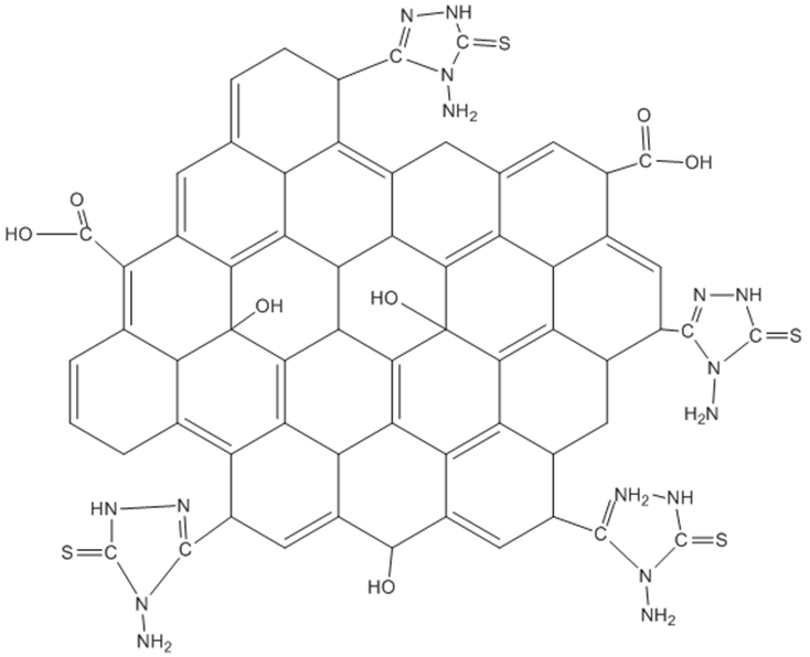
0.3g RGO was mixed with 1.2g TCH in around bottle flask and heated in the sand bath until TCH melted. The mixture was kept on the 168 C (Fusion temperature of TCH) for 10min and stirring with glass rod. The product of RGO functionalized with 5-substituted 4-amino, 3-mercapto 1, 2, 4 Triazole –thion (RGOT) Figure 1, and washed with distilled water to dissolve the non-reacted TCH and dried under vacuum 60oC.
Figure 1. Functionalized RGO with 5-substituted 4-amino, 3-mercapto 1, 2, 4 Triazole –thion

2.6. Modification and Pretreatment of (RGOT- SPCE)

A mixture from 1 mg of RGOT 3μL Nafion was dispersed in 10 ml of double distilled water for 1 h with ultrasonic devices. The WE has been coated with 4 μL of suspended RGOT leaved 2 hours evaporate the solvent in the air. The prepared SPCE was initially pre-treated to increase the sensitivity and to achieve a stable base line for a numerous experiments by sweeping the potential between -0.1 and +1.9 V in 0,1M H2SO4 for 15 times to achieve a repeatable voltammogram [38]. After the activation of the SPCEs were washed and dried, then to the modified and pre-treated SPCEs were added 50μL freshly prepared solution of AMP with PBS as a supporting electrolyte to cover all the reservoir area of the modified WE, CE and RE.

3. Results and Discussion

3.1. FTIR Characterization

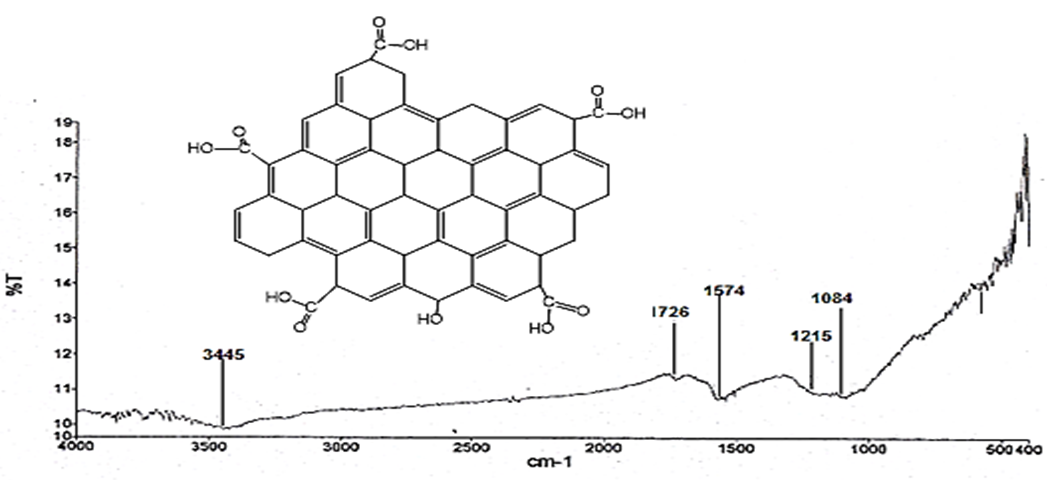
Reduced Graphene oxide FT-IR showed O-H stretching vibration peak 3445 cm-1 was significantly small due to the deoxygenation. The stretching at 1726 cm-1 assigned the C = O (carbonyl / carboxy), (C = C) 1574 cm-1 (aromatics) and 1215 cm-1 to 1084 cm-1 for Co stretching, some residual oxygen groups were present at 3454 cm-1 [39, 40] Figure (2).
Figure 2. FT-IR spectrum of RGO
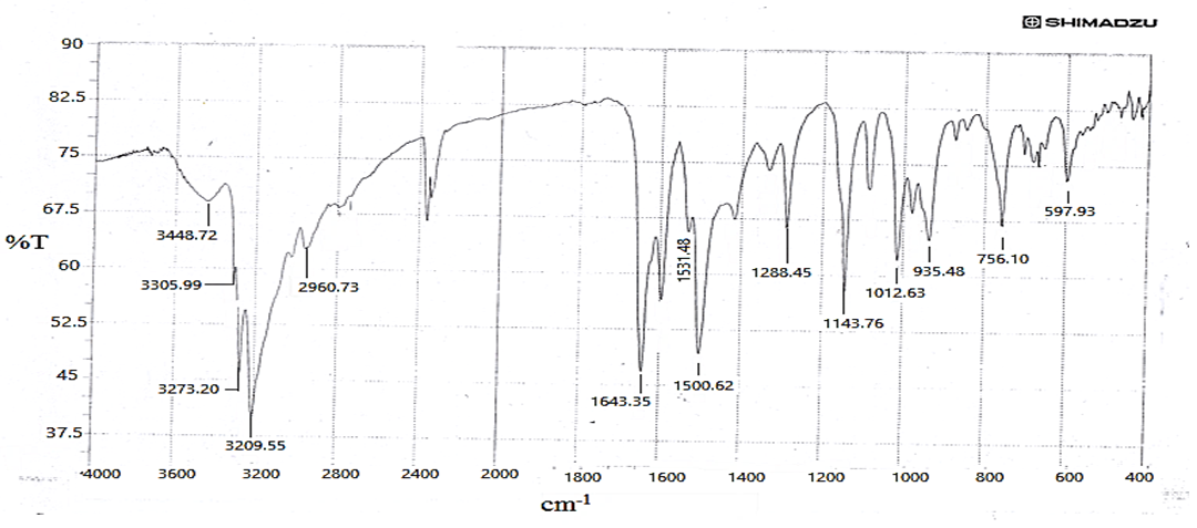
Figure (3) show FT-IR spectrum of prepared TCH, and peaks at 3209.5, 3273.2, 3305.9 cm-1 assigned to NH and NH2 stretching vibrations, The NH2 1643.35cm-1 in 1143, 79 cm-1 respectively, NH in 1531 and 1500 .6 cm-1 assigned to the N-H and C-N stretching vibrations C = S stretching of the two peaks at 1288.45 and 935.48 cm-1 [41].
Figure 3. FT-IR spectrum of thiocarbohydrazide TCH
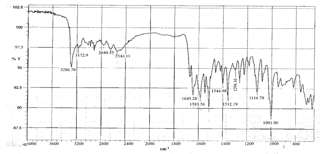
The FT-IR spectrum of RGO functionalized with 5-substituted 4-amino, 3-mercapto 1, 2, 4 Triazole –thion (ASTT) figure (4) was to show a band in 3286, 7 cm-1 and 3172.9 cm-1 attributed to NH2, N-H at 3154 cm-1 and 2430cm-1, C = S at 1325-1288 cm-1.
Figure 4. FT-IR spectrum of RGO functionalized with (5-substituted 4-amino, 3-mercapto 1, 2, 4 Triazole –thion) ASTT
XRD characterization of the prepared RGO
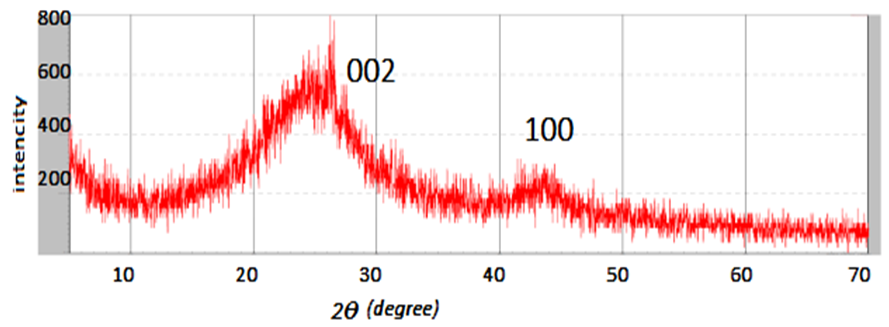
Fig (5) shows XRD patterns of RGO peak at 2Ɵ = 24.5o was appeared like the peak of graphite and agreed with (JCPDS Card no.75 -1621) with plane (002) the d- spacing d002 = 3.3 Å, the constant lattice was a = 2.78Å [35]. The broad peak indicated the amorphous nature of the RGO.
Figure 5. XRD pattern of RGO

3.2. Surface Characterization

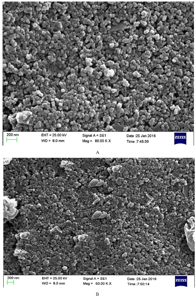
SEM imaging was necessary to reveal the morphology of Graphene-based working electrodes. Fig. 6 (A and B) show typical SEM images of two electrodes utilized throughout this work. There is a clear similarity between bare-SPCE (Figs. 6A) and RGOT-SPCE (Figs. 6B) electrode, where (Figs. 6A) can be seen that the surface is slightly rough and disordered in bare-SPCE, while RGO functionalized with (ASTT) RGOT-SPCE (Figs. 6B) seems to be an overall slightly smoother than the bare-SPCE, which is likely due to the RGOT with binder polymer nafion utilizing the electrode ink of the latter to fabricate a more compacted surface. However, the granule's sizes appear smaller than for the bare electrode on the existence of accumulated RGOT sheets. Fig. 6B appears similar to that previously reported SEM image in [42].
Figure 6. SEM images of the bare-SPCE (A) and RGOT-SPCE

3.3. Voltammetric of Ampicillin and RGOT-SPCE

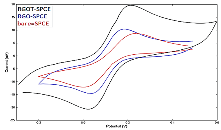
The Fe (CN)6-3 compound is sensitive to the carbon-based electrodes; the Fe (CN)6-3 was chosen to study RGOT-SPCE modified electrodes. Bare and reduces Graphene oxide-SPCE modified (WE) electrodes was coated with 0, 5 m M ferrocyanide solution and the cyclic voltammograms (CVs) shows. Fig.7. the CV cycles display of oxidation and reduction process for the redox of Fe3- / Fe2-. It was evident that the peak current intensity of bare-SPCE was lower than that of modified electrode with the reduced Graphene oxide. It was because the oxygen functional groups reinforce the electric resistance and decreases the charge transfer between the modifying layer and substrate but the functionalized reduced graphene oxide with (ASTT) increased the electroactive surface area and provided the conductive bridges for the electron-transfer of ferrocyanide. Therefore, electro catalytic activity of reduced Graphene oxide was further enhanced by the electrode by introducing various ASTT group in the structure of the RGO. However, RGOT-SPCE modified electrodes show better results than that bare-SPCE.
Figure 7. Cyclicvoltagram in red the bar-SPCE, in blue RGO-SPCE and in black RGOT-SPCE
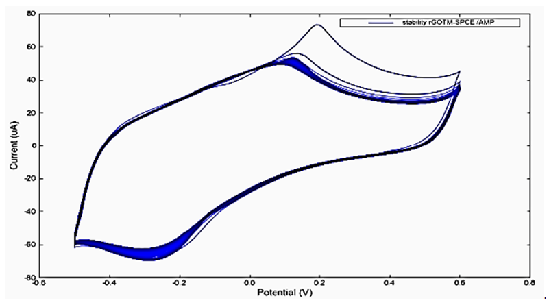
Moreover, the electrochemical stability of the RGOT-SPCE tested. The results show that the highest number of definitions of modified electrode was almost the same as the second CV cycle on the 50th repeated CV cycling fig.8.
Figure 8. The electrochemical stability modified electrode for more than 50 time repeated cyclic voltammetry cycle
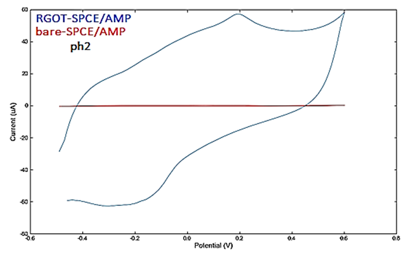
Figure 9. A very well difference appeared between the cyclic voltammograms of RGOT-SPCE composites and bare-SPCE in the presence of ampicillin AMP in PBS at the pH2value the scan was with rates 0.1V.s-1. The obvious oxidation peak was found ca. + 0.19V and the reduction of ca. -0.19V on the modified RGOT-SPCE.
Figure 9. The behavior of RGOT-SPCE and bare-SPCE in the presence of AMP at pH2
The results show that on the application of RGOT-SPCE, the electrochemical redox of AMP is much stronger than in the bare-SPCE, so that the composites electrodes attractive for applications in the field of electrochemical Sensing. The bare-SPCE indicates low conductivity because of the carbon material in the SPCE poor with the covalent double bonds cause an increase in the band gap. However, the conductance of the prepared RGOT-SPCE displayed higher conductivity than the bare-SPCE, In addition, AMP interaction with ASTT and the deprotonation of ASTT induced the changes in the electronic structure of the ASTT and during the deprotonation was added the hydrogen ion to the β-lactam group of AMP in aqueous solution to yield the carboxylic acid group instead of ketone group scheme 2.
Scheme 2. The formation of carboxylic acid group instead of ketone group during the protonation process on the β-lactam group
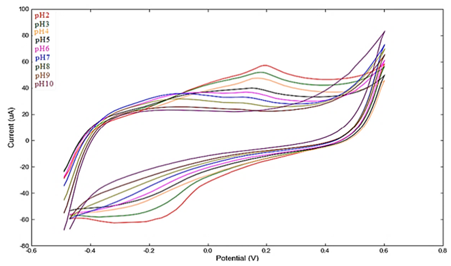
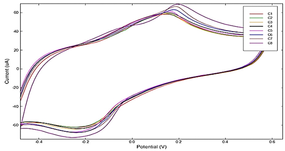
Therefore, we consider that both the deprotonation of ASTT and high conductivity of RGOT-SPCE facilitate better sensing performance for AMP. To show the dependence of the electrocatalytic current concentration of APM was measured cyclic voltammograms of RGOT-SPCE and various PBS solution pH values. The figure10 appeared at pH2 the highest current value, and at this pH value was the best deprotonation of ASTT and at pH2 examined a series concentrations of AMP have been obtained. Fig. 11 depending on the application of AMP the oxidation peak current was increased with the increasing of AMP concentration. At present, there was a slight shift in the peak position of concentration, it may be said that the adsorption of the side-product of the electrochemical oxidation products on the electrode surface.
Figure 10. Cyclic voltammogram of RGOT-SPCE/AMP in different pH
Figure 11. Cyclic voltammogram of AMP (PBS PH2) at different con. (1 × 10⁻⁶ to 5 × 10⁻⁵ mol.) with scan rate 0.1 V.s-1
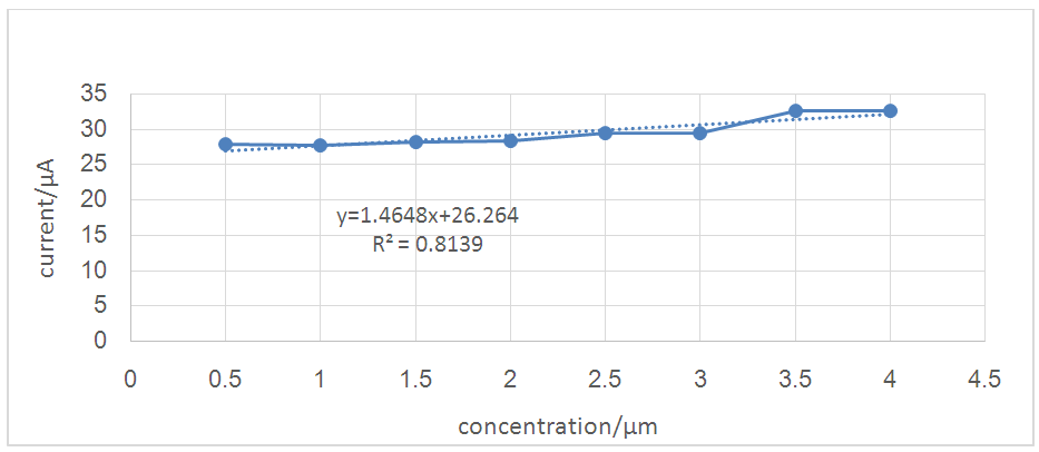
Figure 12 show linear relationships of the oxidation peak at different concentration of AMP (1 × 10⁻⁶ - 5 × 10⁻⁵ mol.). The linear regression equation was I (μA) = 1.4648 (μM) + 26.264, with the correlation coefficients of 0.8139.The limit of detection was calculated according to eqau.1and obtained the result 0.12 µmol/L
(1)
Where Sy/x was the standard deviation of peak current and b is the slope of the related calibration curve.
Figure 12. Oxidation peak at the different concentration of AMP
It was noted that the research's focusing on identifying and determination the LOD of the ampicillin with the CV technique very few. For comparison, kloth et al. were mention to 1.1 μg/L for different antibiotic in raw milk, Aderian et al. were mention to LOD value of 58-190nmol/L and Leyden MR et al. were mention to 0.67 μ mol/L.

4. Conclusions

In summary, we have prepared RGOT SPCEs composites with covalence bonding of ASTT with RGO. The characterization process shows a successful formation of composites. The detection of ferrocyanide was with electrochemical reaction and shows that the signal enhancement of composite, lead to suggest that the application of RGOT in electrochemical sensors. Furthermore, it is clear that the modified RGOT- SPCE display the electro catalytic activity to the oxidation and reduction of AMP.

References

[1]  Novoselov KS, Geim AK, Morozov SV, Jiang D, Zhang Y, Dubonos SV, Grigorieva IV and Firsov AA, 2004,. Electric Field Effect in Atomically Thin Carbon Films. Science. 306, 666-669.
[2]  Sakhaee-Pour A, Ahmadian MT, Vafai A, 2008, Applications of single-layered graphene sheets as mass sensors and Atomistic dust detectors. Solid State Communications. 145, 168-172.
[3]  Stoller MD, Park S, Zhu Y, An J and Ruoff RS. 2008, Graphene-Based Ultracapacitors. Nano Lett, 8, 3498–3502.
[4]  Choi SH, Kim YL, Byun KM. 2011, Graphene-on-silver substrates for sensitive surface plasmon resonance imaging biosensors. Opt Express. 19, 458-466.
[5]  Peigney A, Laurent Ch, Flahaut E, Bacsa RR, Rousset A. 2001, Specific surface area of carbon nanotubes and bundles of carbon nanotubes. Carbon. 39, 507-514.
[6]  Kang X, Wang J, Wu H, Aksay IA, Liu J and Lin Y. 2009, Glucose Oxidase–graphene–chitosan modified electrode for Direct electrochemistry and glucose sensing Biosens. Bioelectron. 25, 901-905.
[7]  Liu Y, Yu D, Zeng C, Miao Z and Dai L. 2010, Biocompatible Graphene Oxide-Based Glucose Biosensors. Langmuir, 26, 6158-6160.
[8]  Zhuang Z, Li J, Xu R, Dan X. 2011,Electrochemical Detection of Dopamine in the Presence of Ascorbic Acid Using Overoxidized Polypyrrole/Graphene Modified Electrodes. J. Electrochem. Sci. 6, 2149 – 2161.
[9]  Du D, Zou Z, Shin Y, Wang J, Wu H, Engelhard MH, Liu J, Aksay IA, Lin Y. 2010, Sensitive Immunosensor for Cancer Biomarker Based on Dual Signal Amplification Strategy of Graphene Sheets and Multienzyme Functionalized Carbon Nanospheres. Anal. Chem. 82, 2989-2995.
[10]  Shao Y, Wang J, Wu H, Liu J, Aksay IA, Lina Y. Graphene Based Electrochemical Sensors and Biosensors. Electroanalysis 22, 1027 – 1036.
[11]  Dan P, Lu Y, Kybert NJ, Luo ZT, Johnson ATC. 2009, Intrinsic Response of Graphene Vapor Sensors. Nano Letters. 9, 1472-1475.
[12]  Schedin F, Geim AK, Morozov SV, Hill EW, Blake P, Katsnelson MI, Novoselov KS. 2007, Detection of individual gas molecules adsorbed on graphene. Nature Materials. 6, 652 – 655.
[13]  Ang PK, Chen W, Wee ATS, Loh KP. 2008, Solution-gated epitaxial graphene as pH sensor. J. Am. Chem. Soc. 130, 14392–14393.
[14]  Dong X, Shi Y, Huang W, Chen P, Li LP. 2010, Electrical Detection of DNA Hybridization with Single-Base Specificity Using Transistors Based on CVD-Grown Graphene Sheets. Adv. Mater. 22, 1–5.
[15]  Mohanty N, Berry V. 2008, Graphene-based single-bacterium resolution biodevice and DNA transistor: interfacing graphene derivatives with nanoscale and microscale biocomponents. Nano Letters. 8, 4469–4476.
[16]  Ohno Y, Maehashi K, Yamashiro Y, Matsumoto K. 2009, Electrolyte-gated graphene field-effect transistors for detecting pH and protein adsorption. Nano Letters. 9, 3318–3322.
[17]  Yang W, Ratinac KR, Ringer SP, Thordarson P, Gooding JJ, Braet F. 2010, Carbon Nanomaterials in Biosensors: Should You Use Nanotubes or Graphene? Angew. Chem. Int. Ed. 49, 2114 – 2138.
[18]  Wang Y, Li Z, Wang J, Li J, Lin Y. Graphene and graphene oxide: biofunctionalization and applications in biotechnology. Trends in Biotechnology. 2011, 29, 205-212.
[19]  Hirsch A, Vostrowsky O. Functionalization of Carbon Nanotubes. Topics in Current Chemistry. 2005, 45, 193-237
[20]  Martinez MT, Tseng YC, Ormategui N, Loinaz I, Eritja R, Bokor . 2009, J. Label-Free DNA Biosensors Based on Functionalized Carbon Nanotube Field Effect Transistors. Nano Letters 9, 530–536.
[21]  Jeng ES, Moll AE, Roy AC, Gastala JB, Strano MS. 2006, Detection of DNA Hybridization Using the Near-Infrared Band-Gap Fluorescence of Single-Walled Carbon Nanotubes. Nano Letters, 6, 371–375.
[22]  Lu C-H, Yang H-H, Zhu CL, Chen X, Guo-Nan Chen Prof. 2009, A Graphene Platform for Sensing Biomolecules. Angew. Chem. Int. Ed, 48, 4785–4787.
[23]  Balavoine F, Schultz P, Richard C, Mallouh V, Ebbesen TW, Mioskowski C. 1999, Helical crystallization of proteins on carbon nanotubes: a first step towards the development of new biosensors. Angew. Chem. Int. Ed38, 1912–1915.
[24]  Loh KP, Bao Q, Ang PK, Yang. 2010, The chemistry of graphene. J. Mater. Chem20, 2277–2289.
[25]  Leyden MR, Schuman C, Sharf T, Kevek J, Remcho VT, Minot ED. 2010, Fabrication and Characterization of Carbon Nanotube Field-Effect Transistor Biosensors. Proc. SPIE, doi:10.1117/12.861329.
[26]  Balasubramanian K. Burghard M. 2005, Chemically functionalized carbon nanotubes. Small, 1, 180-192.
[27]  Dreyer DR, Park S, Bielawski CW and Ruoff RS. 2010, The chemistry of graphene oxide. Chem. Soc. Rev.39, 228-240.
[28]  Balasubramanian K, Burghard M. 2008, Electrochemically functionalized carbon nanotubes for device applications. J.Mater. Chem. 18, 3071-3083.
[29]  Maroto A, Balasubramanian K, Burghard M, Kern K. 2007, Functionalized metallic carbon nanotube devices for pH sensing. ChemPhysChem. 8, 220-223.
[30]  Balasubramanian K, Friedrich M, Jiang C, Fan Y, Mews A, Burghard M, Kern K. 2003, Electrical transport and confocal Raman studies of electrochemically modified individual carbon nanotubes. Adv. Mater. 15, 1515-1518.
[31]  Currie, Janet, Wanchuan Lin, Wei Zhang, 2011. Patient Knowledge and Antibiotic Abuse: Evidence from an Audit Study in China. Journal of Health Economics 30, 933-949.
[32]  Dar-Odeh, Najla Saeed, Abu-Hammad Osama Abdalla, Al-Omiri, Mahmoud Khaled, Khraisat, Ameen Sameh, Shehabi, Asem Ata, 2010. Antibiotic Prescribing Practices by Dentists: a Review. Therapeutics and Clinic Risk Management 6, 301-306.
[33]  Mendham, J.; Denny, R.C.; Barnes, J.D.; Thomas, M (2000). Vogel's textbook of quantitative chemical analysis (5th.ed.). Harlow: Pearson Education.
[34]  Eggleston, Karen, Ling Li, Qingyue Meng, Lindelow Magnus, Wagstaff Adam, 2008. Health Service Delivery in China: A Literature Review. Health Economics 17(2), 149-165.
[35]  I. Jung, (2007), Simple approach for high –contrast optical imaging and characterization of graphene based sheets. Nano Lett.7 3569-3575.
[36]  CK. Chua and M. Pumera. (2014), Chemical reduction of graphene oxide: a synthetic chemistry viewpoint, Chem. Soc. Rev. 43 291-312.
[37]  XH. Sun and YF Liu, study on the synthesis of thiocarbohydrazide chemistry (china) (1999) 62:46-48.
[38]  A. Vasilescu, S. Andreescu, C. Bala, S.C. Litescu, T. Noguer, J.L. Marty, (2003), Screen printed electrodes with electropolymerized Meldola. Blue as versatile detectors in biosensors, Biosensors and Bioelectronics. 18, 781–790.
[39]  Devesh K. M., Subhendu B., Mostafaizur R. Dipak K. "Morphology and Cyclic Voltammetry Analysis of in situ Polymerized Polyaniline /Graphene Composites" J. Electrochem. Sci.Eng., Vol.3 ,no.4,pp.157-166, 2013.
[40]  I. Latif, Taghreed B. Alwan, Ammar H. 2012, Low Frequency Dielectric Study of PAPA-PVA-GR Nanocomposites 2(6): 190-200.
[41]  GR. Burns, (1968) metal complexes of thiocarbohydrazide'' Inorg. Com.7:277-283.
[42]  A. Gomis-Berenguer, M. Go´mez-Mingot, V. Montiel, A. Canals, T. Thiemann, R. O. Kadara, C. E. Banks and J. Iniesta, 2012, RSC Adv., 2, 7735–7742.
[43]  M.E. Swatz, I.S Krull. Analytical Method Development and Validation, Marcel Dekker, New York; 1997.
[44]  M. A. Khalilzadeh, F. Khaleghi, F. Gholami & H. Karimi- Maleh." Electrocatalytic Determination of Ampicillin Using Carbon-Paste Electrode Modified with Ferrocendicarboxylic Acid" J. Analy. LettersVolume 42, Issue 3, 2009, pages 584-599.
[45]  Kloth, K.; Rye-Johnsen, M.; Didier, A.; Dietrich, R.; Martlbauer, E.; Niessner, R.; Seidel, M. A, 2009, regenerable immunochip for the rapid determination of 13 different antibiotics in raw milk. Analyst 134, 1433–1439.
[46]  Adrian, J.; Pinacho, D.G.; Granier, B.; Diserens, J.M.; Sanchez-Baeza, F.; Marco, M.P. A 2008, multianalyte ELISA for immunochemical screening of sulfonamide, fluoroquinolone and beta-lactam antibiotics in milk samples using class-selective bioreceptors. Anal. Bioanal. Chem. 391, 1703–1712.