Nanoscience and Nanotechnology

p-ISSN: 2163-257X    e-ISSN: 2163-2588

2015;  5(2): 30-35

doi:10.5923/j.nn.20150502.02

Synthesis, Characterization and Optical Properties of L-Arginine Stabilized Gold Nanocolloids

A. L. Sunatkari1, S. S. Talwatkar2, Y. S. Tamgadge3, G. G. Muley4

1Department of Physics, Siddhartha College of Arts, Science and Commerce, Fort, Mumbai, India

2Department of Physics, N.G. Aacharya and D.K. Maratha College of Arts, Science and Commerce, Chembur, Mumbai, India

3Department of Physics, Mahatma Phule Arts, Commerce & S.R.C. Science College, Warud, India

4Department of Physics, Sant Gadge Baba Amravati University, Amravati, India

Correspondence to: G. G. Muley, Department of Physics, Sant Gadge Baba Amravati University, Amravati, India.

Email:

Copyright © 2015 Scientific & Academic Publishing. All Rights Reserved.

Abstract

We investigate the effect of surface passivation using L- arginine on Localised surface plasmon resonance (LSPR) and size of colloidal Gold Nanoparticles (AuNPs) synthesized by chemical reduction method. Highly stable colloidal gold nanoparticles (AuNPs) stabilised in L-Arginine were synthesized. Optical absorption spectra of colloidal AuNPs exhibit an absorption peak at 520 nm due to Surface Plasmon Resonance. As the L-arginine concentration increased from 1 mM to 5 mM, blue shift in the LSPR peak position noticed. Fourier transform infrared spectra show the shift in absorption band position due to donor-acceptor interactions between L-arginine functional groups and AuNPs surface suggesting strong encapsulation of AuNPs. The size of the AuNPs is also found to be reduced from 11 to 7.5 nm with the stabilizer concentration as analysed using transmission electron microscopy. The X-ray diffraction study reveals face-centred cubic (fcc) structure of AuNPs.

Keywords: L-arginine capping, UV-Visible, FTIR, TEM, Gold nanoparticles

Cite this paper: A. L. Sunatkari, S. S. Talwatkar, Y. S. Tamgadge, G. G. Muley, Synthesis, Characterization and Optical Properties of L-Arginine Stabilized Gold Nanocolloids, Nanoscience and Nanotechnology, Vol. 5 No. 2, 2015, pp. 30-35. doi: 10.5923/j.nn.20150502.02.

1. Introduction

Metal nanoparticles (MNPs) are an important class of nanomaterial due to their size-dependant properties and potential applications in sensors, catalysis and data storage [1-2]. Generally, Because of their high surface energy, MNPs are unstable and need to be stabilized against aggregation by suitable surface modifying agents. The common choice of functional groups for AuNPs surface modification are cyano (-CN), mercapto(-SH), thiol-mediated binding of ligands are known to have high affinity for gold and expected to produce quite small sized AuNPs [3]. Numerous reports are available on the effect of surface passivation on the properties of MNPs, which would play a vital role in the applications such as biosensors [4], DNA/drug delivery [5], Imaging [6], bio diagnostic and optoelectronics devices [7]. Common choice for MNPs surface modification is thiol-mediated binding of ligands [8-9]. Aniline, long-chain amines, carboxylic compounds have been used as stabilizers in the synthesis of MNPs [10]. Researchers have also studied the role of Poly-Vinyl Pyrrolidone (PVP), polyacrylate and polyacrylamide as protective agents which can effectively alters shape, size, stability and linear optical properties of AgNPs [11]. More recently, researchers have diverted attention to the binding of metal NPs with amino-acids [12]. Amino acids are inherently compatible, and one of the common amino acid is L-arginine, which has zwiterrionic structure. Upon functionalization of MNPs with L-arginine molecules, they can highly facilitate the interaction and hence have a potential to bring drastic changes in optical properties. Joshi et. al. reported the synthesis of L-Lysine capped gold nanoparticles [13]. Bhargava et.al reported the synthesis of gold nanoparticles using the amino acids L-tyrosine, glycyl-L-tyrosine, and L-arginine [14]. To the best of our knowledge, reports are not available on the use of L-arginine as protective agent in the synthesis of AuNPs. Therefore, we have used L-arginine for surface modification in the synthesis of AuNPs.
Various methods such as sol-gel method, laser ablation, and chemical reduction have been used to synthesize gold nanoparticles. Among these, chemical reduction method [15-18] is extensively used because of its simplicity and accuracy.
The objective of this work is to prepare AuNPs in colloidal form with various concentration of L-arginine reduced by sodium Borohydride by wet chemical (reduction) method and to discuss the effect of L-arginine concentration on LSPR, size of AuNPs, and stability of gold colloids.

2. Experimental Section

2.1. Chemicals

Gold (III) chloride hydrate (HAuCl4, 99.999 % purity), sodium borohydride (NaBH4, 98% purity), L-arginine (99% purity) and PVP (MW 10000) purchased from Sigma-Aldrich and used without further purification. De-ionised water was used in synthesis process. L-arginine was used as capping agent and NaBH4 was used as a reducing agent.

2.2. Synthesis of L-arginine Stabilised AuNPs Colloid

Stock solutions of 5 mM HAuCl4, 10 mM NaBH4 were prepared. L-arginine solutions with 1, 2.5, and 5 mM concentrations were prepared by dissolving appropriate amount in double distilled water. All solutions were kept in ice-bath for 20 min. In 200 ml volumetric flask, 30 ml double distilled water, 10 ml NaBH4 and 20 ml L-arginine solutions were taken and stirred at 50°C for 20 min. AuNPs colloidal solution was obtained by drop wise addition of 20 ml gold precursor into the above mixture. Solution was turned dark violet (stable colour) in colour in 10 min indicating formation of AuNPs. AuNPs were precipitated by centrifugation for 20 min. at 4000 rpm. Precipitate washed with methanol to remove free legends and then dried by means of optical heating at 40°C. As-prepared AuNPs colloids were stable for long duration of 3 months.

2.3. Characterizations

Absorption spectra were recorded covering wavelength range 200-900 nm using ultraviolet-visible (UV-vis) spectrometer (Black-C-SR, Stellarnet Inc. USA). Average size and shape of nanoparticles were determined form transmission electron micrograph (TEM) recorded using transmission electron microscope (JEM 2100F, JEOL) with point to point resolution of 0.19 nm and accelerating voltage 200 kV. Structural study of nanoparticles was carried out with X-ray diffractometer (Miniplex-II, Rigaku) with CuKα wavelength 1.54 Å in the angle 2θ ranging from 20 to 80o. Fourier transform infrared (FT-IR) spectra were obtained with machine 3000 hyperion microscope with vertex 80 FTIR system (Bruker, Germany) in the range of wavelength 450 - 7500 cm-1 with resolution of 0.2 cm-1.

3. Results and Discussion

3.1. UV-vis Absorption Spectroscopy

Optical absorption spectra of L-arginine capped gold nanocolloids reduced with sodium borohydride synthesized by chemical reduction method is recorded in the wavelength range of 200 nm - 900 nm at room temperature. Fig. 1 illustrates the results obtained for various concentration of L-arginine.
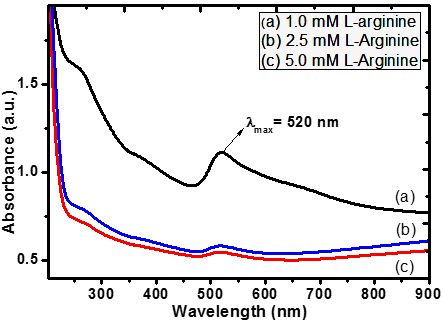
Figure 1. Absorption spectra of AuNPs colloids with (a) 1, (b) 2.5 and (c) 5 mM L-arginine concentrations
The absorption spectra shows Plasmon resonance peak position at 520 nm for 1 mM concentration of L-arginine is the characterstics feature of AuNPs indicating particle size is below 15 nm [19]. As we increased the concentration of L-arginine from 1mM to 5 mM, as the resonance absorption peak is shifted from 520 to 513 nm indicating blue shift. The strong absorption peaks at 520, 518, and 513 nm corrosponds to the excitation of surface plasmon vibrations in the AuNPs [20]. The blue shift in the SPR peak position, accompanied by broadening of peak, is attributed to the decrease in size of AuNPs and hence, electron motion within nanoparticles is confined more and more which leads to decrease in the free electron collision time [16, 21]. The formation of smaller sized AuNPs with increase in L-arginine concentration from 1mM to 5 mM is authenticated from TEM measurements (Fig. 1(a) and 1(b)). As expected from Mie’s Theory, a weak absorption peak appeared at 262, 271 and 276 nm for 1, 2.5 and 5 mM L-arginine concentration, respectively, is because of Plasmon band of small particles (less than 4 nm in diameter) occures as a shoulder originating from electron surface scattering effect [22]. Interestingly, L-arginine capped AuNPs were stable for three months without any aggregation whereas free AuNPs were started aggregation within two weeks at room temperature. S. Aryal et.al [3] also found similar results when they stabilized AuNPS in L-Cysteine. The aggregation of AuNPs is due to the decrease in the surface potential that resulted from transfer of electron to the AuNPs from NaBH4, a reducing agent used in the synthesis process [23]. In some previous reports, aggregation of AuNPs attributed to its different morphology such as decahedron, tetrahedron, truncated tetrahedrons and cubes. These AuNPs have different crystalline facet with different chemical activities. Therefore the strength of binding force of stabilizer (L-Arginine) on different crystalline facet is not uniform. As a result, stabilizer with weak binding force on some facet will start desorbing and starts aggregation [24]. In this sense, Stability of AuNPs up to three months is peculiar since it suggests how strongly L-arginine is bonded to AuNPs.

3.2. FT-IR Study

Fig. 2 shows FT-IR spectra of L-arginine capped AuNPs taken in the wavelength range of 400 cm-1 - 4000 cm-1. Generally, in the synthesis of AuNPs surfactant ligands that binds to their surfaces which stabilize the nuclei and large nanoparticles against aggregation by electrostatic repulsive force. The electrostatic repulsive force is due to the equally charged L-arginine molecules adsorbed on the AuNPs surface through its electron-donating end group. Because of the electrostatic interaction between AuNPs surface (acceptor) and L-arginine ligands (donor), stable nanoparticles can be formed [25]. FT-IR is a powerful tool to investigate the bonding between surface of AuNPs and L-arginine molecule. Previously, L-arginine demonstrated a very good surfactant in the synthesis of CdS and ZnS nanoparticles by Talwatkar et.al. [26].
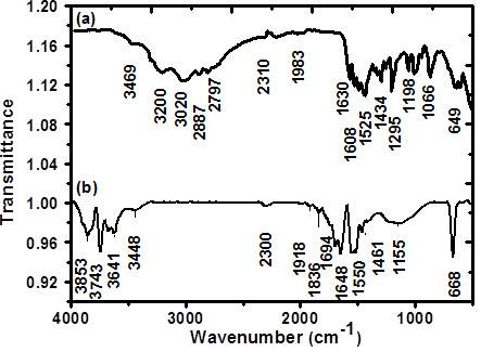
Figure 2. FT-IR spectrum of (a) Pristine L-arginine and (b) AuNPs capped by L-arginine
In general, amino acids exists as zwitterions and exhibits characteristics spectra of carboxylate and primary amine salts hence it shows NH3+ stretching, symmetric/asymmetric N-H bend and symmetric/asymmetric carboxylate COO- stretching as characteristics vibrations when bond with AuNPs surface. Peaks in the high energy region from 3853 cm-1 to 3641 cm-1 are mainly due to O-H stretching absorption of COOH and water molecules present in the sample. N-H stretching band of medium intensity observed at 3448 cm-1 for secondary amines. Peak at 2310 cm-1 is due to N-H stretching vibration spectra, shifted at 2300 cm-1 in L-arginine capped AuNPs. N-H+ stretching vibration in dilute solution spectra is seen at 1918 and 1836 cm-1 and slightly shifted to 1938 cm-1. N-H deformation vibration band shifted from 1648 cm-1 to 1630 cm-1. It is also observed that N-H deformation band is shifted to higher frequency region at 1694 cm-1 because of its reaction with dispersing medium (AuNPs solution). Primary amine group (N-H) generally absorb at 1615 – 1580 cm-1. In Pure L-arginine it is found at 1550 cm-1 due to N-H bending vibrations and shifted to 1525 cm-1 in AuNPs stabilized in L-arginine. Peak at 1461 cm-1 is assigned to asymmetric deformation vibration in CH3. Asymmetric C-N stretching shows band normally in the range of 1130 – 1145 cm-1 which slightly shifted to lower frequency side at 1155 cm-1. The band at 668 cm-1 in L-arginine capped AuNPs is assigned due to COO- plane deformation. The significant shift in COO- and NH3+ stretching is likely due to the change in dipole moment when L-arginine binds to AuNPs surface. The chararacterstics peaks in the FTIR spectra advocate the strong bonding between AuNPs and L-arginine functional groups [27, 28].

3.3. Structural Analysis

X-ray diffraction (XRD) study gives the information about morphology and phase formation of L-arginine capped AuNPs. Fig. 3 shows XRD spectrum of AuNPs embedded in PVP polymer matrix recorded in the range of 20 to 80o for 2θ values.
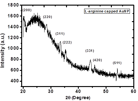
Figure 3. XRD spectrum of 1 mM L-arginine stabilised Au-PVP thin film
The XRD pattern exhibit peaks at 2θ values of 20.11, 28.50, 33.50, 35.27, 44.31, 45.85, and 54.06 which corresponds to the lattice planes (2 0 0), (2 2 0), (3 1 1), (2 2 2), (3 3 1), (4 2 0), and (5 1 1), respectively. The intensity and the shape of the peaks are the consequences of the face centred cubic (fcc) phase of gold Nano crystals. The diffuse nature of curve is due to the existence of PVP matrix [29]. These diffraction peaks suggest the existence of AuNPs in PVP matrix. All peaks in the XRD pattern matches with JCPDS data (JCPDS-PDF No. 01-1172). From full-width at half- maximum (FWHM) of diffraction peaks, the average size of AuNPs is estimated using Debye-Scherer equation 2R = 0.9λ/β cosθ [28], where 2R is size (diameter) of QD, β is full width half maxima (FWHM) of XRD peak in radians, θ is diffraction angle and λ is wavelength of X-ray (1.540598Å). The average size of AuNPs is found about 11.5 nm, 10 nm, 8 nm for 1, 2.5 and 5 mM L-arginine concentration respectively. The trend of reducing size is further authenticated by TEM analysis. Fig.4 (a) and 4(c) displays a TEM images of well dispersed AuNPs in PVP colloidal with uniform size distribution and uniform shape for 1mM L-arginine and 5 mM L-arginine concentration respectively. These images provide good evidence of homogeneity of material. The average sizes found from TEM images are 11 ± 0.4, 9 ± 0.6 and 7.5 ± 0.3 nm for 1, 2.5, and 5mM L-arginine concentration respectively. We noticed the aggregation of AuNPs stabilized L-arginine after three months [Fig. 4(f)]. The size histograms of gold nanoparticles displayed in Fig.4 (b) and 4(d). The selected area electron diffraction (SAED) pattern taken for AuNPs colloidal for 5 mM L-arginine concentration is shown in the Fig.4(e).
The outcome of XRD analysis established the fact that increase in L-arginine (stabilizer) concentration, AuNPs size reduces. Many reports, in consistent with our findings, are available in the literature [30-32]. TEM measurements and XRD analysis is in consistent with UV-vis spectroscopy measurements.
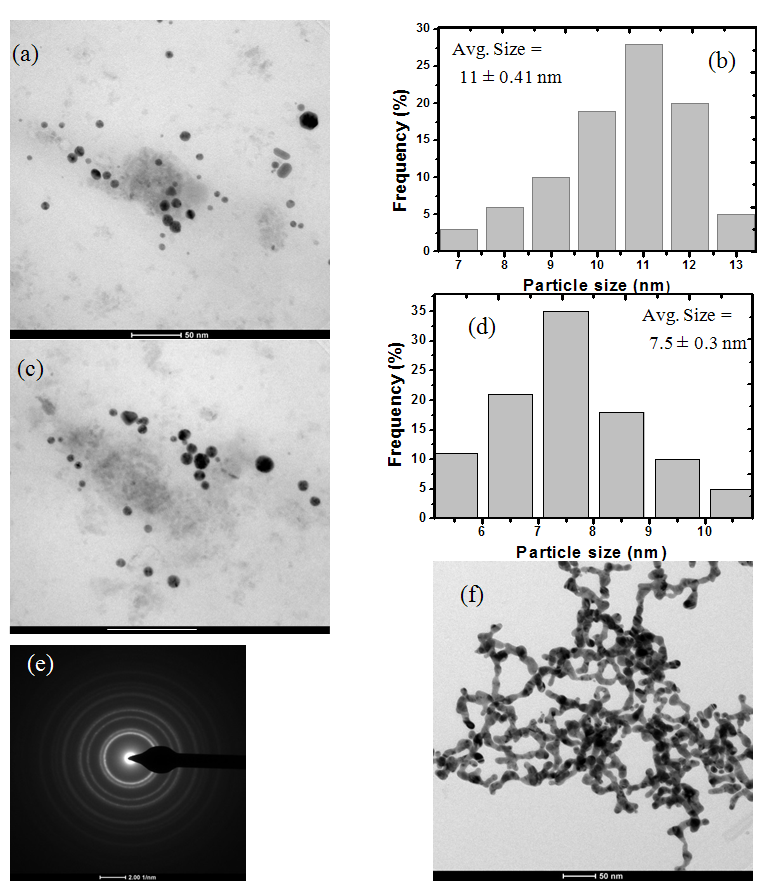
Figure 4. (a) TEM image of Au-PVP colloidal NPs (for 1 mM L-arginine), (b) corresponding histogram of particle size distribution, (c) TEM image of Au-PVP colloidal NPs (for 5 mM L-arginine), (d) represents corresponding particle size histogram (e) SAED pattern of 5 mM L-arginine and (f) showing Agreegation after 3 months

4. Conclusions

In summary, highly stable AuNPs were obtained using L-arginine as a stabilizer. The effect of L-arginine concentration on LSPR, size of AuNPs was studied. The average size of AuNPs, as analysed by TEM, was found to be decreasing with increase in L-arginine concentration. The presence of fcc structured AuNPs was confirmed from XRD studies. The blue shift in the LSPR peak position is noticed. The strong bonding between AuNPs and L-arginine functional groups confirmed from FT-IR study. Results proved that L-arginine is a very good stabilizing agent.

ACKNOWLEDGEMENTS

Author (A. L. Sunatkari) is grateful to the University Grants Commission (UGC), India for providing financial assistance under Minor Research Project (47/1690/10). Authors acknowledge the support of Director, SAIF facility, IIT-Bombay, Mumbai for providing FT-IR and TEM facilities and Chairman, DST-FIST, SGB Amravati University, Amravati for providing XRD facility.

References

[1]  M. C. Daniel and D. Astruc, “Gold nanoparticles: assembly, supramolecular chemistry, quantum-size-related properties, and applications toward biology, catalysis, and nanotechnology”, Chem. Rev.104, 293-346, (2004).
[2]  E. Shahriari, W. M. M. Yunus, and E. Saion, “Effect of particle size on nonlinear refractive index of Au nanoparticle in PVA solution”, Braz. J. Phys. 40, 256-260, (2010).
[3]  S. Aryal, B. Remant, N. Dharmaraj, N. Bhattarai, C. Kim, and H. Kim, “Spectroscopic identification of S-Au interaction in cysteine capped gold nano particles”, Spectrochimica Acta Part A, 63, 160-163, (2006).
[4]  W. C. W. Chan, and S. Nie, “Quantum dots bio conjugates for Ultrasensitive Nonisotopic detection”, Science, 281, 2016 – 2018, (1998).
[5]  J. J. Storhoff, and C. A. Mirkin, “Programme d Materials Synthesis with DNA”, Rev. 99, 1849 – 1862, (1999).
[6]  A. Bielinska, J. D. Eichman, I. Lee, J. R. J. Baker, and L. Balogh, “Imaging Au-0-PAMAM gold dendrimer nanocomposites in cells”, J. Nanopart. Res. 4, 395-403, (2002).
[7]  D. Zare, A. Akbarzadeh, and N. Bararpour, “Synthesis and functionalization of gold nanoparticles by using of poly functional amino acids”, Int. J. Nanosci. Nanotechnol. 6, 223-230, (2010).
[8]  Y. S. Shon, and E. Cutler, “Aqueous synthesis of alkanethiolate-protected Ag nanoparticles using bunte salts”, Langmuir 20, 6626-6630, (2004).
[9]  S. T. He, J. N. Yao, S. S. Xie, H. J. Gao, and S. J. Pang, “Superlattices of silver nanoparticles passivated by mercaptan”, J. Phys. D:Appl. Phys. 34, 3425-3429, (2001).
[10]  H. Hiramatsu, and F. E. Osterloh, “A Simple Large-Scale Synthesis of Nearly Monodisperse Gold and Silver Nanoparticles with Adjustable Sizes and with Exchangeable Surfactants”, Chem. Mater. 16, 2509-2511, (2004).
[11]  M. Chen, L. Wang, J. Han, J. Zhang, Z. Li, and D. Qian, “Preparation and Study of Polyacryamide-Stabilized Silver Nanoparticles through a One-Pot Process”, J. Phys. Chem. B 110, 11224-11231, (2006).
[12]  P. R. Selvakannan, S. Mandal, S. Phadtare, R. Pasricha, and M. Sastry, “Capping of Gold Nanoparticles by the Amino Acid Lysine Renders Them Water-Dispersible”, Langmuir 19, 3545-3549, (2003).
[13]  H. Joshi, P.S. Shirude, V. Bansal, K.N. Ganesh, and M. Sastry, “Isothermal Titration Calorimetry Studies on the Binding of Amino Acids to Gold Nanoparticles”, J. Phys. Chem. B. 108, 11535-11540, (2004).
[14]  S. K. Bhargava, J.M. Booth, S. Agrawal, P. Coloe, and G. Kar, “Gold nanoparticle formation during bromoaurate reduction by amino acids”, Langmuir 21, 5949-5956, (2005).
[15]  S. A Vorobyova, A. I Lesnikovich, and N. S. Sobal, “Preparation of Silver Nanoparticles by Interphase Reduction”, Colloids and Surfaces A: Physicochemical and Engineering Aspects, 152, 375 – 379, (1999).
[16]  S. H. Choi, Y. P. Zhang, A. Gopalan, K. P. Lee, and H. D. Kang, “Preparation of Catalytically Efficient Precious Metallic Colloids by γ-irradiation and Characterization”, Colloids and Surfaces A: Physicochemical and Engineering Aspects 256 pp. 165 – 170, (2005).
[17]  Z. Li, Y. Li, X. F Qian, J. Yin, and Z. K. Zhu, “A Simple Method for Selective Immobilization of Silver Nanoparticles”, Applied Surface Science 250 pp. 109 – 116, (2005).
[18]  T. Tsuji, N. Watanabe, and M. Tsuji, “Laser Induced Morphology Change of Silver Colloids: Formation of Nano-size Wires”, Applied Surface Science 211 pp. 189 – 193, (2003).
[19]  P. R. Selvakannan, S. Mandal, S. Phadtare, R. Pasricha, and M. Sastry, “Capping of gold nanoparticles by the amino acid lysine renders them water-dispersible,” Langmuir,19, 3545-3549, (2003).
[20]  P. Jiang, S. Xie, J. Yao, S. Pang, and H. Gao, “The stability of self-organised 1-nonanethiol-capped gold nanoparticle monolayer”, J. Phys. D: Appl. Phys., 34, 2255-2259, (2001).
[21]  D. Compton, L. Cornish, and E. van der Lingen, “The order nonlinear optical properties of goldnanoparticles in glasses”, Gold Bull., 36:2, 51-58, (2003).
[22]  S. Kumar, and S. B. Rai, “Spectroscopic studies of L-arginine molecule”, Indian J. Pure & appl. Phys. 48, 251-255, (2010).
[23]  H. Joshi, P. S. Shirude, V. Bansal, K. N. Ganesh, and M. Sastry, “Isothermal titration calorimetry studies on the binding of amino acids to the gold nanoparticles”, J. Phys. Chem. B. 108, 11535-11540, (2004).
[24]  S. S. Talwatkar, Y. S. Tamgadge, A. L. Sunatkari, A. Gambhire, and G. G. Muley, “ amino acids (L-arginine and L-alanine) passivated CdS nanoparticles: Synthesis of spherical hierarchical structure and nonlinear properties”, Solid State Sci., 38, 42-48 , (2014).
[25]  A. Giri, N. Goswami, P. Lemmens, and S. Pal, “Synthesis of water soluble L-arginine capped CdSe/ ZnS QDs and their interaction with synthetic DNA: Picosecond resolved FRET study”, Mater. Res. Bull. 47, 1912-1918, (2012).
[26]  G. Socrates, Infrared and Raman Characteristic Group Frequencies: Tables and Charts, Third Ed., John Wiley & Sons Ltd. UK, 2004.
[27]  S. S. Talwatkar, A. L. Sunatkari, Y. S. Tamgadge, V. Pahurkar, and G. G. Muley, “Influence of Li+ and Nd3+ co-doping on structural and optical properties of l-arginine-passivated ZnS nanoparticles” Appl. Phys. A, 118, 675-682, (2015).
[28]  Y. Hamanaka, K. Fukuta, A. Nakamura, L. M. Liz-Marzan, and P. Mulvaney, Appl. Phys. Lett. 84, 4938-4930, (2004).
[29]  U. Kreibig and M. Vollmer, Optical Properties of Metal Clusters, Springer, Berlin, 1995.
[30]  Y.S. Tamgadge, A. L. Sunatkati, S. S. Talwatkar, V. G. Pahurkar and G. G. Muley, “Linear and nonlinear optical properties of nanostructured Zn(1−x)SrxO–PVA composite thin films”, Opt. Mater. 37, 42-50, (2014).
[31]  S. S. Talwatkar, A. L. Sunatkari, Y. S. Tamgadge, V. Pahurkar, and G. G. Muley “Surface passivation by L-arginine and enhanced optical properties of CdS quantum dots co-doped with Nd3+-Li+”, J. Nanostruct. in Chemistry, 5, 205-212, (2015).
[32]  R. A. Ganeev, A. I. Ryasnyansky, A. L. Stepanov, C. Marques, R. C. da Silva, and E. Alves, “nonlinear optical properties of silver nanoparticles prepared by laser ablation in various liquids,” Opt. Commun., 253, 205-213, (2005).