Nanoscience and Nanotechnology

p-ISSN: 2163-257X    e-ISSN: 2163-2588

2015;  5(1): 7-13

doi:10.5923/j.nn.20150501.02

Thermal and Mechanical Properties of Chitosan / Mimosa Tenuiflora / Multiwalled Carbon Nanotubes Composite Developed by Thermally Induced Phase Separation

S. A. Martel-Estrada1, Olivas-Armendáriz2, L. Peón-Prieto1, P. Urquizo-Monrreal1, N. Hernández-Osuna1, C. A. Martínez-Pérez2, J. L. Hernández-Arellano1, E. Santos-Rodríguez3

1Instituto de Arquitectura, Diseño y Arte, Universidad Autónoma de Ciudad Juárez, Chih., México

2Instituto de Ingeniería y Tecnología, Universidad Autónoma de Ciudad Juárez, Chih., México

3UNACH/MCTP, Ciudad Universitaria Carretera Emiliano Zapata Km. 4, Chiapas, México

Correspondence to: E. Santos-Rodríguez, UNACH/MCTP, Ciudad Universitaria Carretera Emiliano Zapata Km. 4, Chiapas, México.

Email:

Copyright © 2015 Scientific & Academic Publishing. All Rights Reserved.

Abstract

The inclusion of multiwall carbon nanotubes (MWCNTs) into polymeric matrices is expected to improve their mechanical and thermal properties. In this work, biodegradable composites of chitosan/mimosa tenuiflora/multiwalled carbon nanotubes have been prepared through thermally induced phase separation method, in order to evaluate their thermal and mechanical properties for applications in tissue engineering. Different compositions were analyzed. Variation in porous morphology of the scaffold was studied using a field emission scanning electron microscopy (SEM). The morphology of the composites developed indicates a highly interconnected porous structure with a diameter from 1-100 µm. Presence of MWCNT improves the thermal stability and mechanical properties of the chitosan/mimosa tenuiflora scaffold. The results from IR, TGA, DSC and mechanical properties suggest that there is a chemical interaction between the constituents of the composite.

Keywords: Chitosan, MWCNT, Mimosa Tenuiflora, TGA, DSC, SEM

Cite this paper: S. A. Martel-Estrada, Olivas-Armendáriz, L. Peón-Prieto, P. Urquizo-Monrreal, N. Hernández-Osuna, C. A. Martínez-Pérez, J. L. Hernández-Arellano, E. Santos-Rodríguez, Thermal and Mechanical Properties of Chitosan / Mimosa Tenuiflora / Multiwalled Carbon Nanotubes Composite Developed by Thermally Induced Phase Separation, Nanoscience and Nanotechnology, Vol. 5 No. 1, 2015, pp. 7-13. doi: 10.5923/j.nn.20150501.02.

1. Introduction

Biological systems perform complex macroscale tasks. The first level of cellular organization is composed of macromolecules with lengths at the nanoscale. Nanoscale have demonstrated regulatory effects on cell behavior, including adhesion, migration, proliferation, signaling, genetic expression and stem cell fate. For these reasons, biomaterials are being designed with nanoscale details to improve cellular behavior [1].
Chitosan is one of the most popular natural polymers used in tissue engineering applications [2]. Chitosan can be obtained from chitin by deacetylation [2, 3]. It is a polysaccharide composed of glucosamine and N-acetyl glucosamine linked with a β1-4 glucosidic linkage [2]. It is biocompatible and biodegradable [2]. Nevertheless, some of its properties, such as bioactivity [2, 4], mechanical properties, etc., need to be improved. Chitosan can form porous structure by thermally induced phase separation [4, 5].
Carbon nanotubes (CNTs) are graphitic tubular structures with extraordinary physical, electrical and chemical properties [1, 6]. They have an excellent potential to reinforce polymeric matrices because of their small dimensions, specially high aspect ratio, strength and stiffness [7]. They can improve mechanical properties and biocompatibility, modulate cell shape, regulate stem cell differentiation and improve proliferation [1, 8]. There are two subsets of CNT: single-walled carbon nanotubes (SWCNTs) and multi-walled carbon nanotubes (MWCNTs).
Mimosa Tenuiflora (Willd) Poiret is a species of the Mimosoideae, a subfamily of the Fabaceae botanical family in the south of Mexico, Venezuela and Brazil [4]. Mimosa Tenuiflora contains alkaloids, mainly N-N- dimethyltryptamine, serotonin, triperpenoid glycosides, and steroidal saponins [4, 9].
Previously, an effort has been devoted to evaluate the potential use of chitosan/mimosa tenuiflora composite as a bioactive material [4]. Although this blend showed good bioactivity, the mechanical properties must be improved. So, in this work, biodegradable composites of chitosan/ mimosa tenuiflora/multiwalled carbon nanotubes have been prepared through thermally induced phase separation method, in order to evaluate their thermal and mechanical properties for application in tissue engineering.

2. Experimental

2.1. Materials

Chitosan (75-85% deacetylated) with molecular weight of 310,000-376,000 Da (75 -85% deacetylated) was purchased from Sigma-Aldrich (United States). The bark of M. Tenuiflorawas obtained directly from the Jiquipilas Valley (Chiapas, Mexico). It was ground to powder using a grinder. without any preparation. Glacial acetic acid (Mallinckrodt, United States) was used as a solvent for the blends. The MWCNT were fabricated in our laboratory by spray pyrolysis method according with a previous research [10].

2.2. Preparation of Nanocomposite

Chitosan/M. Tenuiflora/Multiwalled carbon nanotubes (MWCNT) composite was prepared using the thermally induced phase separation (TIPS) technique. It was prepared 80/20 Ch/M. Tenuiflora composites to which it was added 1% and 2% of MWCNT. First, measured percentage of the purified MWCNT was dispersed in 1% (v/v) aqueous glacial acetic acid by sonication for 5 min at room temperature. Then, Chitosan and M. Tenuiflora were added to the MWCNT solution and mixed together at an 80/20 ratio. Finally, the composite was frozen at -20℃ for 4 h and then the solvent was extracted by a freeze-drying in Labconco Free Zone 2.5 for 48 hours.
The composites were neutralized by immersion in ethanol at -20°C during 12 hours. It was prepared an 80% ethanol aqueous solution, to which it was added 0.5(w%/w%) of NaOH. The composites were immersed into the NaOH-CH3CH2OH solution at the same temperature during 12 hours more. Then, the samples were washed and rinsed with distilled water.

2.3. Characterization

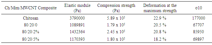
Chemical characterization of the composites was made using a FTIR-ATR spectrometer (Nicolet 6700). All spectra were recorded at 100 scans and 16 cm−1 resolution. X-ray diffraction patterns of the samples were obtained at 2θvalues between 5° and 80° with a step size of 0.02° using an X-ray diffractometer using (PANanalyticalX’Pert PRO) operated in continuous mode. The morphology of the composite was characterized using a Field Emission Scanning Electron Microscope (JEOL JSM-7000F).
Thermogravimetric analysis (TGA) and Differential Scanning Calorimetry (DSC) of the composite was carried out using TG-DSC analyzer (SDT Q600 from TA Instruments, United States). Dry samples were heated from 30℃ to 750℃at 10℃/min under nitrogen atmosphere with a flow rate of 80 mL/min. The first derivative of the mass-change with respect to time (DTG) was calculated and plotted as a function of the temperature.
The average pore size and pore size distribution of the composite was measured from the SEM micrographs. For this purpose, it was used the Scandium Universal SEM Imaging Platform software (Soft Imaging System) in the original magnification. Three different cross-sections of each scaffold were used to estimate the pore size and at least 120 pores were measured to estimate the distribution.
Finally, RSA III (Rheometrics Analyzes System) TA was used to elaborate a stress-strain analysis under a compression clamp in a frequency of 6.2832 rad/s and in a temperature 37℃, for all samples. Disks of approximately 5 mm in thickness and 15 mm in diameter were used for this test.

3. Results and Discussion

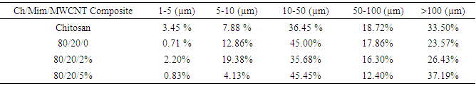
The microstructural properties of the scaffold play an important role to tissue repair, including pore diameter that influence cell attachment, proliferation and migration [11]. Pore size desired for optimal cellular activity and viability to interact with proteins and bioactivity and cellular development is 20-120 μm [5, 12]. The morphology of the composites as shown in Figure 1 indicates a highly interconnected porous structure. The porous structure consists of pores ranging in diameter from 1-100 µm. Table 1 shows those ranges of poresizes for the composites developed in this study.
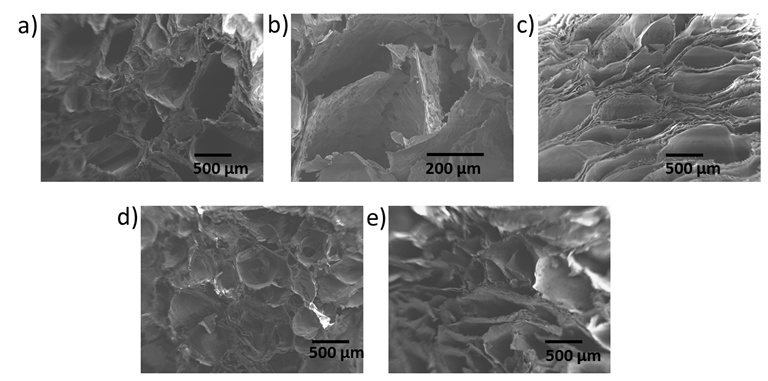
Figure 1. SEM micrographs of composites a) 100/0 Chitosan/M. Tenuiflora, b) 80/20 Chitosan/M. Tenuiflora, c) 70/30 Chitosan/M. Tenuiflora, d) 80/20/2% Chitosan/M. Tenuiflora/ MWCNT and e) 80/20/5% Chitosan/M. Tenuiflora/ MWCNT
Table 1. Pore size of the composites
     
The optimum pore size required for different tissue engineering applications depends on the specific cells, in which the scaffold will be used. Some researchers found that pores greater than 100 μm but less than 400 μm are optimal for osteoconduction [13, 14]. Therefore, from the perspective of pore size, all the composites developed in this study are suitable for bone engineering.
In this context, according to the results, the 80/20/5% Ch/Mim/MWCNT composite shows the higher pore size, even in the range that has been used for bone engineering. Also, the composite includes important percentage of microporosity for nutrient delivery. It the current study, porous scaffolds were fabricated for use as a scaffold in tissue engineering applications. Previously, Chitosan/ Mimosa Tenuiflora composites were developed [4], but the scaffolds showed poor mechanical properties. So, for the purposes of this study, it was very important that the inclusion of MWCNT does not affect the pore size.
Thermal properties of the composites were assessed by thermogravimetrical analysis (TGA). The results (Figure 2) indicate that there are two phases of degradation for all the composite samples. The thermal stability of the composites improved by addition of 2% MWCNT to the 80/20 Ch/Mimosa Tenuiflora composite. Chitosan decomposed between 232℃ and 326℃ before melting due to dehydration of the saccharide rings and decomposition of chitosan [5]. The initial weight lost around 100-150℃ is associated with the loss of water in chitosan [15]. The second severe weight loss occurred around 232℃ to 326℃, and is called the main region of decomposition (Table 2). All composites exhibited a single peak of fast thermal degradation (Figure 3). The temperature at the maximum degradation rate (Tmax) increased with increasing MWCNT content in the composites, suggesting that combination of the substances enhances thermal stability of the composites [5]. This is supported by the DTG profiles, indicating that the chemical interaction among the components resulted in a relatively homogenous composition. Thermal decomposition of the composites was greatly improved with the inclusion of MWCNT. From the thermal gravimetric curves, it can be seen that the thermal degradation of composites started at the higher temperature compared to chitosan. Results show that the degradation temperature varies between 239℃ and 326℃, showing that no degradation events occur during the scaffold fabrication.
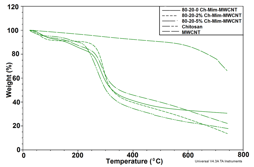
Figure 2. TGA curves showing the thermal degradation of the composites
Figure 3. DTG curves showing the thermal degradation of the composites
Table 2. Thermal degradation of the composites
     
The blend miscibility of the composites was determined from their glass transition temperature (Tg). The heat flow curves for the composites are presented in Figure 4. The glass transition temperatures obtained through these curves for the samples were 274.97℃, 287.80℃, 281.39℃, for the compositions 80/20/0%, 80/20/2% and 80/20/5% Chitosan/ M. Tenuiflora/ MWCNT, respectively. The change of heat capacity corresponding to the specific volume of chitosan might be small at the glass transition temperature, because the chitosan consist of 2-amino-2-deoxy-D-glucopyranose. As a consequence, the small baseline step is not clear in DSC curve.
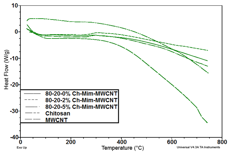
Figure 4. DSC curves of the composites
The Tg was observed to move to a higher value with increasing percentage of mimosa tenuiflora in the composite. Nevertheless, it was found that 2% of MWCNT permits higher Tg than 5%, due possible to agglomeration of the nanotubes at 5%. These results show evidence of partial miscibility among the composite components.
Scaffolds materials for bone engineering should possess sufficiently high mechanical properties to mimic the structure and performance of the bone. Therefore, we evaluated the morphology and compression strength of the scaffolds. Figure 1 shows the SEM images of the scaffolds. The composites exhibited open pores with diameters in the range needed to mineralize bone, and interconnected architecture for vascularization [16]. Table 3 exhibits the mechanical properties for the chitosan and the Chitosan/M. Tenuiflora/ MWCNT composites. The 80/20/2% composite exhibited the biggest module indicating that it is a more resistant material than the80/20/5% Chitosan/M. Tenuiflora/ MWCNT composite, but not chitosan. Nevertheless, as the MWCNT increases, the elastic module varies from 1.43 x10⁶ Pa to 1.17 x 10⁶ Pa, corresponding to 80/20/2% and 80/20/5% Chitosan/M. Tenuiflora/MWCNT respectively. These results exhibited improved mechanical properties for the composite using 2% of MWCNT for use in tissue engineering. Increase in MWCNT content did not improve the mechanical properties, possibly because of poor dispersion of the nanotubes [17]. When CNT is agglomerated, it acts to increase the local stress in the composite [1]. Although it was proved previously that there is an interaction between the amino group of chitosan and carboxyl groups in Mimosa Tenuiflora or hydroxyl groups in Chitosan and carboxyl groups of the Mimosa Tenuiflora [4], the mechanical properties of the composite is decided predominantly by the poor mechanical properties of Mimosa Tenuiflora.
Table 3. Mechanical properties of Chitosan/M. Tenuiflora/ MWCNT composites
     
In Figure 5, the IR spectra of the developed composites are shown, the chitosan spectrum shows the characteristic bands at 1543 cm-1which can be assigned to N–H bending vibration (amide II) [5]. The bands at 3500-3000 cm-1 correspond to O-H stretching vibration associated with free, inter and intra-molecular hydroxyl groups. The peaks in the region 3000-2800 cm-1 are a result of C-H stretching [18]. The peak at 1650 cm-1 corresponds to the stretching C O (amide I), and the peak at 1418cm-1 appeared to –C–O stretching of primary alcoholic group in chitosan [5]. The spectrum also shows peaks at 1154 cm-1that could be assigned to the saccharide structure. The absorption band for MWCNT- COOH is observed at 1741cm−1. The band at 1652 cm−1 corresponds to the stretching vibration of the C–O group. The absorption band around 1557 cm1 is related to –C=C group [19].
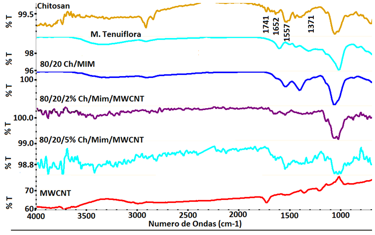
Figure 5. FTIR spectra of Chitosan/M. Tenuiflora/MWCNT composites
The M. Tenuiflora spectrum shows the characteristics bands at 1614 cm−1 and 1511 cm−1, which can be assigned to amide I and amide II, respectively. The peak at 1449 cm-1 appeared to –C–O stretching of primary alcoholic group in M. Tenuiflora. Also, the spectrum shows evidence of ether group at 1029 cm-1 and the absorption band for the p-xylene group of M. Tenuiflora at 843 cm-1 [4].
On the other hand, the 80/20/2% and 80/20/5% Ch/M. Tenuiflora/MWCNT shows that the N-H band was affected and shifted from 1543 cm-1 to 1557 and 1556 for the compositions with 2% and 5% of MWCNT content, respectively. It is possible to observed that these peaks appeared more pronounced in the spectrum of composites due possible to the interaction of the amide I of Chitosan and M. Tenuiflora. The peak that corresponds to the primary alcohol groups (-C-O stretching) was shifted from 1418 cm-1 to 1456 cm-1 for composite with 5% of MWCNT content. Also, Figure 5 shows that the ether peak became broader with lower intensity, as the MWCNT content increase, reflecting evidence of interaction between ether groups of chitosan and Mimosa Tenuiflora with the MWCNT. All these events could imply that an interaction of amide I bond and primary alcoholic groups of chitosan with the M. Tenuiflora occurred. Moreover, these events suggest that it was formed hydrogen bond interaction with MWCNT.

4. Summary

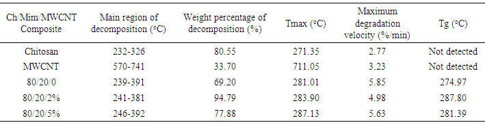
The present study provides more insight into thermophysical properties of Chitosan/Mimosa Tenuiflora/ MWCNT scaffolds. The composite specimens were successfully fabricated by thermally induced phase separation (TIPS) method. The microstructural properties of the scaffold play an important role to tissue repair. The morphology of the composites developed, indicates a highly interconnected porous structure with a diameter from 1-100 µm. So, according with the morphology obtained, all the composites developed in this study could be suitable for bone engineering. Presence of MWCNT was found to improve the thermal stability and mechanical properties of the chitosan/mimosa tenuiflora scaffold.Thermal decomposition of the composites was greatly improved with the inclusion of MWCNT. The thermal degradation of composites fabricated started at the higher temperature compared to chitosan. The obtained results from IR, TGA, DSC and mechanical properties suggest a chemical interaction between the polymers. It is believed that an interaction occurred between the amino group (mainly in chitosan) and carboxyl groups in MimosaTenuiflora or hydroxyl (mainly in chitosan) and carboxyl groups in the blends. Also, these events suggested that it was formed hydrogen bond interaction with MWCNT. The results show improved mechanical and thermal properties of the composite compared to Chitosan/Mimosa Tenuiflora composites, which can provide better stability of the scaffold. However, biocompatibility studies are required to confirm the suitability of the composites for bone engineering.

ACKNOWLEDGEMENTS

The authors acknowledge the financial support of the Mexican Public Education Secretary and the Mexican National Council for Science and Technology (CONACyT) through Projects SEP-CONACyT CB 2012-01-180909 and PROMEP/103.5/13/2347. Also, the authors appreciate the support of Mr. Armando Cardona-Torres during the edition of this document.

References

[1]  P. Newman, A. Minett, R. Ellis-Behnke, H. Zreiqat, Carbon nanotubes: their potential and pitfalls for bone tissue regeneration and engineering, Nanomedicine: nanotechnology, biology, and medicine, 9 (2013) 1139-1158.
[2]  L. Kong, Y. Gao, G. Lu, Y. Gong, N. Zhao, X. Zhang, A study on the bioactivity of chitosan/nano-hydroxyapatite composite scaffolds for bone tissue engineering, European Polymer Journal, 42 (2006) 3171-3179.
[3]  I.B. Leonor, E.T. Baran, M. Kawashita, R.L. Reis, T. Kokubo, T. Nakamura, Growth of a bonelike apatite on chitosan microparticles after a calcium silicate treatment, Acta biomaterialia, 4 (2008) 1349-1359.
[4]  S.A. Martel-Estrada, I. Olivas-Armendáriz, E. Santos- Rodríguez, C.A. Martínez-Pérez, P.E. García-Casillas, J. Hernández-Paz, C.A. Rodríguez-González, C. Chapa- González, Evaluation of in vitro bioactivity of Chitosan/ Mimosa tenuiflora composites, Materials Letters, 119 (2014) 146-149.
[5]  S.A. Martel-Estrada, C.A. Martínez-Pérez, J.G. Chacón-Nava, P.E. García-Casillas, I. Olivas-Armendariz, Synthesis and thermo-physical properties of chitosan/poly (dl-lactide-co- glycolide) composites prepared by thermally induced phase separation, Carbohydrate Polymers, 81 (2010) 775-783.
[6]  X. Hu, S. Cook, P. Wang, H.M. Hwang, X. Liu, Q.L. Williams, In vitro evaluation of cytotoxicity of engineered carbon nanotubes in selected human cell lines, The Science of the total environment, 408 (2010) 1812-1817.
[7]  F. Gholami, S.H.S. Zein, L.-C. Gerhardt, K.L. Low, S.H. Tan, D.S. McPhail, L.M. Grover, A.R. Boccaccini, Cytocompatibility, bioactivity and mechanical strength of calcium phosphate cement reinforced with multi-walled carbon nanotubes and bovine serum albumin, Ceramics International, 39 (2013) 4975-4983.
[8]  A.O. Lobo, H. Zanin, I.A. Siqueira, N.C. Leite, F.R. Marciano, E.J. Corat, Effect of ultrasound irradiation on the production of nHAp/MWCNT nanocomposites, Materials science & engineering. C, Materials for biological applications, 33 (2013) 4305-4312.
[9]  J. Zippel, A. Deters, A. Hensel, Arabinogalactans from Mimosa tenuiflora (Willd.) Poiret bark as active principles for wound-healing properties: specific enhancement of dermal fibroblast activity and minor influence on HaCaT keratinocytes, Journal of ethnopharmacology, 124 (2009) 391-396.
[10]  S.A. Martel-Estrada, E. Santos-Rodríguez, I. Olivas- Armendáriz, E. Cruz-Zaragoza, C.A. Martínez-Pérez, The effect of radiation on the thermal properties of chitosan/ mimosa tenuiflora and chitosan/mimosa tenuiflora/ multiwalled carbon nanotubes (MWCNT) composites for bone tissue engineering, (2014) 55-64.
[11]  O. Qutachi, Vetsch, J., Gill, D., Cox, H., Scurr, D., Hofmann, S., Müller, R., Quirk, R., Shakesheff, K., Rahman, C., Injectable and porous PLGA microspheres that form highly porous scaffolds at body temperature, Acta biomaterialia, 10 (2014) 5090-5098.
[12]  C.M. Murphy, M.G. Haugh, F.J. O'Brien, The effect of mean pore size on cell attachment, proliferation and migration in collagen-glycosaminoglycan scaffolds for bone tissue engineering, Biomaterials, 31 (2010) 461-466.
[13]  L.A. Cyster, D.M. Grant, S.M. Howdle, F.R. Rose, D.J. Irvine, D. Freeman, C.A. Scotchford, K.M. Shakesheff, The influence of dispersant concentration on the pore morphology of hydroxyapatite ceramics for bone tissue engineering, Biomaterials, 26 (2005) 697-702.
[14]  P.B. Malafaya, T.C. Santos, M. van Griensven, R.L. Reis, Morphology, mechanical characterization and in vivo neo-vascularization of chitosan particle aggregated scaffolds architectures, Biomaterials, 29 (2008) 3914-3926.
[15]  Y. Wan, X. Lu, S. Dalai, J. Zhang, Thermophysical properties of polycaprolactone/chitosan blend membranes, Thermochimica Acta, 487 (2009) 33-38.
[16]  K. Lin, L. Chen, H. Qu, J. Lu, J. Chang, Improvement of mechanical properties of macroporous β-tricalcium phosphate bioceramic scaffolds with uniform and interconnected pore structures, Ceramics International, 37 (2011) 2397-2403.
[17]  I. Olivas-Armendariz, S.A. Martel-Estrada, M.E. Mendoza- Duarte, F. Jiménez-Vega, P. García-Casillas, C.A. Martínez-Pérez, Biodegradable Chitosan/Multiwalled Carbon Nanotube Composite for Bone Tissue Engineering, Journal of Biomaterials and Nanobiotechnology, 04 (2013) 204-211.
[18]  J. Antoniou, Liu, F., Majeed, H., Zhong, F., Characterization of tara gum edible films incorporated with bulk chitosan and chitosan nanoparticles: a comparative study, Food Hydrocolloids, 44 (2015) 309-319.
[19]  M. Theodore, M. Hosur, J. Thomas, S. Jeelani, Influence of functionalization on properties of MWCNT–epoxy nanocomposites, Materials Science and Engineering: A, 528 (2011) 1192-1200.