Nanoscience and Nanotechnology

p-ISSN: 2163-257X    e-ISSN: 2163-2588

2015;  5(1): 1-6

doi:10.5923/j.nn.20150501.01

Synthesis of Zinc Oxide Nanoparticles via Sol – Gel Route and Their Characterization

Riyadh M. Alwan, Quraish A. Kadhim, Kassim M. Sahan, Rawaa A. Ali, Roaa J. Mahdi, Noor A. Kassim, Alwan N. Jassim

National Center for Packing and Packaging, Corporation of research and industrial development, Iraqi Ministry of industry and minerals

Correspondence to: Alwan N. Jassim, National Center for Packing and Packaging, Corporation of research and industrial development, Iraqi Ministry of industry and minerals.

Email:

Copyright © 2015 Scientific & Academic Publishing. All Rights Reserved.

Abstract

In this work, zinc oxide nanoparticles were readily synthesized through sol-gel method using zinc acetate as a precursor. The crystalline structure, morphology of synthesized ZnO nanoparticles were observed using powder X-ray diffraction (XRD), FTIR analysis, scanning electron microscopy (SEM) and their optical properties characterized using UV -visible spectroscopy.XRD results revealed that the prepared ZnO sample is highly crystalline, having wurtzite crystalstructure. FT-IR spectra peak at 417.52 cm-1 indicated characteristic absorption bands OF ZnO nanoparticles. UV-Vis absorption spectrum showed a typical spectrum for ZnO nanoparticles. The SEM image shows that ZnO nanoparticles prepared in this study are spherical in shape with smooth surface.

Keywords: ZnO nanoparticles, Sol-gel, SEM & XRD

Cite this paper: Riyadh M. Alwan, Quraish A. Kadhim, Kassim M. Sahan, Rawaa A. Ali, Roaa J. Mahdi, Noor A. Kassim, Alwan N. Jassim, Synthesis of Zinc Oxide Nanoparticles via Sol – Gel Route and Their Characterization, Nanoscience and Nanotechnology, Vol. 5 No. 1, 2015, pp. 1-6. doi: 10.5923/j.nn.20150501.01.

1. Introduction

Due to novel properties like high refractive index, binding energy, high thermal conductivity, antibacterial and UV- protection of ZnO, it could be used in many materials and products. The products include medicine, cosmetics, rubber, solar cells and foods [1].
Zinc oxide has high biocompatibility and fast electric transfer kinetics, such phenomena encourage the use of this material as a biomimic membrane to immobilize and modify the biomolecules [2].
In many literatures, it can be learned that nano ZnO offers better performance compared to that of bulk size [3]. Zinc is a necessary element to our health and ZnO nano particles also have good biocompatibility to human cells [4].
Recently ZnO is listed as generally documented as safe material by FDA (food and drug administration, (US A) [5, 6].
Ceramic powders like MgO, CaO, TiO2 and ZnO were found to inhibit strongly bacterial growth [7]. Many methods have been used to prepare ZnO nanoparticles like sol-gel method [8-15], thermal decomposition, chemical vapor decomposition (CVD) and alloy evaporation-deposition [16-22].
A simple, fast wet chemical route based on cyclohexyl amine for synthesizing zinc oxide nanoparticles in aqueous and ethanolic media was established by Abdul-Aziz (2013).
Particles of polyhedra morphology were obtained for ZnO prepared in ethanol, while spherical and some chunky particles were obtained for zinc oxide prepared in water. [23].
Bari (2009) [24], has observed that when NH4OH is used as the solvent for zinc acetate to synthesis nano ZnO particles, the particles are spherical, while the particles are wire like when sodium hydroxide is used as solvent. Also, the results of Zaborski (2010) [25] revealed the morphology of ZnO which was prepared in the presence of the ionic liquids is spherical while it changes to plate-like without ionic liquids.
It demonstrated that ZnO with different morphologies such as flowers and rods can be controllable obtained by simply varying the basicity in the solution. [26]
Eric (1998), found that ZnO nanoparticles continue to grow after synthesis, even when stored at 0C°. The ability to obtain various particle sizes is based on this phenomenon. Also, it was found that the solution composition and temperature have a marked influence on the rate of the particle growth. [27]
In brief, the solvents, temperature and media of experiment affect the particle size and particle morphology of synthesized ZnO nanoparticles. The aim of this research was to find a simple route to prepare nano ZnO particles viaSol- Gel method and characterize the final product using several techniques.

2. Experimental Section

All the chemical reagents in this experiment were obtained from commercial sources as guaranteed – grade, and were used as received without further treatment.
In our experiment, the sol - gel method was used for preparation of zinc oxide nanoparticles (ZnO-NPs). In a typical procedure 12.6g of zinc acetate dihydrate was added to 400 ml of double distilled water with continuous stirring to dissolve zinc acetate completely. Then the solution was heated to 50℃ and 600 ml of absolute alcohol was added slowly with stirring. After this, 6ml of H2O2 (% 47) was added dropwise to the vessel and mixed it using a magnetic stirrer to get an almost clear solution. This solution was incubated for 24 hours and the solution was dried at 80℃ for several hours to obtainwhite nano zinc oxide.
Nano zinc oxide was washed several times with double distilled water to remove the byproducts. After washing, the ZnO nanoparticles were dried at 80℃ in hot air oven. Complete conversion of zinc oxide will occur during the drying process.

3. Physical and Physico – chemical Characterization

Morphology of the sample was investigated using scanning electron microscope (SEM). Specimens were prepared by dispersing ZnO nanoparticles in absolute ethanol under ultrasonic stirring, dropping some of the solution onto a glass slide, and evaporating the solvent naturally in air. Then these specimens were sputter coated with a thin gold layer of about 3 nm thick in vacuum.
The crystallinity was determined by XRD powder diffraction. Analysis was performed by using an XRD SHIMADZU 6000 diffractometer equipped with a Cukα (K=1.54 A°) source, maintaining applied voltage of 40 kV and current at 30 mA. About 0.3 g of dried ZnO particles were deposited as a randomly oriented powder into a plexiglass sample container, and the XRD patterns were recorded between 5°and 50° angles, with speed of 5.0 deg /min.
The crystalline domain diameter (D) was obtained from XRD peaks using the following Scherrer's equation [28]: D=K * λ /β*cosθ
Where λ is the wavelength of the incident X-ray beam; Ɵ the Bragg's diffraction angle; β the width of the X-ray pattern line at half peak – height in radian and the dimensionless shape factor (K) has a typical value of 0.89, but varies with the actual shape of the crystalline [14].
Inductive coupled plasma (ICP-OES spectrometer 725 series-Agilent Technologies) was used to determine the concentration of Zn.
The reaction yield was calculated by measuring the concentration of Zn in the solution before and after the completion of the reaction.
The UV- Vis absorption of the samples was recorded using an automated spectrometer (Spectro UV-VIS Double beam UVD-3500) in the wavelength range 190nm -900nm.
ZnO powder was analyzed using FTIR (model Jasco-4200). A disk of 1:3 ratio of KBr was prepared with a mixture of dried ZnO and then examined under IR Spectrometer. Infrared spectra were recorded in the region of 400to 4000 cm-1.

4. Results and Discussion

X-ray diffraction analysis
The phase purity and composition of the particles obtained by a sol - gel process examined by XRD. Figure (1) shows a typical XRD pattern of ZnO nanoparticles, prepared in this work.
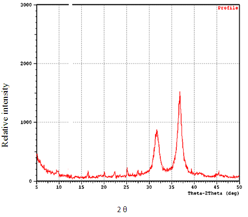
Figure 1. Shows Typical XRD pattern of ZnO nanonanoparticls
A number of Bragg reflections with 2θ values of 31.74°, 36.83° and 47.62° are observed corresponding to (100), (101) and (102) planes, shows a typical XRD pattern of ZnO nanoparticles in the range of 5°-50° at a scanning step of 0.01 (ICPDS card No.89.1397). Almost similar values have also been reported by Yadav 2006 [29].
Average size of the zinc oxide nanoparticles was determined as 58.3 nm from the width of dominate peaks (100) and (101) reflections according to the Debye - Scherrer equation.
All diffraction peaks are indexed according to the hexagonal phase of ZnO. No characteristic peaks of impurity phases except ZnO are found which revealed that good crystalline in nature of the samples.
The broadining of the peaks in the above XRD pattern can be atributed to the small particle size of the synthesized ZnO [30-31].
FT-IR
FT-IR is an effective method to reveal the composition of products. Figure (2) is a typical FTIR spectrum of pure ZnO nanoparticls, the peak at 417.52 cm-1 is the characteristic absorption of Zn-O bond and the broad absorption peak at 3438 cm-1 can be attributed to the characteristic absorption of hydroxyl. These data are similar to the results observed by others [32-37]. Anyhow, the FTIR and XRD results show high purity of the obtained ZnO nanoparticles.
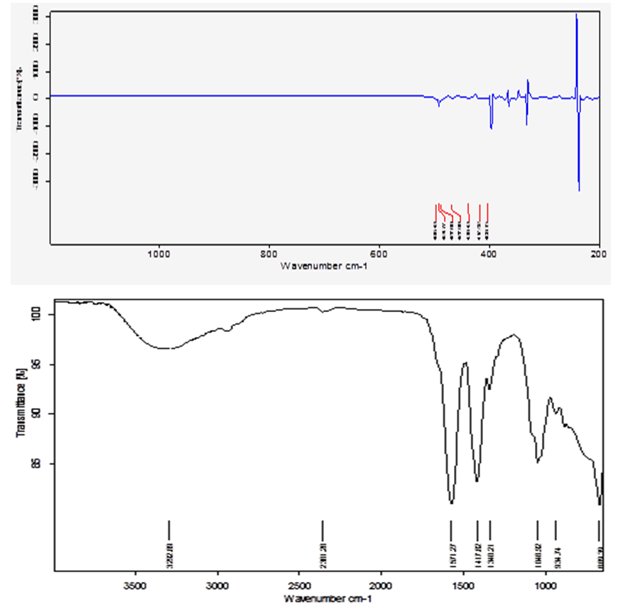
Figure 2. FTIR Transmition spectra of pure ZnO nanoparticls
SEM
Figure 3 shows the SEM image of ZnO nanoparticles. The SEM image was taken at X25,000 magnification. The image shows ZnO particles are spherical in shape with smooth surface and the size of the particles around 100-200 nm.
Figure 3. Shows the SEM image of ZnO nanoparticles
In another experiment, freshly prepared ZnO sol-gel was coatedonto polyethylene thin film and dried at 80℃. The particle size of ZnO nanoparticles prepared via this method was about 50-60 nm (image not shown here). We can clearly conclude that ZnO nanoparticles continue to grow after synthesis, even when stored at room temperature. These results agreed fairly well with those of Radyum Ikono (2012) [3] and Saptashi Ghosh (2014) [38].
ICP
Inductively coupled plasma (ICP –OES) elemental analysis was carried out for the determination the concentration of Zn [39-45]. The yield of ZnO was about %98.2. This result consistent with other published work [23].
UV
Figure 4 shows the UV-Vis optical obsorption spectrum of ZnO nano particles dried in air at 80℃. The absorption spectrum shows a sharp absorbance onset at 345 nm, which indicates an almost uniform size of the nanoparticles.
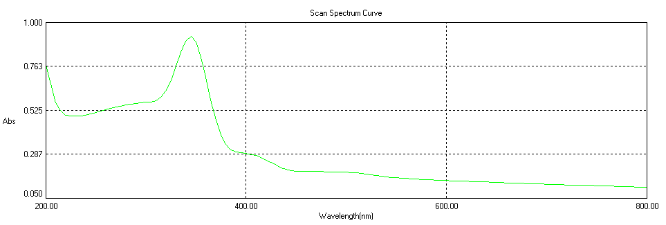
Figure 4. UV-Vis optical absorption spectrum of ZnO nano particles
However, upon change in particle size or particle shape, a slight shift in the absorption was observed. Our result is in agreement with those of other authors [46-50].
The optical band gap of sol samples is estimated from the UV-Vis Diffuse Reflectance Spectroscopic (UV-Vis DRS) studies. Reflection and scattering produce a characteristic reflectance spectrum, providing information about the structure and composition of themedium. The direct band gap of ZnO is estimated from the plot of (αhט)2 versus hט, where hט is the photon energy and α is the ratio of the absorption coefficient to the scattering coefficient, and found to be 2.935 ev. As calculated from figure 5.
Figure 5. DRS plot for finding the band gap of ZnO

5. Conclusions

ZnO nanoparticles have been successfully synthesized by simple Sol-Gel method. The prepared ZnO nanoparticles were spherical in shape and were characterized using XRD, UV-Vis absorption, FT-IR and SEM techniques. The average particle size was found to be 58.3 nm using Scherrer's equation and 100-200 nm obtained from SEM measurement for ZnO nano particles dried at 80℃.
ZnO nanoparticales offer tremendous potential in future applications of electronic and magneto–electric devices. Also, maybe, applied for photocatalysis, gas sensing, biomedical device and sun sucreen applications. The method has a high yield and can be used for large scale synthesis of ZnO nano particles [51].

ACKNOWLEDGEMENTS

This work was gratefull supported by the Iraqi ministry of industry and minerals – Directory of planning in the frame work of grant No. 197 in October 2012.

References

[1]  Klingshirn, 2007, Zno: Material, Physics and Application. Cheme Phy Chem 8: 782-803.
[2]  Chitra, K. and Annadurai G., Antimicrobial activity of wet chemically engineered spherical Zno nano particles on food borne pathogen, International food Research Journal 20 (1): 59-64 (2013).
[3]  Radyum, I. , Putri, R. I., and siswanto, S., Effect of PH variation on particle size and purity of Nano zinc oxide synthesized by sol-gel method ; International Journal of Engineering and Technology, 2012, Vol:12, No. 6 pp 5-9.
[4]  He H., Yang V., and Ye Z. 2011b. Layer-structured Zno nanowire arrays with domina surface and acceptor-related emissions- Materials Letters 65: 1351-1354.
[5]  Emamifar A., Kadivar M., and Zad S.S. 2010. Evaluation of nanocomposite packaging containing Ag and ZnO on shelf life of fresh orange juice innovative-food science and Emerging Technology 11: 742-748.
[6]  Sawai J. (2003), Qualtitative evaluation of Antimicrobial activities of metalic oxide powders (ZnO, MgO and CaO) by concluctimetric assay; J.Microbial methods: vol,54:177-182.
[7]  Jin T., Sun D., Su J. Y and Sue H. J. 2009. Antimicrobial efficacy of Zinc Oxide quantum dots against Listeria monocyto genes, salmonella enteritidis and Escherichia coli, 0157: H7. Journal of food science, Vol. 74 No.1 pp M46-M52.
[8]  Ranvir S. P., Prepartion of modified ZnO nanoparticles by sol -gel process and their characterization, Master of Technology, 2009, Thapar university, Punjab.
[9]  Haung X. H, Guo R. Q. and WuI. B.; Mesoporous ZnO nanosheets for lithium ionbatries, Materials Letters, Volume 122, 2014, pp 82-85.
[10]  Hua Li, Jain C.D. and HuiR.D.; Synthesis and Characterization of chitosan ZnO nanoparticles composite membranes, carbohydrate research, Vol. 345, No. 8,2010, pp. 994-998.
[11]  Boundifa A., Zhang C. and Lahem M.; Highly sensetive and rapid NO2 gas sensors based on ZnO nanostructures and the morphology effect on their sensing performances, the 14th international meeting on chemical sensor (IMCS) 2012.
[12]  Kolekar T.V., Yadav H.M. and Bandgar S.S.; Synthesis by sol- gel (method and characterization of ZnO nanoparticles), Indian streams research Journal, Vol. 1No. 12011, pp. 1-4.
[13]  Khorsand A. Z., Razali R., Majid W. H.; Synthesis and characterization of narrow size distribution of zinc oxide nanoparticles, International journal of nanomedicine, Vol. 6, 2011, pp. 1399-1403.
[14]  Surye p.G.; synthesis and characterization of zinc oxide nanoparticles by sol-gel process, Master of science in physics, National instituete of technology Rourkela, Orissa, India, 1-36(2012).
[15]  Sreetama D. and Bichitra N. G.; Charaterization of ZnO nanoparticles grown in presence of Folic acid template, Journal of nanobiotechnology, 2012, vol. (10), pp. 29-34.
[16]  WuH., and W. J. Amer. Ceram. Soc. 2006, 89(2) 699.
[17]  Qin X. and Wang S. Journal of Applied Polymer science 2006, 102, 1285.
[18]  Liu., J. J., Yu. M. H., and Zhou, W. L. Apply. Phys. lett. 2005, Vol. 87, 172505.
[19]  Yang Y., Li X. and Bao X. 2003. ZnO nanoparticles prepared by thermal decomposition of b-cyclodextrincoated Zinc acetate, Chemical physics letters 373. 22-27.
[20]  Tonto P.;Phatanasri S. and Praserthdam, P. 2008 preparation of ZnO nanorods by solovthermal reaction of zinc acetate in various alchohols. Ceramics International 34: 57-62.
[21]  Wu B.J. and liu S. C.; 2002. low –Temperature Growth of well – Aligned ZnO Nanorods by chemical Vapor Deposition. Advanced materials 14 ;215 -218 .
[22]  Baruha S. and Dutta J.2009 .Hydrothermal growth of ZnO nanostructure. Science and Technology Advanced .Materials 10: 01 3001.
[23]  Abdulaiz Bagatas, Ahmad Alshammari and Hendrik kossliek; Room temperature synthesis of zinc oxide nanoparticles in different media and their application in cyanide photodegredation; Nanoscale research letters (2013), 8:516.
[24]  Bari A.R., shindeM. D. and patil L. A.; Effect of solvents on the particle morphology of nanostructured ZnO; Indian Journal of pure and applied physics, vol:47 (2009), pp24-27.
[25]  Zaborski; Effect of ionic liquids and surfactants on zinc oxide nanoparticle activity in cross linking of acrylonitrite butadiene elastomers, Journal of applied polymer science, Vol.116, issue,pp 155-164 (2010) .
[26]  Yuxin wang, Xinyong Li and Yongying Chen; Controllable synthesis of ZnO nanoflowers and their morphology – dependent photocatalytic activities; Separation and purification technology, Vol.62, Issues 3,2008, pp 727-732.
[27]  Eric A. Meulenkamp; Synthesis and growth of ZnO nanoparticles; J.phys. Chem. B1998, Vol.102,pp. 5566-5572.
[28]  Kumar S. A. and Chen. S.M. 2008. Nanostructured Zinc Oxide particles in chemically modified electrodes for biosensors applications, Analytical Letters 41 (2): 141-158.
[29]  Yadav A., Prasad V, katheA. and Yadav D.; 2006 ; Functional finishing in cotton fabrics using zinc oxide nanoparticles; Bulletin of material science,Vol.29, No.6, pp.641-645 .
[30]  Sangeetha N. and Kumaragura L.; Extracelluler synthesis of zinc oxide nanoparticles using seaweeds of gulf of manar, Indani, Journel of nanobiotechnology, 2013, Vol.11(39) pp.1-11.
[31]  KathirveluS. and Louis D.; UV protection finishing of textiles using ZnO nanoparticles, Indian journal of fiber and textile research Vol. 34, 2009, pp 207 -273 (R2).
[32]  Sher Bahadar Khan, Mohamed Rahman and Khalid Alamry; An assessment of zinc oxide nanosheets asa selective adsorbent for cadmium; nanoscale research letters, 2013,8; 377.
[33]  Khan, S. B. and Rahman Mohamed; Low temperature growth of ZnO nanoparticles, photocatalyst and acetone sensor ; Talanto 2011, 85:943
[34]  Faisal M. khan, Rahman M. Jamal; Role of ZnO–GeO2 nanostructures as a photocatalyst and chemical sensor, J. material science techuology 2011, 27: 594.
[35]  Sunil Bajpai, Navin Chard and Ruchi Lodhi; Water sorption properties and antimicrobial action of zinc oxide nanoparticles loaded sago starch film, Journal of microbiology, Biotechnology and food sciences, 2013, Vol (4) pp 2368 – 2387.
[36]  Zhanhu G., Suying W. and Brian S.; Particle surface engineering effect on the mechanical, optical and photoluminescent properties of ZnO / vinyl – ester resin nanocomposites, journal of material chemistry , 2007, 17,806 -813.
[37]  Thilagavathi Thirugnanam; Effect of polymers (PEG on PVP) on sol –gel synthesis of microsized zinc oxide; Journal of nanomaterials volume 2013 (2013), Article ID 362175, 7pages.
[38]  Saptashi Ghosh, Debline Majumder and Somenath Roy; Facil sonochimical synthesis of zinc oxide nanoflaskes at room temperatur; Materials letter, volume 130, 2014 pages 215-217.
[39]  Aneesh P.M, Vanaja A.V. and Jayarj M. K.; Synthesis of ZnO nano particals by hydrothermal method; Nanophotonic materials IV, edited by Zeno Gaburro, Stefano Cabrini, Proc. of SPIE Vol. 6639 (2007).
[40]  Seyed mohammad Majedi, Hiankeelee. and Barry c. Kelly; Chemometric analytical approach for the cloud point extraction and inductively coupled plasama mass spectrometric determntion of zinc oxide nanoparticals in water sample; analytical Chemistry, 2012, 84(15), pp 6546 – 6552.
[41]  Xiandeng Hou and Bradley jones; Inductively coupled plasma optical emission spectroscopy, in Encyclopeida of analytical chemistry, R.A .Meyers (Ed.), pp 9468-9485, 2000.
[42]  Jake Andrew Farlow; Chronic toxicity of nano metalics on red swamp crayfish in laboratory and mesocosm studies, master of sciense; Louisiana state university 2014.
[43]  Carmen Silivia kira and Vera Akiko Maihara; Determination of major and minor elementsin dairy product through inductively coupled plasma optical emission spectrometry after wet partial digestion and neutron activation analysis; food chemistry 100 (2007) 390-395.
[44]  Mahajan pramod, DhokeS. K and Trafdar J.C. Effect of nano – ZnO on growth of mung bean and chicpea seedlings using plant agar method; Applied biological research (2011), Vol. 13(2) pp 54-61.
[45]  Amy Rosen and Gary M. Hieftje; Inductivity coupled plasma mass spectrometry and electrospray massspectrometry for speciation analysis: applications and instrumentation; Spectrochimical Acta part B 59 (2004) 135-146.
[46]  Guo L., Cheng X. Y., Yan Y. J. and Ge W. K.; Synthesis and optical properties of crystalline polymer-capped ZnO nanorods, Materials science and engineering, C16(2001) 123-127.
[47]  Marcos R.M., Daniel S. V. and Antonio M. N.; Zinc oxide composites prepared by in situ process: UV barrier and luminescence properties, Material letters, 125(2014) 75-77.
[48]  Shingo T., Atsushi N., Takeharu T. and Hiroyuki Wada; Optical properties of ZnO nanoparticles capped with polymers, Materials 2011, 4, 1132-1143.
[49]  Tanujjal B., Karthik K. L., Soumik S. and Joydeep D.; Modulation of defect – mediated energy transfer from ZnO nanoparticles for the photocatalytic degradation of bilirubin, Beilstein Journal of nanotechnology, 2013, 4, 714-725.
[50]  A. O. Fosirail, C. Chigbo, S. L. Mammah and F. L. Ezema; The influence of reaction time on deposited ZnO thin flim by successive ionic layer adsorption and reaction; International research jounal of scientific findings, Vol. 1(4), PP. 127-131 (2014).
[51]  Robina Shahid, Hesham M-A. Soliman and Mamoun Muhmmed; Novel low temperature route for large scale synthesis of ZnO quantum dots; International journal of science, 2012, Vol.1 pp. 153-161.