-
Paper Information
- Next Paper
- Previous Paper
- Paper Submission
-
Journal Information
- About This Journal
- Editorial Board
- Current Issue
- Archive
- Author Guidelines
- Contact Us
Nanoscience and Nanotechnology
p-ISSN: 22163-257X e-ISSN: 2163-2588
2012; 2(4): 125-128
doi: 10.5923/j.nn.20120204.06
Green Synthesis of Silver Nanoparticles byMulberry LeavesExtract
 Abstract
 Reference
 Full-Text PDF
 Full-Text HTML
Akl M. Awwad 1, Nidà M. Salem 2
1Royal Scientific Society, Princess Sumaya University for Technology, El Hassan Science City, Amman, Jordan
2Plant Protection Department, Faculty of Agriculture, University of Jordan, Amman Jordan
Correspondence to: Akl M. Awwad , Royal Scientific Society, Princess Sumaya University for Technology, El Hassan Science City, Amman, Jordan.
| Email: |  |
Copyright © 2012 Scientific & Academic Publishing. All Rights Reserved.
Utilizing the reduced property of mulberry leaves extract and silver nitrate, silver nanoparticles (AgNPs) were synthesized at room temperature. Silver nanoparticles were characterized using UV-visible absorption spectroscopy, scanning electron microscopy (SEM) andX-ray diffraction (XRD).Further, silver nanoparticles showed effective antibacterial activity toward StaphylococcusaureusandShigella sp..
Keywords: SilverNanoparticles,Mulberry Leaves Extract, Characterization, Antibacterial Activity
Article Outline
1.Introduction
- The growing need of environmental friendly nanoparticles, researchers are using green methods for the synthesis of various metal nanoparticles. But nowadays, plant extract has been used as reducing and capping agent for the synthesis of nanoparticles which could be advantageous over photochemical reduction, heat evaporation, electrochemical reduction, and chemical reduction methods. Several biological systems including bacteria, fungi, and yeast have been used in synthesis of nanoparticles. Silver nanoparticles have attracted intensive research interest because of their important applications as antimicrobial, catalytic, textile fabrics and plastics to eliminate microorganisms. Because of such a wide range of applications, numerous methods concerning the fabrication of silver nanoparticles, as well as various silver-based compounds containing ionic silver (Ag+) or metallic silver (Ag0) have been developed. The synthetic methods used for the preparation of silver nanoparticles, some toxic chemical used as a reducing agent such as NaBH4, citrate, or ascorbate is most commonly used. In recent years, plant-leaf extracts synthesis of nanoparticles is gaining importance due to its simplicity and eco-friendliness. Although green synthesis of silver nanoparticles by plant leaves extract such as mangosteen[1], Rosa rugosa[2],Stevia rebaudiana[3], Chenopodium album[4],Macrotylomauniforum[5], Acalypha indica[6],Ficus benghalensis[7], Trianthemadecandra[8], Cycas[9],Catharanthusroseus[10],Piper longum[11], Nicotiana tobaccum[12] , anddifferent leaf plants[13-15]. This study was designed with a simple, cost-effective and environmentally synthesis method of silver nanoparticles (AgNPs) at ambient conditions using mulberry leavesextract as a reducing and stabilizing agent. The AgNPs synthesized in this method has the efficient antimicrobial activity against pathogenic bacteria.
2. Experimental
- Silver nitrate (AgNO3) was obtained from Aldrich Chemicals. All glassware have been washed with sterile distilled water and dried in an oven before use.
2.1. Preparation Mulberry Leaves Extract
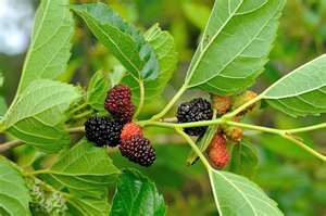 | Figure 1. Picture of Mulberry leaves |
2.2. Synthesis of Silver Nanoparticles
- In a typical reaction procedure, 5 ml of mulberry leaves extract was added to50 mL of 1x10⁻³ M aqueous AgNO3 solution at room temperature, the resulting solution become grey-black in colour after 60 minutes, indicating the formation of AgNPs. The concentrations of AgNO3 solution and mulberry leaves extract were also varied at 1–4 mM and 5–10% by volume, respectively. UV-vis spectra showed strong SPR band at 425 nm and thus indicating the formation of silver nanoparticles The silver nanoparticles (AgNPs) obtained by mulberry leaves extract were centrifuged at 35,000 rpm for 10 min and subsequently dispersed in sterile distilled water to get rid of any uncoordinated biological materials.
2.3. Characterization Techniques
- UV-Vis absorption spectra were measured using Shimadzu UV-1601 spectrophotometer. Crystallinemetallic silver nanoparticles were examined by X-ray diffractometer (Shimadzu XRD-6000) equipped with Cu Κα radiation source using Ni as filter and at a setting of 30 kV/30 mA. All XRD data were collected under the same experimental conditions, in the angular range 3≤ 2θ ≤ 50. FTIR Spectra for mulberryleaves extract powderwasobtained in the range 4000–400 cm−1 with IR-Prestige-21 Shimaduz FTIR spectrophotometer, using KBr pellet method. Scanning electron microscopy (SEM) analysis of silver nanoparticles analysis was done using Hitachi S-4500 SEM machine. Thin films of the silver nanoparticles were prepared on a carbon coated copper grid by just dropping a very small amount of the sample on the grid, extra solution was removed using a blotting paper and then the film on the SEM grid were allowed to dry by putting it under a mercury lamp for 5 minutes.
3. Results and Discussion
3.1. FT-IR Spectrum
- To investigate the functional groups of mulberry leaves extract, a FT-IR study was carried out and the spectra are shown in Figure 2. Themulberryleaves extract displays a number of absorption peaks, reflecting its complex nature. A peak at 3313cm−1 results due to the stretching of the N–H bond of amino groups and indicative of bonded hydroxyl (-OH) group. The strong absorption peak at 2913cm−1 could be assigned to –CH stretching vibrations of –CH3 and –CH2 functional groups. The shoulder peak at 1701 cm-1 assigned for C=O group of carboxylic acids. The peak at 1635cm−1 indicates the fingerprint region of CO, C–O and O–H groups, which exists as functional groups of oliveleaves extract.. The absorption peaks at 1361 cm−1 could be attributed to the presence ofC–O stretching in carboxyl. The intense band at 1018 cm-1 can be assigned to the C-N stretching vibrations of aliphatic amines.FTIR study indicates that the carboxyl (-C=O), hydroxyl (-OH) and amine (N-H) groups of mulberry leaves extract are mainly involved in reduction of Ag+ to Ag nanoparticles.
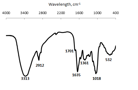 | Figure 2. FT-IR of Mulberry leaves powder |
3.2. Visual Observation AndUV-Vis Spectral Study
- Formation and stability of AgNPs in sterile distilled water is confirmed using UV-vis spectrophotometer in a range of wavelength from 200 to 800 nm.As soon as, mulberry leavesextract was mixed in aqueous solution of silver ion complex, the reduction of pure Ag+ ions to Ago was monitored by measuring UV–vis spectrum of the reaction media at regular intervals. UV–vis spectra were recorded as function of reaction time, Fig. 3 We observe that there is no peak showing no sign for the synthesis of silver nanoparticles but after 30min the surface plasmon resonance of silver occur at 425nm and steadily increasing with the time of reaction without much change in the peak wavelengthFig.3. After 60 min, the increase in the number and size of the AgNPs came to an end, may be due to the depletion of the silver ions (Ag+)in the mulberryleaves extract.
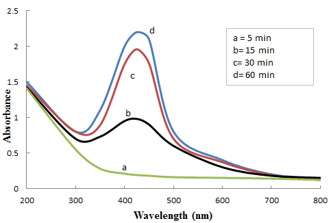 | Figure 3. Effect of contact time on AgNPs synthesis by mulberry leaves extract |
3.3. X-ray diffraction (XRD) Studies
- Analysis through X-ray diffraction was carried out to confirm the crystalline nature of the particles, and the XRD pattern showed numbers of Braggs reflections that may be indexed on the basis of the face cantered cubic structure of silver. A comparison of our XRD spectrum with the standard confirmed that the silver particles formed in our experiments were in the form of nanocrystals, as evidenced by the peaks at 2θ values of 38.02 θ, 43.58, and 64.32, and 77.22 corresponding to (111), (200), (220) and (311), respectively Bragg reflections of silver. The X-ray diffraction results clearly show that the silver nanoparticles formed by the reduction of Ag+ ions by the mulberry leaves extract are crystalline in nature. As mentioned in the method section, the silver nanoparticles once formed were repeatedly centrifuged and redispersed in sterile distilled water prior to XRD analysis, thus ruling out the presence of any free biological material that might independently crystallize and giving rise to Bragg reflections. It was found that the average size from XRD data and using Debye-Scherer equation was 20 ± 2.8 nm. The presence of structural peaks in XRD patterns and average crystalline size around 20 nm clearly illustrates that AgNPs synthesized by our green method were nanocrystalline in nature. FT-IR analysis of mulberry leaves extract was carried out to identify possible presence of functional groups that might have contributed to the process of bio-reduction of silver ions (Ag+) to silver nanoparticles (AgNPs).The XRD pattern of the dried AgNPs obtained by mulberry leaves extract is shown in Fig. 4. A number of Bragg reflections with 2θ values of 38.02o, 43.58o, 64.32o, 77.22o correspond to the (111), (200), (220), and (311) sets of planes are observed which may be indexed as the band for face center cubic structures of silver. The XRD patterns thus clearly illustrates that the AgNPs synthesize by the present green method are crystalline in nature. The average particle size of silver nanoparticles synthesized by the present green method can be calculated using Debye-Scherrer equation [16-17].D = Kλ / β cos θWhere D = the crystallite size of AgNPs particles λ = the wavelength of x-ray source (0.1541 nm) used in XRDβ = the full width at half maximum of the diffraction peak.K = the Scherrer constant with value from 0.9 to 1.θ = the Bragg angle.
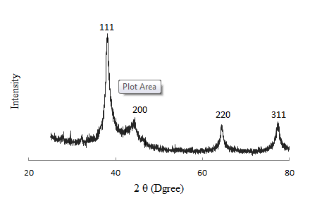 | Figure 4. XRD pattern of AgNPS synthesized by mulberry leaves extract |
3.4. SEM Analysis of Silver Nanoparticles (AgNPs)
- The suspended silver nanoparticles in sterile distilled water were used for scan electron microscope analysis by fabricating a drop of suspension onto a clean electric stubs and allowing water to completely evaporate. The SEM image of silver nanoparticles, Fig. 5 showed cubical and relatively uniform shape of nanoparticle formation with diameter range 20-40 nm. The larger silver particles may be due to the aggregation of the smaller ones, due to the SEM measurements.
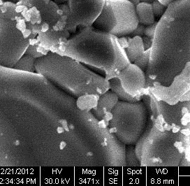 | Figure 5. SEM of silver nanoparticles |
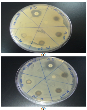 | Figure 6. Activity of silver nanoparticlesagainst (a)Staphylococcus aureus and (b)Shigella sp |
3.5. Antibacterial Activity Study of Silver Nanoparticles (AgNPs)
- From the preliminary screening by disc diffusion method, it was observed that silver nanoparticles have antibacterial activities at concentration of 2µg/disc. This was observed on Staphylococcus aureus and Shigella sp. bacteria. The zone of inhibition ranged from 9 to 10 mm, Fig. 6.
4. Conclusions
- Green chemistry approach towards the synthesis of nanoparticles has many advantages such as, ease with which the process can be scaled up and economic viability. We have developed a fast, eco-friendly and convenient method for the synthesis of silver nanoparticles using mulberry leaves extract with a diameter range of 20nm. These particles are monodispersed and spherical. No chemical reagent or surfactant template was required in this method, which consequently enables the bioprocess with the advantage of being environmental friendly. Color change occurs due to surface plasmon resonance during the reaction with the ingredients present in the plant leaves extract results in the formation of silver nanoparticles which is confirmed by UV–vis, XRD and SEM, having average mean size of 20nm had fcc structure. The antibacterial activity of biologically synthesized silver nanoparticles was evaluated againstStaphylococcus aureus and Shigella sp.showed effective bactericidal activity.
ACKNOWLEDGEMEBNTS
- Authors are thankful to Royal Scientific Society, USAID/SABEQ program and Jordan University for financial support and having given feasibilities to carry out the research work.