-
Paper Information
- Next Paper
- Previous Paper
- Paper Submission
-
Journal Information
- About This Journal
- Editorial Board
- Current Issue
- Archive
- Author Guidelines
- Contact Us
Nanoscience and Nanotechnology
p-ISSN: 22163-257X e-ISSN: 2163-2588
2012; 2(4): 99-103
doi: 10.5923/j.nn.20120204.02
Synthesis of Silver Nanoparticles in DNA Template and Its Influence on Nonlinear Optical Properties
 Abstract
 Reference
 Full-Text PDF
 Full-Text HTML
B. Nithyaja 1, 2, H. Misha 1, V. P. N. Nampoori 1
1International School of Photonics, Cochin University of Science and Technology, cochin, India
2Sree Narayana College for Women Kollam, Kerala, India
Correspondence to: B. Nithyaja , International School of Photonics, Cochin University of Science and Technology, cochin, India.
| Email: |  |
Copyright © 2012 Scientific & Academic Publishing. All Rights Reserved.
We have synthesized highly stable silver nanoparticles in aqueous solution at room temperature by standard reduction method using de-oxyribo nucleic acid (DNA) as a stabilizing agent. The nonlinear optical properties of above nanoparticles were investigated using Z-scan technique at 532nm.The obtained nonlinear absorption and refraction coefficient is found to be large. The imaginary part of third order susceptibility depends on the concentration of DNA at low pump power of 50MW/ cm2.It is observed that at high pump power of175MW/ cm2, the imaginary part of third order susceptibility does not depend on the concentration of the DNA. The imaginary parts of third order nonlinear optical susceptibility measured by Z-scan technique revealed that silver nanoparticles in DNA template are potential candidate for optoelctronic device applications.The silver nanoparticles in DNA template show good optical limiting property.
Keywords: Silver Nanoparticles, De-oxy Ribonucleic Acid, Z-scan, Nonlinear Absorption
Article Outline
1. Introduction
- Synthesis of nanoparticle is an emerging field due to its potential application in catalysis, optical,magnetic and electronic devices[1-4]. Among biological molecules de-oxyribo nucleic acid (DNA) has been used extensively as a biotemplate to grow inorganic quantum confined structure and to organize non biological building blocks into extended hybrid materials because of its physicochemical stability, well defined sequence of DNA base and a variety of super helix structures[5-8].DNA template has also been described as a smart glue for assembling nano particles. In general, synthesis of nano particles based on DNA template has been done by incubating metal ion coated DNA in a reduction agent solution. The ion DNA complexes controlled the release of metal ions, slowed down their reduction and effectively inhibited metal ions from growing into big clusters. Nano-structures of metals such as silver, gold, palladium, platinum, copper, and nickel have been successfully synthesized by using DNA network templates[5-9].In recent years, a number of Ag nanoparticles with different shapes have been widely synthesized by using DNA as the template because of its self-assembly and mechanical properties, as well as the high affinity between DNA bases with Ag+ cations. Silver dendrites have been synthesised by an electro chemical technique in an aqueous solution of AgNO3 in the presence of DNA[9]. Lanlan Sun et.aldescribed the preparation of silver nanoparticles ring on DNA template. The silver nanoparticles ring which was about 1.5 μm in length, and about 2.2 nm in height can be obtained by adjusting the reaction time[10 ]. A large-scale
 -DNA network on a mica surface was successfully fabricated with a simple method. Silver nanoparticles capped with the cationic surfactant cetyl trimethyl ammonium bromide (CTAB) were self-assembled on to a two-dimensional DNA network template by electrostatic interaction and formed nano porous silver films, which can be used as active surface-enhanced raman scattering (SERS) substrates[8]. DNA was found to be an excellent template for the growth of metallic nanoparticles. Since nanoparticles are capped with DNA, they are environmentally friendly, non-toxic and renewable also.Nonlinear optical and electronic properties of nano sized metal particles have drawn special attention because of their strong and size-dependent plasmon resonance absorption and their applications as nonlinear materials for optical switching, optical limiting,beam flattening and computing because of their relatively large third-order nonlinearity and ultra fast response time. Silver nanoparticles have received increased attention in the past few years due to its large third order nonlinearity. The nonlinear optical properties of the metal nanoparicles depend on the host material which support them. A large number of investigations have been carried out to study nonlinear optical properties of metal nano particles dispersed in different optically transparent solid matrices and liquids[11-16]. However, seldom research has been done on the nonlinear optical property of the metal nanoparticles in DNA template. An advantage of colloidal suspensions compared with fabrication of structures in solid matrix is that the nanoparticles in suspension are easily reconfigurable. The nanoparticle environment in colloidal system is composed of the solvent and stabilizing agents adsorbed on the nanoparticles surface to avoid aggregation. The solvent and the stabilizing agent may change the optical properties of the nanoparticles in different ways[15, 16].In this paper we report the results obtained from investigation of nonlinear optical properties of silver nanoparticles prepared in aqueous solution of DNA.
2. Experimental
- Silver nano particle is synthesized in aqueous solution of DNA (derived from Herring sperm) by standard reduction technique. A 20mL solution with a concentration of 0.25mM AgNO3 and DNA with different concentration (0.05wt%, 0.1wt%, 0.15wt %,) in water is prepared. While stirring vigorously 0.6ml of 10mM NaBH4 was added at once. Stirring was stopped after 30 s. The silver nanoparticles are characterized by UV-VIS absorption spectroscopy and scanning electron microscopy method. A mode-locked Nd:YAG laser having 10 ns pulses at a repetition rate of 10 Hz giving second harmonic at 532 nm was used in the z-scan experiment to study the optical nonlinearity. The sample is moved along the beam axis of light focussed with a lens of focal length 200 mm. The radius of the beam waist w0 is calculated to be 42.56μm. The Rayleigh length
 /
/ , is estimated to be 10.6mm, which is much greater than the thickness of the sample cuvette (1mm), which is an essential prerequisite for z-scan experiments. The transmitted beam energy, reference beam energy, and their ratio are measured simultaneously by an energy ratio meter having two identical pyroelectric detector heads. The effect of fluctuations of laser power is eliminated by dividing the transmitted power by the power obtained at the reference detector. The data are analyzed by using the procedure described by Bahae et al.[22] and the nonlinear coefficients are obtained by fitting the experimental z-scan plot with the theoretical plots.
3. Results and Discussions
- a. Characterization of silver nano particles:Silver nanosol in DNA template is synthesized as described above. The addition of DNA results stable solutions of silver nano particles at room temperature and found to be stable for several months. Two major challenges often faced in the preparation of nano crystals in solution are Oswald ripening and agglomeration. Use of capping agents prevent these by effectively shielding the surface of the nuclei as soon as they are formed and thus preventing them from coming into direct contact with solution. Often molecules with longer chains are used as capping agents[12].DNA acts as a good stabilizing agent capping agents as well as template for nano particles using the double helix nature of DNA. Per helix turn of the DNA can produce one major groove and one minor groove. The diameter of the double helix is 2.0 nm. Based on this structural mode, nanoparticles with size of 2-3nm will go into the major groove of the DNA where the space is big enough to accommodate them. Average particle size of the prepared nanoparticle is found to be around 60nm. In our case the nanoparticles may go into the space among the DNA chain network in solution[9]. Due to the long polymeric chain of the DNA molecule there may be the formation of mesh like net work in solution. The nucleation may be occurs first starting from the Ag+ bound on the DNA base or the phosphate groups, and then the silver ions are slowly reduced to the metallic silver on the nucleation site. The growth of the silver nanoparticles are finally blocked by limited space in network[13].Before carrying out the characterization of the optical nonlinearities, the linear absorption spectra of silver nano sol in different concentrations of DNA template was obtained. Optical absorption measurement is an initial step to ob-serve the nano sol behavior. Absorption spectra of silver nanosol in three concentrations of DNA (0.05wt%, 0.1wt%, 0.15wt% respectively) are shown in fig.1.It is clear from the figure that, the Ag plasmon resonance lies around 415nm and absorption is less for higher concentration of DNA. This indicates silver nano particle is formed in all concentrations of DNA even though the number of silver nano-particles formed in higher concentration is less. The SPR peak of silver nanoparticles mentioned in many other previous report is around 420nm[4,5] which is consistent with our observation. Figure(2) shows SEM picture of the silver nanoparticles formed on DNA template.
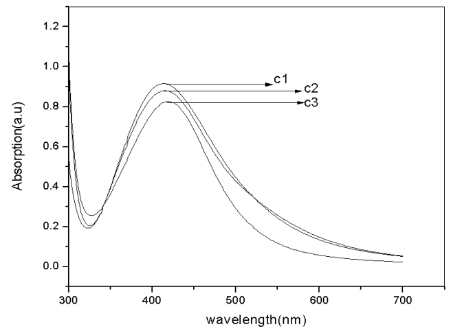 | Figure 1. Absorption spectra of silver nanoparticles in aqueous solution three concentrations of DNA. (Where C1, C2, C3 represents .05wt%, 0.1wt%, 0.15wt% of DNA respectively) |
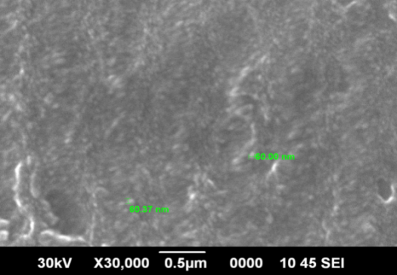 | Figure 2. SEM image of of silver nanoparticles in aqueous solution of DNA (0.05wt%) |
 is given by the equation
 | (1) |
 is the low intensity absorption coefficient and
  is nonlinear absorption coefficient.The normalized transmittance for open aperture Z-scan is given by the equation
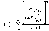 | (2) |
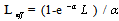 , z is the longitudinal displacement of the sample from focus (Z=0), I0 is the peak intensity at focus, Leff is the effective interaction length, L is the sample length0 is the Rayleigh length.Imaginary part of the
  is given by the equation
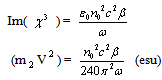 | (3) |
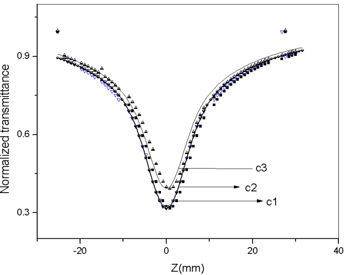
 ) at an intensity of 50 MW/cm2 are shown in Table 1. In this fluence these values are found to be comparable with those synthesized through other procedures[13-17]. Also we have observed that as concentration of DNA increases the nonlinear absorption is found to be decreasing. This is due to the effect of DNA on silver nano sol. As concentration of DNA increases the number of silver nanoparticles formed is decreased (absorption spectra). In effect the non linear optical property of the silver nanosol is decreased. Nonlinear optical properties of nanoparticles are strongly influenced by the surface plasmon resonance (SPR) absorption. It is observed that a laser pulse can cause interand or intraband electron transition in the metal nanoparticle system depending on the excitation wavelength and intensity. The excited electrons are free carriers and leads to transient absorption. This also leads to strong nonlinear absorption. Nonlinear scattering of nanoparticle also contribute to nonlinear absorption. From table 1.and absorption spectra, it is clear that the nonlinear absorption decreases by approximately 30% when going from C1 to C3 while the height of absorption the plasmon resonance changes by 10% only. This discrepancy is may be due to contribution of nonlinear scattering. As concentration of DNA changes number of silver nanoparticle in solution is decreased so that nonlinear scattering is also decreased. Here enhanced absorption of the silver nanoparticles may be due to the interaction between the nanoparticles with DNA matrix which support them. The surface plasmon resonance associated with collective oscillations of electrons in the nanoparticles is strongly influenced by the host dielectric function. Optical response of the nanoparticles may vary when dielectric function of the surface layer of the nanoparticles changes with stabilizing agent[15]. Here DNA acts as a good stabilizing agent for the nanoparticles.
 | Figure 3. Open aperture Z-scan curves of silver nanoparticles in aqueous solution with different concentration of DNA at a typical fluence of 50 MW/cm2. (where C1, C2 ,C3 represents 0.05wt%,0.1wt%,0.15wt% of DNA respectively) |
 ] for silver nanoparticles in aqueous solution with different concentration of DNA at a typical fluence of 50 MW/cm2
 and Im
  at an intensity of 175 MW/cm2 are shown in Table 2.At high pump power the value of the
 is found to be decreased compared to low pump power even though it shows high optical nonlinearity. The increase in the laser intensity induces bleaching in the ground state absorption, which results in a transmittance increase (SA process). This may be the reason for the decreased non linearity at high pump power. It is clear from the figure4 and table 2 that at high pump power the nonlinearty of the prepared samples do not depend on the concentration of the DNA. This is due to the effect of DNA on nonlinear optical properties of silver nano particles. At high pump power DNA shows third order optical nonlinearity[17]. So at high pump power as concentration of the DNA increases, the no:of silver nanoparticle formed is less, at the same time nonlinearity of the DNA increases with concentration. These two effects compensate each other. In effect at high pump power the concentration of DNA does not effect the nonlinearity of silver nano sol.
|
 ] for silver nanoparticles in aqueous solution with different concentration of DNA at a typical fluence of 175MW/cm2
 | Figure 4. Open aperture Z-scan curves of siver nanoparticles in aqueous solution with different concentration of DNA at a typical fluence of 175 MW/cm2. (where C1, C2 ,C3 represents 0.05wt%,0.1wt%,0.15wt% of DNA respectively) |
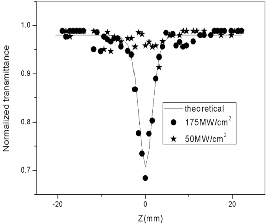 | Figure 5. Open aperture Z-scan curves of aqueous solution of DNA(0.05wt%) at a typical fluence of 50 MW/cm2and 175 MW/cm2 |
 =12cm/GW). Marek Samoc et al. reported DNA shows moderate nonlinear absoption at 532nm[17].Nonlinear optical property of the DNA depends on the polymerisation of DNA strand, molecular weight, host material etc. To examine the optical limiting property of the silver nanopartcles in DNA matrix the nonlinear transmission of the silver nano sol is studied as a function of input fluence (Figure .6).The fluence level for optical limiting at 532nm for different concentration of DNA is found to be 88 MW/cm2. An important term in the optical limiting measurement is the limiting threshold. It is obvious that the lower the optical limiting threshold, the better the optical limiting material. Optical limiters are devices that transmit light at low input fluences or intensities, but become opaque at high inputs. The optical limiting property occurs mostly due to mechanisms like excited state absorption, two-photon absorption and nonlinear scattering as well.[19].
 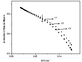 | Figure 6. Optical limiting performance of siver nanoparticles in aqueous solution with different concentration of DNA. (Where C1, C2, C3 represents0.05wt%,0.1wt%,0.15wt% of DNA respectively) |
4. Conclusions
- In conclusion, we have synthesized highly stable silver nanoparticles in aqueous solution at room temperature by standard reduction method using DNA. The linear and nonlinear optical properties of silver nanoparticles at three concentration (.05wt%, .1wt%, .15wt %) of DNA is studied. Absorption spectra shows the Ag plasmon resonance lies around 410nm and silver nano-particles formed in higher concentration is less. The nonlinear absorption coefficient
 and imaginary part of third order susceptibility depends on the concentration of DNA at low pump power of 50MW/ cm2.It is observed that at high pump power of 175MW/ cm2 the nonlinear absorption coefficient(Beff) and imaginary part of third order susceptibility does not depend on the concentration of the DNA. The imaginary parts of third order nonlinear optical susceptibility measured by Z-scan technique revealed that silver nano particle synthesized in aqueous solution of DNA have good nonlinear optical response and could be chosen as ideal candidate with potential applications for nonlinear optics. The optical nonlinearity and limiting properties of these Ag nanoparticles are comparable or superior to those synthesized through other procedures. The fluence level for optical limiting at 532nm for different concentration of DNA is found to be 88 MW/cm2. The nonlinear absorption of silver nanoparticles at high intensity were attributed to the influence of DNA on nanoparticles.
ACKNOWLEDGEMENTS
- The authors B.Nithyaja is thankful to UGC (Govt of India) for their financial support. H.Misha is thankful to Kerala state council for Science Technology and Environment (KSCSTE) for financial support.
References
| [1] | S.Forster,M.Antonietti, ,“Amphiphilic Block Copolymers in Structure-Controlled Nanomaterial Hybrids” Adv,Mater.10(1998)195-217 |
| [2] | M.Moffit,A.Eisenberg , “Size Control of Nanoparticles in Semiconductor-Polymer Composites.1.Control via Multiplet Aggregation Numbers in Styrene-Based Random Ionomers” Chem.Mater.7 (1995) 1178-1184 |
| [3] | R.P.Andres,J.D.Bielefeld.J.I.Henderson,D.B.Janes,R.Kolagunta,C.P.Kubuiak,W.J.Mahoney,R.J.Osifchin, “Self-Assembly of a Two- Dimensional Superlattice of Molecularly Linked Metal Cluster” Science 273(1996)1690-1693 |
| [4] | C.Petit,P.Lixon,M.P.Pileni, “In Situ synthesis of silver nanocluster in AOT reverse micelles”,J.Phys.Chem.97(1993)12974-12983 |
| [5] | Braun E, Eichen Y,Sivan U, Ben-Yoseph,G , “DNA-templated assembly and electrode attachment of a conducting silver wire” Nature 1998,391,775 |
| [6] | Zhiguo Liu, Yuangang Zu , Yujie Fu, Yuliang Zhang, Huili Liang“Growth of the oxidized nickel nanoparticles on a DNA template in aqueous solution” Materials Letters 62 (2008) 2315–2317 |
| [7] | Yong Yao, Yonghai Song and Li Wang, “Synthesis of CdS nanoparticles based on DNA network templates”Nanotechnology 19 (2008) 405601 (8pp) |
| [8] | Gang Wei,Li Wang,Zhiguo Liu,Yonghai Song,Lanlan Sun,Tao,Yang,and Zhuang Liz , “DNA-Network-Templated Self Assembly of Silver Nanoparticles and their application in Surface enhanced Raman scattering” J Phys Chem. B. (2005)109(50)23941-7. |
| [9] | Zhu,Xuehong Liao,Hong-Yuan Chen Junjie, “Electrochemical preparation of silver dendrited in the presence of DNA” Material Research Bulletin 36 (2001) 1687-1692. |
| [10] | Lanlan Sun, Gang Wei, Yonghai Song, Zhiguo Liu, Li Wang and Zhuang Li “Fabrication of silver nanoparticles ring templated by plasmid DNA” Applied Surface ScienceVolume 252, Issue 14, 15 May (2006) 4969-4974 |
| [11] | R.A.Ganeev,M.Baba,A.I.Ryasnyanskii,M.Suzuki,andH.Kuroda, , “Laser Ablation of Silver in Different Liquids: Optical and Nonlinear Optical Properties of Silver Nanoparticles”,Opt.Spectosc.99,(694(2005) |
| [12] | C.Wang,Y.Fu,Z.Zhou,Y.Cheng,and Z.Xu, “Femtosecond filamentation and supercontinuum generation in silver-nanoparticle-doped water” Appl Phys. Lett. 90,181119(2007) |
| [13] | D.Rativa,R.E.de Araujo,and A.S.L.Gomes, “One photon nonresonant high-order nonlinear optical properties of silver nanoparticles in aqueous solution Opt.Express 16,19244(2008) |
| [14] | R.A.Ganeev,A.I.Ryasnyansky, “Nonlinear optical characteristics of nanoparticles in suspensions and solid matrices,” Appl.Phys.B 84,295(2006) |
| [15] | Luis A.Gomez,Cid B.de Araujo,A.M.Brito Silva, and Andre Galembeck“Influance of stabilizing agents on the nonlinear susceptibility of silver nanoparticles” J.Opt.Soc.Am.B/Vol.24,No.9/2007 |
| [16] | L. A. Gomez, C. B de Araujo, A. M. Brito-Silva, A.Galembeck; “Solvent effect on the linear and nonlinear optical response of Silver nanoparticles”, Appl. Phys. B 92,61(2008) |
| [17] | Marek Samoc,Anna Samoc James G Grote, “Complex nonlinear refractive index of DNA” Chemical Physics Letters 431(2006) 132-134 |
| [18] | M. Sheik Bahae, A. A. Said, T.Wei, D. J. Hagan, E. W. Van Stryland ; “Sensitive measurements optical nonlinearities using a single beam”, IEEE,26 760 (1990) |
| [19] | Zhengle Mao, Lingling Qiao, Fei He, Yang Liao, Chen Wang, Ya Cheng; “Thermal-induced nonlinear optical characteristics of ethanol solution doped with silver nanoparticles”, Chinese Optics Letters, 7, 949 (2009) |