Research in Neuroscience

p-ISSN: 2326-1226    e-ISSN: 2326-1234

2014;  3(1): 22-28

doi:10.5923/j.neuroscience.20140301.03

Protective Role of Either Ginger or Lipoic Acid in Senile Female Rats

Nadia M. S. Arafa1, Elham H. A. Ali2

1Faculty of Science, Biology Department, Jazan University, KSA & National Organization for Drug Control and Research, Department of Physiology

2Zoology Department, Faculty of women for Arts, Science and Education, Ain Shams University, Cairo, Egypt

Correspondence to: Nadia M. S. Arafa, Faculty of Science, Biology Department, Jazan University, KSA & National Organization for Drug Control and Research, Department of Physiology.

Email:

Copyright © 2014 Scientific & Academic Publishing. All Rights Reserved.

Abstract

Aging associated with neurodegenerative disorders. The dysregulation of brain lipid metabolism might contribute to the mechanisms of aging and Alzheimer's disease. The study aimed to evaluate the change in free fatty acids and free calcium ion contents, as well as the total antioxidant activity in senile female rat's brain. Moreover, the possible protective effect of either ginger or alpha lipoic acid. The experimental groups (n=6/group) administered orally for four weeks and classified into: 1- Control adult female rats group and 2- Senile female group (both groups) received one ml/100 g b.wt. 0.5 g/100 ml CMC. 3- Senile ginger group received 250 mg/kg ginger. 4- Senile lipoic treated group administered 65 mg/kg lipoic acid. The results suggested protective effect of ginger and alpha lipoic acid in senile female rats through modulation of, saturated, unsaturated fatty acids and the total antioxidant capacity in cortex and hippocampus. In addition, the cortical free calcium ions content that could protect the senile rats from ageing diseases.

Keywords: Ginger, Alpha Lipoic Acid, Cortex, Hippocampus, Free Fatty Acids, Total Antioxidant Capacity

Cite this paper: Nadia M. S. Arafa, Elham H. A. Ali, Protective Role of Either Ginger or Lipoic Acid in Senile Female Rats, Research in Neuroscience , Vol. 3 No. 1, 2014, pp. 22-28. doi: 10.5923/j.neuroscience.20140301.03.

1. Introduction

The central nervous system is particularly vulnerable to oxidative injury. It contains high concentrations of readily oxidizable poly-unsaturated fatty acids and has a high rate of oxygen consumption per unit volume. Also possesses relatively low levels of antioxidant defense system (Koenig and Meyerhoff, 2003; Freitas, 2009).In the central nervous system, lipids are one of the main targets of reactive oxygen species, because their cell membranes are rich in polyunsaturated fatty acids (Cini and Moretti, 1995). Studies suggesting that dysregulation of brain lipid metabolism might contribute to the mechanisms of aging and Alzheimer's disease (AD); lipid metabolism has not evaluated extensively in aging brain (Snigdha et al. 2012). Epidemiological studies implicate that the high-fat diet confers a significant risk for development of AD and the degree of saturation of fatty acids is critical in determining the risk for AD (Mattson et al., 2002). The saturated free fatty acids (FFAs), palmitic and stearic acids, caused increased amyloidogenesis and tau hyperphosphorylation in primary rat cortical neurons. These FFA-induced effects observed in neurons mediated by astroglial FFA metabolism (Patil et al., 2007). Fatty acid is required for both the structure and function of every cell in the body and forms an important component of cell membranes. It undergoes changes during the process of injury, repair and cell growth (Cameron and Cotter 1997; Murugan and Pari, 2007). These FFA-induced effects observed in neurons mediated by astroglial FFA metabolism (Patil et al., 2007). Fatty acid is required for both the structure and function of every cell in the body and forms an important component of cell membranes. It undergoes changes during the process of injury, repair and cell growth (Cameron and Cotter 1997; Murugan and Pari, 2007).
Deficiencies in polyunsaturated essential fatty acids (PUFA) implicated in mood disorders (Sublette et al., 2009). Linoleic acid, eicosapentaenoic acid (EPA, 20:5) and docosahexaenoic acid (DHA, 22:6) are especially important during human brain development. Maternal deficiency of omega-3-fatty acids related to deficits in neurogenesis, neurotransmitter metabolism, and altered learning and visual function in animals (Shahidi et al., 2005; Innis, 2008). Dietary omega-3-fatty acids are certainly involved in the prevention of some neuropsychiatric disorders, particularly depression, as well as in dementia, notably Alzheimer’s disease (Calon and Cole, 2007). Alpha linoleic acid and DHA is essential for nervous tissue growth and function (Bourre, 2004).
Ginger (Zingiber officinale) used in the Indian traditional system of medicine for digestive disorders, common cold, and rheumatism (Sharma and Singh, 2012). The nonvolatile pungent compounds namely gingerols, shogaols, paradols, and zingerone) are some of the extensively studied phytochemicals. They account for the antioxidant, anti-inflammatory, antiemetic, and gastroprotective activities (Kubra and Jaganmohanrao, 2012).The nonvolatile pungent compounds (namely gingerols, shogaols, paradols, and zingerone) are some of the extensively studied phytochemicals and account for the antioxidant, anti-inflammatory, antiemetic, and gastro protective activities (Kubra and Jaganmohanrao, 2012).
Alpha lipoic acid (ALA, DL-6, 8-dithiooctanoic acid) is a naturally occurring compound synthesized in small quantities by most plants and animals (Perera et al., 2011). Considered as endogenous thiol antioxidant that quench ROS, regenerate glutathione (GSH). It chelate metals such as iron, copper, mercury, and cadmium, which known to mediate free-radical damage in biological systems (Akpinar et al., 2007; Applegate et al., 2008).
The study aimed to declare the possible protective effect of ginger or alpha lipoic acid on senile female rats as antioxidants through studying the change in metabolic brain free fatty acids in cortex and hippocampus.

2. Material and Methods

Twenty-four female rats, 18 senile (30 months old) and 6 adults (6 months) obtained from the National Organization for Drug Control and Research (NODCAR). Animals housed plastic cages each cage contained six rats. Animals were maintained under controlled temperature of 25±2℃ and 12 hours light/12 hours dark cycle throughout the experiment. The animals allowed to food, as commercial pelleted diet and water were available ad libitum.
Chemicals: Ginger purchased from Arab Co. For Pharmaceuticals & Medicinal Plants MEPACO- EGYPT as tablet contains treated ginger (Zingiber officinale) 400mg. Lipoicacid supplied as tablets containing 300 mg manufactured by EVA Company, Egypt.Saturated fatty acids (SFAs), lauric (C12:0), myristic (C14:0), palmitic (C16:0), stearic (C18:0) and arachidic (C20:0). Unsaturated fatty acids (USFAs), oleic (C18:1ω–9), linoleic (C18: 2ω–6),), cis-5.8.11.14.17-eicosapentaenoic (C20:5ω–3) (EPA) and cis-4.7.10.13.16.19-docosahexaenoic acid (DHA) (22:6ω-3). All FAs purchased from Sigma-Aldrich Co.(St. Louis, USA). All other chemicals and solvents used were of the High Performance Liquid Chromatography and analytical grade.
Animal Grouping: The animals divided into four main groups; each one contained six rats as follows: 1- control normal adult female rats (C) received 1 ml/100 g.b.wt. Carboxymethylcellulose sodium salt (CMC) orally. 2-Control senile female rats (S) received CMC as group 1. 3-Senile ginger group (S+G) received 250 mg/kg in CMC. 4-Senile lipoic acid group (S+L) received 65 mg/kg lipoic acid in CMC. Treatments extended for four weeks with daily oral administration. Doses calculated as equivalent to the human recommended dose according to Reagan-Shaw et al. (2008).
Biochemical Assay: Following the completion of the experiments, the rats sacrificed after 12 hours from the last dose by rapid decapitation. Brain cortex and hippocampus excised for the determination of free fatty acids as methyl estersby GC according to the method previously described in Firląg et al., (2013). Using column HP-5 (Methyl siloxane). Cortex also used for the determination of Ca+ contents according to the method in Arafa (2010) and the total antioxidant capacity according to the method of Koracevic et al. (2001).
Statistical Analysis: Reported values represent means ± SE. Statistical analysis evaluated by one-way ANOVA. Once a significant F test obtained, LSD comparisons performed to assess the significance of differences among various treatment groups. Statistical Processor System Support "SPSS" for Windows software, Release 21.0 (SPSS, Chicago, IL) was used.

3. Results and Discussion

The data depicted for senile female group showed in cortex (Figure 1 (A-I)) significant decrease in SFAs (palmitic, stearic and arachidic) and USFAs (oleic, linoleic, EPA and DHA) and a significant increase in myristic acid. While showed, significant decrease in SFAs (lauric, stearic and arachidic) and USFAs (oleic, linoleic, EPA and DHA), but significant increase in SFAs, myristic and palmitic in hippocampus (Figure 2 (A-I)) as compared to the adult control values. The data in Figure 3 (A-D) demonstrated the significant decrease in cortex and hippocampus total saturated (SFAs) and unsaturated (UFAs) fatty acids in senile female rats as compared to the control values. In cortex, TAC and Ca+2 contents decreased significantly in senile group as compared to the control values (Figure 4(A and B)).
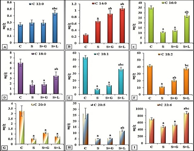
Figure 1. (A-I): Cortex free fatty acids content. Data expressed as mean ± SE. Superscript represent significance at p≤0.05 as (a) from control(c), (b) from senile group (S), from senile+ginger group (S+G)
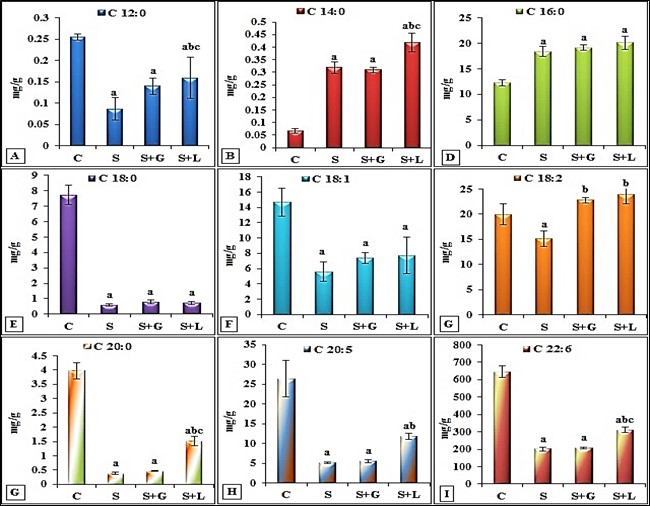
Figure 2. (A-I): Hippocampus free fatty acids content. Data expressed as mean ± SE. Superscript represent significance at p≤0.05 as (a) from control(c), (b) from senile group (S), from senile+ginger group (S+G)
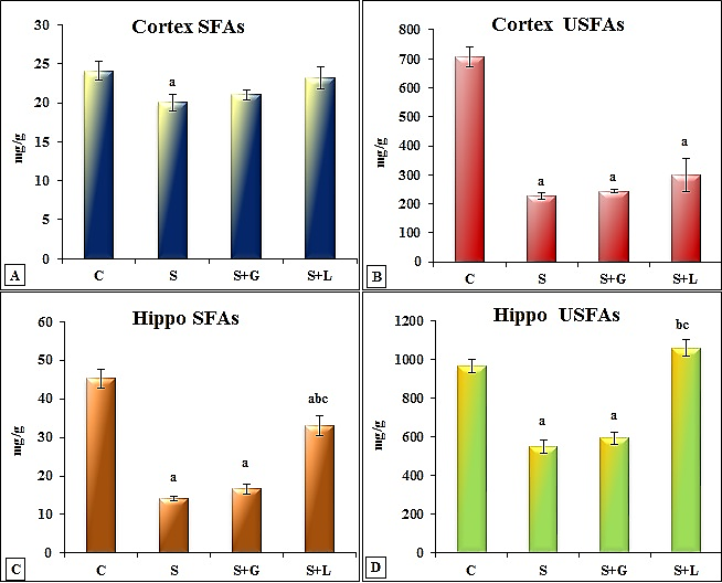
Figure 3. (A-D): Cortex and hippocampus saturated fatty acids (SFAs) and unsaturated fatty acids (USFAs) content. Data expressed as mean ± SE. Superscript represent significance at p≤0.05 as (a) from control(c), (b) from senile group (S), from senile+ginger group (S+G)
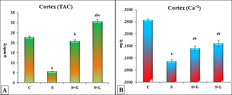
Figure 4. (A & B): Cortex total antioxidant capacity (TAC) and calcium ions contents (Ca+2) content. Data expressed as mean ± SE. Superscript represent significance at p≤0.05 as (a) from control(c), (b) from senile group (S), from senile+ginger group (S+G)
The results confirmed the age-related metabolic changes in lipids, resulted in variations in lipid content that also reflected changes in the proportions of gray and white matter as well as the increase in the oxidative stress results in a lower concentration of n-3 PUFA in the nervous system (Ledesma et al., 2012). The ratio of saturated to unsaturated fatty acids is one of the key factors influencing cell membrane fluidity. Thenperturbations in the fatty acid ratio have implications for neurological diseases (Hazel and Williams, 1990; Yehuda et al., 2002). The essential fatty acids linoleic acid and alpha-linolenic acid (SC-PUFAs) (-3 fatty acid) are required for normal nerve impulse transmission and brain function and serve as precursors of other PUFAs. The levels of two of these metabolites, DHA and arachidonic acid decreased in aged rats. Asignificant age-dependent decrease in the levels and turnover of other PUFAs observed in the hippocampus and cortex (Yehuda et al., 2005; Bazan et al., 2011; Ledesma et al., 2012; Fabelo et al., 2014). Oleic acid and palmitoleic acid are the major monounsaturated fatty acids in fat depots and membrane phospholipids. These fatty acids synthesized by the stearoyl-CoA desaturase. The ratio of stearic acid to oleic acid is one of the factors influencing membrane fluidity and cell–cell interaction. Abnormal alteration of this ratio play a role in several physiological and disease states including diabetes, cardiovascular disease, obesity, hypertension, neurological diseases, immune disorders, cancer, and aging (Ntambi, 1999). Rioux et al., (2008), suggested the contribution of high dietary myristic acid and the increased tissue storage ofα-linolenic acid and the overall bioavailability of (n-3) polyunsaturated fatty acids in the brain. Lin et al., (2012), indicated through pre-dementia studies that decrease in EPA level is a risk factor for cognitive impairment.
Different studies discussed the age-related variations in the total antioxidant defenses in cortex and hippocampus. Also the oxidative stress-related neurodegenerative disorders (Siqueira et al., 2005; Muthuswamy et al., 2006; Murali and Panneerselvam, 2007; Badr El-Din et al., 2010).
Previously, Rogasevskaia and Coorssen, (2006) tried to explore potential dysfunctional changes in regulatory mechanisms that may contribute to a breakdown in calcium homeostasis in aging. The study of Tanaka et al., (1996) provided evidence for that age-related decrease in presynaptic functions. Reduction in calcium influx via voltage-dependent calcium channels followed by decreased acetylcholine (Ach) release from synapses in spite of the abundance of ACh within the synapses. Wainwright (2002) reported that deficiencies of EFA influenced specific neurotransmitter systems in animals, particularly the dopamine systems of the frontal cortex. As well as associated behaviors and also the age- related reduced contents of cortex and hippocampus neurotransmitters which influenced by the FFA composition (Hegazy & Ali, 2011).
Ginger treatment exhibited the significant increase compared to the senile group values. In the cortex, myristic and linoleic acids (Figure 1 (A-I)), and in hippocampus linoleic acid (USFA), (Figure 2 (A-I). The data in Figure 3 (A-D) demonstrated not statistically different increase in cortex and hippocampus SFAs and UFAs in ginger treated group as compared to the senile group values. Cortex TAC and Ca+2 contents significantly increased in ginger group as compared to the senile group values (Figure 4(A and B). Results confirmed the antioxidant potential of the ginger due to its active ingredients especially essential oils (Yeh et al., 2014).Brain tissue has 60% lipid, and it has a remarkable high-energy consumption (Crowford, 1993). In addition, these tissues are more susceptible to oxidative damage than other tissues (Foloyd et al., 2001). It is in line with the neuroprotective effect of ginger that may affect neurotransmitters which playing crucial role in learning and memory. In addition to, interaction with inhibitory and excitatory system and calcium channel inhibition. Beyond the antioxidant effect (Stoilova et al., 2007; Waggas, 2009; Jittiwat and Wattanathorn, 2012; Hosseini and Mirazi, 2014).
Lipoic acid treatment represented the significant increase in cortex SFAs (lauric, myristic, palmitic, stearic and USFAs, oleic, linoleic, EPA and DHA) (Figure 1 (A-I)), Also in hippocampus SFAs (lauric, myristic and arachidic and USFAs, linoleic acid, EPA and DHA) (Figure 2 (A-I)) as compared to the senile group values. The data in Figure 3 (A-D) demonstrated a significant increase in hippocampus SFAs and UFAs in lipoic treated group as compared to values of senile and ginger treated groups. Cortical TAC significantly increased in lipoic group as compared to the values of senile and ginger treated groups and Ca+2 contents increased significantly compared to senile group value (Figure 4(A and B).Alpha-lipoic acid treatment previously proved impact in reducing oxidant production in rat brain regions in arsenic and stressed rats (Shila et al., 2005; Akpinar et al., 2008).Its beneficial effect in both preventing and reversing abnormalities in an aging brain associated with normalization of lipid peroxidation and partial restoration in the activities of various enzymatic antioxidants. Suggesting it could improve brain antioxidant functions in the elderly (Arivazhagan et al., 2002). Celik and Ozkaya (2002) suggested that the lipoic acid had a therapeutic effect via treatment guinea pig oxidative stress- model by H2O2. Suggesting action by limiting damage from the oxidation reaction in unsaturated fatty acids.

4. Conclusions

The results suggested protective effect of ginger and alpha lipoic acid in senile female rats. That through modulation of fatty acids in cortex and hippocampus, as well as the cortical free calcium ions content. Such could protect the senile rats from aging diseases and their supplementation as complimentary could improve brain antioxidant functions in the senile rats.

ACKNOWLEDGEMENTS

The authors gratefully acknowledge the help of the National Organization for Drug Control and Research (NODCAR).

References

[1]  Akpinar, D., P. Yargiçoğlu, N. Derin, Y. Alicigüzel, A. and Ağar. 2008."The effect of lipoic acid on antioxidant status and lipid peroxidation in rats exposed to chronic restraint stress" Physiol Res., 57(6):893-901.
[2]  Akpinar D, P. Yargiçoğlu, N. Derin, Y. Alicigüzel, M. Sahin, A. and Ağar. 2007."The effect of lipoic acid on lipid peroxidation and visual evoked potentials (VEPs) in rats exposed to chronic restraint stress" Int J Neurosci., 117(12):1691-1706.
[3]  Applegate, M.A., K.M. Humphries, and L.I. Szweda. 2008." Reversible inhibition of alpha-ketoglutarate dehydrogenase by hydrogen peroxide: glutathionylation and protection of lipoic acid". Biochemistry, 47:473–478.
[4]  Arafa N.M.S. 2010. "Efficacy of Echinacea on the action of cyproterone acetate in male rats". Pakistan Journal of Biological Sciences, 13(20): 966-976.
[5]  Arivazhagan, P., S. Shila, S. Kumaran, and C. Panneerselvam. 2002."Effect of DL-alpha-lipoic acid on the status of lipid peroxidation and antioxidant enzymes in various brain regions of aged rats". Exp Gerontol., 37(6):803-811.
[6]  Badr El-Din, N.K., E. Noaman, S.M. Fattah, and M. Ghoneum. 2010."Reversal of age-associated oxidative stress in rats by MRN-100, a hydro-ferrate fluid". In Vivo, 24(4):525-33.
[7]  Bazan, N.G., M.F. Molina, and W.C. Gordon. 2011." Docosahexaenoic acid signalolipidomics in nutrition: significance in aging, neuroinflammation, macular degeneration, Alzheimer's, and other neurodegenerative diseases." Annu Rev Nutr., 31:321-351.
[8]  Bourre, J.M. 2004."Roles of unsaturated fatty acids (especially omega-3 fatty acids) in the brain at various ages and during ageing". J Nutr Health Aging, 8: 163–174.
[9]  Calon, F. and G. Cole. 2007."Neuroprotective action of omega-3 polyunsaturated fatty acids against neurodegenerative diseases: evidence from animal studies". Prostaglandins Leukot Essent Fatty Acids, 77:287–293.
[10]  Cameron, N.E., and M.A. Cottter.1997."Effects of antioxidants on nerve and vascular dysfunction in experimental diabetes". Diabetes Res. Clin. Pract., 45:137–146.
[11]  Celik, S., A. Ozkaya. 2002. "Effects of intraperitoneally administered lipoic acid, vitamin E, and linalool on the level of total lipid and fatty acids in guinea pig brain with oxidative stress induced by H2O2". J Biochem Mol Biol., 35(6):547-552.
[12]  Cini M. and A. Moretti. 1995. "Studies on lipid peroxidation and protein oxidation in the aging brain". Neurobiology of Aging, 16(1): 53-57.
[13]  Crowford, M.A. 1993."The role of essential fatty acids in neural development: implications for prenatal nutrition". Am. J. Clin. Nutr., 57(5): 703-710.
[14]  Fabelo, N., V. Martín, R. Marín, D. Moreno, I. Ferrer, M. Díaz. 2014."Altered lipid composition in cortical lipid rafts occurs at early stages of sporadic Alzheimer's disease and facilitates APP/BACE1 interactions". Neurobiol Aging., 35(8):1801-1812.
[15]  Firląg, M., M. Kamaszewski, K. Gaca, D. Adamek, and B. Bałasińska. 2013."The neuroprotective effect of long-term n-3 polyunsaturated fatty acids supplementation in the cerebral cortex and hippocampus of aging rats". Folia Neuropathol., 51(3):235-242.
[16]  Foloyd, R. A., M. West, and K. Hensley. 2001. "Oxidative biochemical markers; clues to understanding aging in longlived species". Exp. Gerontol., 36: 619-640.
[17]  Freitas, R. M. 2009. "The evaluation of effects of lipoic acid on the lipid peroxidation, nitrate formation and antioxidant enzymes in the hippocampus of rats after pilocarpine-induced seizures". Neuroscience Letters. 455: 140-144.
[18]  Hazel, J. R., and E. E. Williams. 1990. "The role of alterations in membrane lipid composition in enabling physiological adaptation of organisms to their physical environment". Prog Lipid Res., 29:167–227.
[19]  Hegazy, H. G., and E. H. A. Ali. 2011. "Modulation of monoamines and amino-acids neurotransmitters in cerebral cortex and hippocampus of female senile rats by ginger and lipoic acid". African Journal of Pharmacy and Pharmacology, 5(8): 1080-1085.
[20]  Hosseini, A., and N. Mirazi. 2014. "Acute administration of ginger (Zingiber officinale rhizomes) extract on timed intravenous pentylenetetrazol infusion seizure model in mice". Epilepsy Res., 108(3):411-419.
[21]  Innis, S. M. 2008. "Dietary omega 3 fatty acids and the developing brain". Brain Res., 1237:35–43.
[22]  Jittiwat, J., and J. Wattanathorn. 2012. "Ginger Pharmacopuncture Improves Cognitive Impairment and Oxidative Stress Following Cerebral Ischemia". J Acupunct Meridian Stud., 5(6):295-300.
[23]  Koenig, M. L., and J. L. Meyerhoff. 2003. "In vitro neuroprotection against oxidative stress by pre-treatment with a combination of dihydrolipoic acid and phenyl-butyl nitrones". Neurotox Res., 5(4):265-272.
[24]  Koracevic, D., G. Koracevic, V. Djordjevic. S. Andrejevic, and V. Cosic. 2001."Method for measurement of antioxidant activity in human fluids". J. Clin. Pathol., 54:356-361.
[25]  Kubra, I. R., andL. Jaganmohanrao. 2012. "An Overview on Inventions Related to Ginger Processing and Products for food and Pharmaceutical Applications". Recent Pat Food Nutr Agric., 4(1):31-49.
[26]  Ledesma, M. D., M. G. Martin, and C. G. Dotti. 2012. "Lipid changes in the aged brain: effect on synaptic function and neuronal survival". Prog Lipid Res., 51(1):23-35.
[27]  Lin, P. Y., C. C. Chiu, S. Y. Huang, and K. P. Su. 2012. "A meta-analytic review of polyunsaturated fatty acid compositions in dementia". J Clin Psychiatry., 73(9):1245-1254.
[28]  Mattson, M. P., S. L. Chan, and W. Duan. 2002. " Modification of brain aging and neurodegenerative disorders by genes, diet, and behavior". Physiol Rev., 82(3): 637-672.
[29]  Murali, G., and C. Panneerselvam. 2007. "Age-associated oxidative macromolecular damages in rat brain regions: role of glutathione monoester". J Gerontol A Biol Sci Med Sci., 62(8):824-830.
[30]  Murugan, P. and L. Pari. 2007. "Protective role of tetrahydrocurcumin on changes in the fatty acid composition in streptozotocin-nicotinamide induced type 2 diabetic rats". J. Appl. Biomed., 5: 31–38.
[31]  Muthuswamy, A. D., K. Vedagiri, M. Ganesan, and P. Chinnakannu. 2006. "Oxidative stress-mediated macromolecular damage and dwindle in antioxidant status in aged rat brain regions: role of L-carnitine and DL-alpha-lipoic acid". Clin Chim Acta., 368(1-2):84-92.
[32]  Ntambi, J. M. 1999."Regulation of stearoyl-CoA desaturase by polyunsaturated fatty acids and cholesterol". J Lipid Res., 40(9):1549-1558.
[33]  Patil, S., J. Melrose, and C. Chan. 2007. "Involvement of astroglial ceramide in palmitic acid-induced Alzheimer-like changes in primary neurons". Eur J Neurosci., (8):2131-2141.
[34]  Perera, J., J. H. Tan, S. Jeevathayaparan, S. Chakravarthi, and N. Haleagrahara. 2011. "Neuroprotective effects of alpha lipoic Acid on haloperidol-induced oxidative stress in the rat brain". Cell Biosci., 1:12.
[35]  Reagan-Shaw, S., M. Nihal, and N. Ahmad. 2008. "Dose translation from animal to human studies revisited". The FASEB Journal, 22(3): 659–661.
[36]  Rioux, V., D. Catheline, E. Beauchamp, J. Le Bloc'h, F. Pédrono, and P. Legrand. 2008."Substitution of dietary oleic acid for myristic acid increases the tissue storage of α-linolenic acid and the concentration of docosahexaenoic acid in the brain, red blood cells and plasma in the rat". Animal, 2(4):636-644.
[37]  Rogasevskaia, T., and J. R. Coorssen. 2006. "Sphingomyelin- enriched microdomains define the efficiency of native Ca(2+)-triggered membrane fusion". J Cell Sci., 119(13): 2688-2694.
[38]  Shahidi, F., and H. Miraliakbari. 2005. "Omega-3 fatty acids in health and disease. Part 2. Health effects of omega-3 fatty acids in autoimmune diseases, mental health, and gene expression". J Med Food., 8: 133–148.
[39]  Sharma, P., and R. Singh. 2012."Dichlorvos and lindane induced oxidative stress in rat brain: Protective effects of ginger". Pharmacognosy Res., 4(1):27-32.
[40]  Shila, S., V. Kokilavani, M. Subathra, and C. Panneerselvam. 2005. "Brain regional responses in antioxidant system to alpha-lipoic acid in arsenic intoxicated rat". Toxicology, 210(1):25-36.
[41]  Siqueira, I. R., C. Fochesatto, A. de Andrade, M. Santos, M. Hagen, A. Bello-Klein, and C. A. Netto. 2005. "Total antioxidant capacity is impaired in different structures from aged rat brain". Int J Dev Neurosci., 23(8):663-671.
[42]  Snigdha, S., G. Astarita, D. Piomelli, and C. W. Cotman. 2012. "Effects of diet and behavioral enrichment on free fatty acids in the aged canine brain". Neuroscience, 202:326-333.
[43]  Stoilova, I., A. Krastanov, A. Stoyanova, P. Denev, and S. Gargova. 2007. "Antioxidant activity of a ginger extract (Zingiber officinale)". Food Chemistry, 102: 764–770.
[44]  Sublette, M.E., M. S. Milak, J. R. Hibbeln, P. J. Freed, M. A. Oquendo, K. M. Malone, R. V. Parsey, and J. J. Mann. 2009."Plasma polyunsaturated fatty acids and regional cerebral glucose metabolism in major depression". Prostaglandins Leukot Essent Fatty Acids, 80(1):57-64.
[45]  Tanaka, Y., A. Hasegawa, andS. Ando. 1996. "Impaired synaptic functions with aging as characterized by decreased calcium influx and acetylcholine release". J Neurosci Res., 43: 63–76.
[46]  Waggas, A. M. 2009."Neuroprotective evaluation of extract of ginger (Zingiber officinale) root in monosodium glutamate-induced toxicity in different brain areas male albino rats". Pak J Biol Sci., 12(3): 201-212.
[47]  Wainwright, P. E. 2002."Dietary essential fatty acids and brain function: a developmental perspective on mechanisms" Proceedings of the Nutrition Society, 61: 61–69.
[48]  Yeh, H., C. Chung, H. Chen, andC. Wan. 2014."Bioactive components analysis of two various gingers (Zingiber officinale Roscoe) and antioxidant effect of ginger extracts". Food Science and Technology, 55: 32-334.
[49]  Yehuda, S., S. Rabinovitz, R. L. Carasso, and D. I. Mostofsky. 2002."The role of polyunsaturated fatty acids in restoring the aging neuronal membrane". Neurobiol Aging, 23:843–853.
[50]  Yehuda, S., S. Rabinovitz, andF. I. Mostofsky. 2005." Essential fatty acids and the brain: from infancy to aging". Neurobiol Aging., 26 (l):98-102.