
-
Paper Information
- Paper Submission
-
Journal Information
- About This Journal
- Editorial Board
- Current Issue
- Archive
- Author Guidelines
- Contact Us
Journal of Microbiology Research
p-ISSN: 2166-5885 e-ISSN: 2166-5931
2026; 16(1): 1-8
doi:10.5923/j.microbiology.20261601.01
Received: May 13, 2026; Accepted: Jun. 10, 2026; Published: Jun. 29, 2026

Prevalence of Rhizobium radiobacter Among Children with Suspected Sepsis
 Abstract
 Reference
 Full-Text PDF
 Full-text HTML
M. A. Christopher1, J. U. Umoh1, E. E. Owowo1, M. E. Bassey1, V. F. Nyoyoko2
1Department of Microbiology, Faculty of Biological Sciences, Akwa Ibom State University, Ikot Akpaden, Nigeria
2Department of Biological Sciences, Faculty of Computing and Applied Sciences, Topfaith University, Mkpatak, Nigeria
Copyright © 2026 The Author(s). Published by Scientific & Academic Publishing.
This work is licensed under the Creative Commons Attribution International License (CC BY).
http://creativecommons.org/licenses/by/4.0/

Rhizobium radiobacter is an opportunistic Gram-negative bacterium occasionally implicated in bloodstream infections, yet its involvement in pediatric sepsis remains uncommon and poorly characterized. In this study involving 180 children under five years of age assessed for sepsis across three hospitals in Akwa Ibom State, Nigeria, Rhizobium radiobacter was identified as one of the Gram-negative bacterial pathogens isolated from blood cultures. Out of the 123 culture positive cases, R. radiobacter accounted for 8 isolates, representing a prevalence of 4.4% among all confirmed bacterial isolate. The detection of R. radiobacter in pediatric sepsis cases is clinically noteworthy, as the organism is traditionally considered an opportunistic pathogen more frequently associated with immunocompromised individuals. Its presence in children with clinical signs of sepsis suggests that R. radiobacter may play a role in bloodstream infections in this population. R. radiobacter exhibited 100% susceptible to amikacin, gentamicin and ciprofloxacin and 100% resistance to cefotaxime, ceftriaxone and meropenem. This highlights the importance of comprehensive microbial surveillance, as unusual or less frequently encountered organisms may contribute significantly to sepsis etiology and complicate diagnosis and treatment. Early recognition and appropriate antimicrobial therapy remain essential for optimal outcomes in children presenting with sepsis.
Keywords: Children, Gram-negative bacteria, Resistance pattern, Rhizobium radiobacter, Sepsis
Cite this paper: M. A. Christopher, J. U. Umoh, E. E. Owowo, M. E. Bassey, V. F. Nyoyoko, Prevalence of Rhizobium radiobacter Among Children with Suspected Sepsis, Journal of Microbiology Research, Vol. 16 No. 1, 2026, pp. 1-8. doi: 10.5923/j.microbiology.20261601.01.
Article Outline
1. Introduction
- Neonatal and childhood sepsis remains a major global public health challenge and a leading cause of childhood morbidity and mortality, with significant disparities across regions and countries [1,2]. Nigeria, the most populous country in Africa, bears one of the world’s highest neonatal sepsis incidence and mortality rates [3,4]. It is the second-largest contributor to global under-five mortality due to sepsis [3,5]. Across Africa, the prevalence of severe sepsis is estimated at 23.1% [6]. Gram-negative bacteria remain the most common etiologic agents of sepsis. Studies show that 62.2% of culture-proven sepsis cases involve Gram-negative organisms, while 46.8% involve Gram-positive pathogens; these proportions may overlap in cases of polymicrobial infection [7]. Similar findings revealed Gram-negative bacteria as predominant isolates (67.6%) [8]. Hospital-acquired septic shock is most frequently associated with Gram-negative bloodstream infections (47.3%), compared to 30.9% caused by Gram-positive bacteria, while the remaining cases were attributed to other or unspecified pathogens [9]. Sepsis during the neonatal period is associated with life-threatening complications such as respiratory failure, pulmonary hypertension, cardiac failure, renal and liver dysfunction, shock, cerebral edema, and long-term neurodevelopmental impairment [10,11].Rhizobium radiobacter is primarily known as a plant pathogen responsible for tumorigenic diseases in plants. Although it has long been considered non-pathogenic to humans, reports over the past decade show that it can cause opportunistic infections, particularly in immunocompromised patients and those with chronic illnesses. A major risk factor for infection is the use of invasive medical devices, especially central venous catheters and peritoneal dialysis catheters [12]. Microbiologically, R. radiobacter is a Gram-negative, aerobic, motile bacillus with peritrichous flagella. It grows on blood agar and MacConkey agar and is catalase-positive, oxidase-negative, and urease-positive. In high-carbohydrate media, it produces extracellular polysaccharide mucus. Although it shares similarities with Pseudomonas, Alcaligenes and Bordetella, it can be distinguished through biochemical testing and flagellar morphology. Identification commonly involves the 3-ketolactose production test, with carbohydrate oxidation tests (mannitol, lactose, maltose) serving as alternatives. Based on phytogenic effects, these bacteria are classified into species such as Agrobacterium tumefaciens [12]. Rhizobium radiobacter belongs to a group of bacteria that includes Agrobacterium tumefaciens, Agrobacterium rhizogenes and Agrobacterium rubi. Unlike A. tumefaciens, R. radiobacter lacks tumorigenic Ti plasmids. The exact mechanism of transmission to humans remains unclear. Initially, infections caused by R. radiobacter were considered rare and of low clinical significance, with early reports in the 1960s suggesting it was mainly a contaminant with weak pathogenicity. The first well-documented human infection was reported in 1980, involving endocarditis in a patient with a prosthetic aortic valve. Since then, R. radiobacter has increasingly been recognized as an opportunistic pathogen, particularly in adults and children with invasive medical devices such as central venous catheters (CVCs). Studies indicate that more than half of reported infections are catheter-associated bacteremias [13]. The organism is generally sensitive to multiple antibiotics, including gentamicin, ceftazidime, amikacin, carbapenems, piperacillin–tazobactam, cefepime and polymyxin. Preventive strategies focus on minimizing the use of invasive devices, selecting appropriate antimicrobial therapy based on sensitivity testing, using cuffed or tunneled CVCs or catheters impregnated with chlorhexidine and silver sulfadiazine, and reducing exposure to soil-related activities.
2. Materials and Methods
2.1. Study Area
- This study was conducted over an 18-month period, from June 2023 to December 2024, across the three Senatorial Districts of Akwa Ibom State, Nigeria: Akwa Ibom North-West Senatorial District (Ikot Ekpene), Akwa Ibom South Senatorial District (Eket), Akwa Ibom North-East Senatorial District (Uyo). Three hospitals were purposively selected based on accessibility and proximity to the research laboratory. These included General Hospital Ikot Ekpene, Immanuel Hospital Eket, and the University of Uyo Teaching Hospital (UUTH), Uyo. Akwa Ibom State is located in the South-South geopolitical zone of Nigeria. It is bordered by Cross River State to the east, Rivers and Abia States to the west, and the Atlantic Ocean to the south. The three selected study sites offered suitable clinical settings for patient recruitment and sample collection, as well as close geographical proximity to the diagnostic laboratory for timely specimen processing.
2.2. Study Population
- The study population consisted of male and female children under five years of age (0-5 years) who were seen in the Special Care Baby Unit (SCBU) and Sick Baby Unit (SBU) of the selected hospitals. Newborns delivered within the facilities were admitted into the SCBU, while out-born infants referred from other locations were admitted into the SBU. The study included an equal number of participants by sex, with 30 males and 30 females recruited from each senatorial district, giving a total of 60 participants per district.
2.3. Study Design
- A cross-sectional study design was employed to assess neonates and children under five years with and without clinical features of sepsis. Participants presenting with signs and symptoms of sepsis, such as high fever, chills, weakness, extreme discomfort or pain, hypotension or hypertension, confusion, disorientation, poor feeding, shortness of breath, hypothermia, apnea, pallor or jaundice, poor peripheral perfusion, vomiting, abdominal distension, diarrhoea, seizures and jitteriness, were enrolled. Blood samples were collected for microbiological analysis.
2.3.1. Inclusion Criteria
- Children under five years of age (0-5 years), of either sex, whose parents or guardians provided informed consent, and who attended any of the selected hospitals were included.
2.3.2. Exclusion Criteria
- Children older than five years and children whose parents or guardians did not consent to participate were excluded from the study.
2.3.3. Ethical Considerations
- Ethical approval for the study was obtained from the Akwa Ibom State Ministry of Health, authorizing sample collection in public and private healthcare facilities within the state. Written or verbal informed consent was obtained from parents or guardians before sample collection.
2.4. Sample Size
- A total of 180 children were recruited for the study, comprising 60 participants each from General Hospital Ikot Ekpene, University of Uyo Teaching Hospital (UUTH) and Immanuel Hospital Eket, representing the three senatorial districts of Akwa Ibom State.
2.5. Sampling
- Between 10 and 25 samples were collected daily across the three senatorial districts, depending on patient availability and clinical presentation. A total of 180 blood samples were collected from children with and without signs of sepsis. Using a sterile 2 ml syringe, 2 ml of peripheral venous blood was drawn aseptically. Each sample was labeled with the participant’s name, date, age, and hospital number and transported to the clinical microbiology laboratory within 24 hours for processing. All blood collections were performed by the attending nurse in charge of the unit.
2.6. Isolation of Gram-negative Bacteria from Blood Specimens
2.6.1. Culture of Blood Specimens
- Freshly prepared thioglycollate broth was cooled to room temperature, and 2 ml of blood was inoculated into the broth. The culture bottles were incubated at 37°C for 7 days and inspected twice daily for turbidity, indicated by foamy or whitish growth at the upper broth layer. Broths showing no turbidity after 7 days were reported as negative and discarded.
2.6.2. Subculture on Selective and Enrichment Media
- A loopful of broth from turbid thioglycollate bottles was streaked onto blood agar, MacConkey agar and chocolate agar. Blood agar and chocolate agar plates were incubated anaerobically (CO₂-enriched environment) at 37°C for 48 hours. MacConkey agar plates were incubated aerobically at 37°C for 24 hours. Anaerobic conditions were generated using a McIntosh and Filde’s jar.
2.6.3. Pure Culture Isolation and Stock Preparation
- Non-lactose fermenting colonies from MacConkey agar were subcultured onto nutrient agar to obtain pure colonies. Stock cultures were prepared on nutrient agar slants and stored at 8°C. Cultures were transferred to fresh slants weekly. Plates without visible growth were re-incubated before being discarded.
2.6.4. Identification and Characterization of Gram-negative Bacteria
- Presumptive isolates were subjected to Gram staining and identified using the bioMérieux VITEK 2® system (bioMérieux, Marcy-Ī Ētoile, France).
2.6.5. Antimicrobial Susceptibility Testing
- Antimicrobial susceptibility profiles were determined using the Kirby Bauer disc diffusion method on Mueller Hinton agar. A bacterial colony was inoculated into Mueller Hinton broth and adjusted to the turbidity of a 0.5 McFarland standard. The standardized suspension was spread to form a lawn culture. Twelve antibiotics were tested: tetracycline (TET, 10 µg), cotrimoxazole (COT, 25 µg), gentamicin (GEN, 10 µg), cefuroxime (CRX, 30 µg), chloramphenicol (CHL, 10 µg), ceftriaxone (CTR, 30 µg), cefotaxime (CTX, 30 µg), ciprofloxacin (CIP, 5 µg), amikacin (AMK, 30 µg), vancomycin (VAN, 30 µg), ceftazidime (CPZ, 30 µg) and meropenem (MEM, 10 µg). A maximum of six antibiotic discs were placed per plate with 24-30 mm spacing between discs. Plates were left for 1 hour for prediffusion before incubation at 37°C for 24 hours. Zones of inhibition (mm) were measured and interpreted as susceptible, intermediate or resistant according to the clinical and laboratory standards institute (CLSI) guidelines [14]. Multidrug resistance (MDR) was defined as resistance to at least one antibiotic in three or more antibiotic classes. The Multiple Antibiotic Resistance Index (MARI) was calculated using: MARI = X/Y where: -X = number of antibiotics to which an isolate is resistant and Y = total number of antibiotics tested.
3. Results
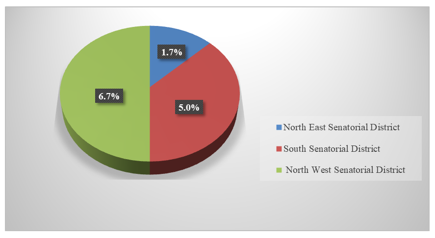 | Figure 1. Prevalence of Rhizobium radiobacter among children according to senatorial districts in Akwa Ibom State |
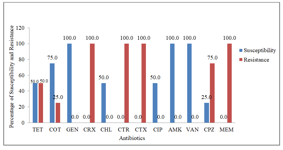 | Figure 2. Percentage of antibiotic susceptibility and resistance of R. radiobacter, isolated from children under five years of age in General Hospital Ikot Ekpene |
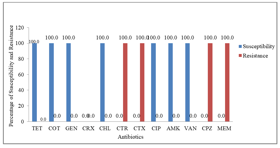 | Figure 3. Percentage of antibiotic susceptibility and resistance of R. radiobacter, isolated from children under five years of age in Uyo Teaching Hospital Uyo |
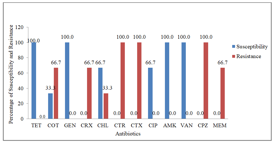 | Figure 4. Percentage of antibiotic susceptibility and resistance of R. radiobacter, isolated from children under five years of age in Immanuel Hospital Eket |
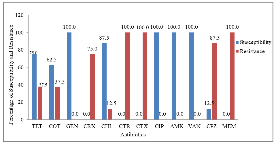 | Figure 5. Percentage of antibiotic susceptibility and resistance of R. radiobacter, isolated from children under five years of age in General Hospital Ikot Ekpene, Uyo Teaching Hospital Uyo and Immanuel Hospital Eket |
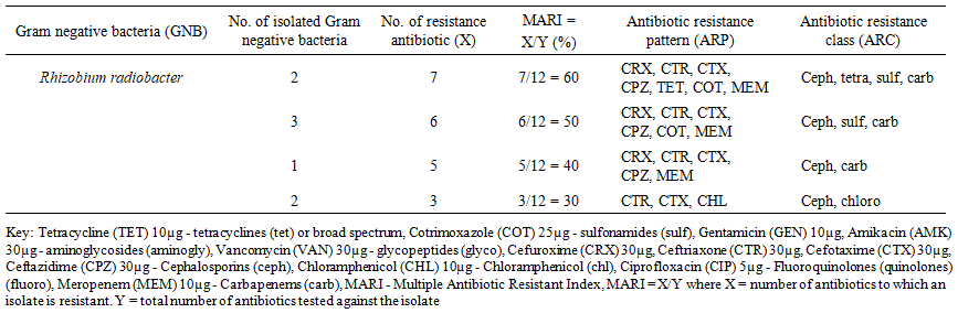 | Table 1. Antibiotic Resistance Pattern of R. radiobacter Obtained from Sepsis in Children under Five Years of Age in General Hospital Ikot Ekpene, Uyo Teaching Hospital Uyo and Immanuel Hospital Eket |
4. Discussion
- The occurrence of Rhizobium radiobacter is likely associated with hospital-related risk factors, particularly neonatal exposure to contaminated medical equipment such as incubators, apnea monitors, and invasive devices including catheters [15]. The reason for the isolation of R. radiobacter could be: immature immune systems and early exposure to several microbial agents, overuse of antibiotics in children which makes them more vulnerable to infections, environmental factors like medical devices that disrupt the mucosa and increase the chance of ascending microorganisms from birth canal into the amniotic sac of the foetus thereby exposing to infection [16]. R. radiobacter infection in a newborn with central venous access underscores this organisms’ rare yet important capacity to cause severe infections in vulnerable pediatric populations [13]. In sepsis, multidrug resistant organisms are crucially involved in causing high mortality in neonates as compared to non-multidrug resistant bacteria [17]. However, Nigeria has been reported as one of the countries with high resistance to antibiotics by Gram negative organisms [18]. In this study, Rhizobium radiobacter was 100% susceptible to amikacin, gentamicin and ciprofloxacin. The high activity of AMK and GEN (aminoglycosides) against the extended spectrum beta (β)-lactamase (ESBL) producers in this study might be due to the less use or less abuse of these antibiotics in treating bacterial infections especially with its strong prescription policy and the stress of its parenteral preparation unlike other drugs which are usually available to be taken orally. Aminoglycosides bind to the bacteria, causing misreading of t-, leaving bacteria unable to synthesize proteins vital to their growth [19]. Other studies also reported high susceptibility of the Gram negative bacteria isolated from sepsis to aminoglycosides [20]. Also ciprofloxacin (fluoroquinolones) remain effective therapeutic option, often recommended as first line agent. It mechanism of action involve, DNA gyrase, causing bacterial DNA fragmentation and cell death. Rhizobium radiobacter was 100% resistance to cefotaxime (CTX), ceftriaxone (CTR) and meropenem (MEM) among all the cephalosporins and carbapenems. The antimicrobial resistance to β-lactam antibiotics primarily occurs due to the carriage of ESBL resistance plasmids in Gram-negative bacteria (GNB) [21]. The exceptions observed in this study may be attributed to loss of porin (reduced drug entry), efflux pumps, carbapenemase enzymes (for meropenem resistance), target site mutations (penicillin binding proteins) and plasmid-independent chromosomal resistance. Cefotaxime resistance to Rhizobium radiobacter was attributed to the production of enzyme β-lactamase which breaks down the β-lactam ring in CTX, rendering the antibiotic ineffective [22]. Also mutation in target proteins changes in the penicillin-binding proteins (PBPs) which are the target sites of CTX can reduce the antibiotics binding affinity and effectiveness [23]. In contrast, 50% resistance of R. radiobacter to CTX was reported [24]. The reason for MEM resistance to R. radiobacter could be due to acquisition of resistance through mutations that result in the loss or reduction of outer membrane porins, limiting the entry of meropenem into the bacterial cell [25]. R. radiobacter has high adhesion properties, producing extracellular mucus that allows it to form biofilms on catheters which increases resistance to antibiotics [26]. R. radiobacter exhibited 60% resistance pattern to cephalosporins [26]. The high resistance rate of the isolates against many antibiotics could be attributed to inappropriate and overuse of antibiotics and sometimes clinicians initiating antibiotic therapy before performing blood culture [27]. It could also be due to the fact that, these organisms develop antibiotic resistance through efflux pumps (over expression of proton transporter AcrAB and protein ToIC), beta-lactamase production and outer membrane alteration mechanisms [28]. Βeta-lactam antibiotics are used widely worldwide against infections caused by Gram-negative bacteria [29]. Resistance is known to be due to various mechanisms among which production of extended-spectrum β-lactamases (ESBLs) has been reported among bacterial isolates from the neonates [30].
5. Conclusions
- The presence of R. radiobacter underscores the importance of strict infection control practices and careful management of invasive devices to reduce the risk of multidrug-resistant Gram-negative infections in neonatal settings. Proper antibiotics screening should be incorporated before antibiotics administration in clinical practice in order to detect bacteria that harbor multi-drug resistance traits and curtail the abuse of antibiotics. All emergency departments should have a screening tool and sepsis bundle to aid early identification of the septic child with timely management and appropriate acceleration.
ACKNOWLEDGEMENTs
- For their collaboration and support, we thank and appreciate all the laboratory staff at Akwa Ibom State University, Ritman University Ikot Ekpene, General Hospital Ikot Ekpene, Uyo Teaching Hospital, Uyo and Immanuel Hospital Eket, Akwa Ibom State.