Journal of Microbiology Research

p-ISSN: 2166-5885    e-ISSN: 2166-5931

2022;  12(1): 1-12

doi:10.5923/j.microbiology.20221201.01

Received: Dec. 24, 2021; Accepted: Jan. 8, 2022; Published: Jan. 21, 2022


Phenotypic Characteristics and Molecular Identification of Virulence Genes in Beta Lactamase Producing Pseudomonas aeruginosa Isolates in Leafy Vegetables and Clinical Sources from North West Ethiopia

Cherinet Yigrem1, Broderick Eribo2, Bewketu Mengesha3

1Department of Biology, Howard University, 400 Bryan St. NW Washington DC, USA

2Department of Biology, Howard University, 415 College St. NW Washington DC, USA

3Department of Immunology and Molecular Biology, University of Gondar, Ethiopia

Correspondence to: Cherinet Yigrem, Department of Biology, Howard University, 400 Bryan St. NW Washington DC, USA.

Email:

Copyright © 2022 The Author(s). Published by Scientific & Academic Publishing.

This work is licensed under the Creative Commons Attribution International License (CC BY).
http://creativecommons.org/licenses/by/4.0/

Abstract

Pseudomonas aeruginosa is an opportunistic pathogen commonly found in soil water and animals. These pathogenic bacteria cause severe nosocomial infections in the blood, lungs (pneumonia), or other parts of the immunocompromised people and chronic infections in cystic fibrosis patients. The aim of this study was, phenotypic characterization and molecular identification of virulence determinants in beta lactamase producing Pseudomonas aeruginosa isolated in leafy vegetables sold in Gondar retail markets and clinical sources from Gondar University Hospital. The study further aimed to identify the plasmid profile, among the bacterial population. All the isolates (n=25) were screened for biofilm formation and the result confirmed that 88% (n = 22) were biofilm producers. The antibiotics susceptibility indicated that 99.3% of the isolates were resistant. One-way ANOVA statistical test (P = 0.097 > 0.05) confirmed that there were no significant differences in antibiotic resistance between vegetables and clinical isolates. Phylogenetic tree showed similar ancestorial relationship between the vegetable and the clinical isolates. Among beta-lactamase-producing antibiotic resistant isolates (n = 25), about 88% (n = 22) were biofilm producers. Pigment production was observed in 96% (n = 24) isolates. Twelve representative isolates were screened for detection of virulence factors. Among them, 75% (n = 9) isolates were found to be LasB positive, while 100% (n = 12) were ExosA producers, 75% (n = 9) were PlcH producers and 100% (n = 12) were ToxA producers. Furthermore, all the virulence screened isolates coproduced more than two type of virulence factors. Out of the total seven screened for plasmid profile six of them were determined positive.

Keywords: Antibiotic resistant, Biofilm formation, P. aeruginosa, Plasmid, Virulence factors

Cite this paper: Cherinet Yigrem, Broderick Eribo, Bewketu Mengesha, Phenotypic Characteristics and Molecular Identification of Virulence Genes in Beta Lactamase Producing Pseudomonas aeruginosa Isolates in Leafy Vegetables and Clinical Sources from North West Ethiopia, Journal of Microbiology Research, Vol. 12 No. 1, 2022, pp. 1-12. doi: 10.5923/j.microbiology.20221201.01.

1. Introduction

Pseudomonas aeruginosa is an encapsulated, Gram-negative, rod-shaped opportunistic pathogen which causes nosocomial infections, particularly in immunocompromised patients [27,49,31]. The treatment of infections caused by P. aeruginosa is usually complicated since the organism is intrinsically immune to many classes of antibiotics and may acquire resistance to all or any effective antibiotics [40,50,70]. Pseudomonas aeruginosa can survive in several environmental conditions by utilizing their diverse metabolic and virulence patterns [43,45,46]. Virulence genes have a special level of intrinsic expression, which results in a variable level of pathogenicity in infected individuals [58]. Mainly, these infections arise due to the development of antibiotic resistance patterns, biofilm formation, and virulence factors production [5]. Biofilm, a source of chronic and protracted infection, presents strong resistance to the system and antibiotics [45,54]. Biofilms are sessile populations of microorganisms which are enclosed by the self-secreted extracellular polysaccharide matrix, or slime, which acts as efficient barriers against antibiotics [15,39,57]. Growth of Pseudomonas aeruginosa in varying environments can enhance pigment production. Moreover, fluctuation in environmental conditions can increase elastase expression in these pathogens [16,25,67]. P. aeruginosa can produce two sorts of phospholipase of which the hemolytic type (PlcH) hydrolyzes sphingomyelin alongside phosphatidylcholine [14,19,27]. Virulence factor ToxA was reported from patients having lung infection, while ExoS is more prevalent in CF patients [9,48]. The expression virulence factors of P. aeruginosa at the cellular and extracellular level is regulated through cell signaling pathways [31,33,52]. The strong correlation between virulence genes and source of infection can help control these infections within the community [20,22,41]. The primary effective mechanism that regulates their expression was quorum sensing, and thus targeting these key regulators will improve therapeutic success within the future [37,76]. The current study documented the extent of pigment production, biofilm formation, and presence of virulence factors in antibiotic resistance P. aeruginosa isolates of vegetable and clinical sources from Gondar Ethiopia. Based on our investigation of previous articles, this study is the first report to reveal the molecular identification of P. aeruginosa virulence factors in Ethiopia.

2. Materials and Methods

2.1. Sample Collection

During the summer of 2015 and 2016, a total of 110 retail vegetable samples including 30 cauliflower, 30 spinach, 28 lettuces, 28 cabbage were collected from four different retail markets in Gondar Ethiopia. The vegetable samples were placed in a cold box at a temperature approximately 4°C, tightly sealed with sterile plastic wrap, and transported to Gondar University Microbiology laboratory and subjected to microbiological analysis within 24 hrs. Forty clinical samples were collected from patients who were admitted to a hospital of Gondar University of Medical Science college. The clinical strains were isolated from different specimens and no duplicate isolates from the same patient were included in this study.
In each city, seven samples were randomly collected from two retail markets and two farmers' markets. The samples were placed in separate sterile plastic bags and then immediately transported to the laboratory in a cooler with ice packs (below 4°C) and processed within 4–6 hrs.
From each vegetable sample 25 g was enriched in 225 mL nutrient broth (Huankai Ltd., Guangzhou, China) for 18-24 hrs. at 37°C on plate count agar. then for P. aeruginosa confirmation the enrichment was streaked onto MacConkey agar (Huankai Ltd., Guangzhou, India), followed by incubation at 37°C for 18-24hrs. From MacConkey agar, three pink, mucoid colonies were picked up and cultured onto nutrient agar at 37°C for 24 h, followed by biochemical identification using API 20 E (BioMe′rieux, Marcy I′Etoile, France). Finally, 25 P. aeruginosa isolates were recovered from all the samples. Among these isolates, 15 were from leafy vegetables, 10 were in clinical source from Gondar University Hospital. Confirmed P. aeruginosa cultures were preserved in Luria-Bertani broth containing 20% glycerol and stored at −80°C for further study.

2.2. Antimicrobial Susceptibility

Antimicrobial susceptibility was determined according to the Clinical and Laboratory Standards Institute (CLSI) guideline [9,20], using the Kirby Bauer disk diffusion assay on Mueller-Hinton agar. The susceptibility profiles were determined for twenty five antibiotics classified into 11 different groups [54] amoxycillin (AMC, 30μg), ampicillin (AMP, 10 μg), cefepime (FEP, 10μg), Cefoxitin (FOX, 30 μg), Cefazolin (CFZ) (30 μg), ceftazidime (CAZ, 30 μg), amikacin (AK, 30 μg), gentamicin (CN, 10 μg), kanamycin (K, 30 μg), Cefaclor (CEC) (25μg), Cefotaxime (CTX) (30μg), clindamycin (DA, 2 μg), Doripenem (DOR) (30 μg), Imipenem (IPM) (30 μg), ciprofloxacin (CIP, 5 μg), Norfloxacin (NOR, 10 μg), Meropenem (MEM) (30μg), Linezolid (LZD, 30 μg), rifampicin (RD, 5 μg), Trimethoprim/sulphamethoxazole (SXT, 25 μg), Quinupristin/dalfopristin (QD, 15 μg), Ceftazidime (CAZ 30μg), Ertapenem(ETP) (30μg), Nalidixic acid (NAL) (30 μg) and Colistin (CST) (30 μg), (Mast Diagnostics, Mast Group Ltd, Merseyside, UK). Pseudomonas aeruginosa ATCC 27835/27853 was used as quality control in each antimicrobial susceptibility assay. The results were interpreted as susceptible, resistance and intermediate according to the criteria recommended by the CLSI and the manufacture’s protocols (Mast Companies, UK). Pseudomonas aeruginosa ATCC 27853 were used as a quality control.

2.3. Plasmid Profile and 16s rRNA Sequencing of Bacteria Isolates

Plasmid extraction of Pseudomonas aeruginosa was performed using the alkaline lyses method by the Omega kit [7,30,62]. The plasmid extraction process begins with labelling Eppendorf tubes according to number of isolates and filled with 1ml of sterile water. The Pseudomonas aeruginosa culture was mix by vertexing. 1.5ml ml of the broth containing the bacterial solution was aliquoted into the labelled tubes and centrifuged at 13,000rpm for 2mins [13]. The supernatant of the centrifuged solution was removed by decanting gently leaving a little of the bacterial solution in the broth. The decanted solution was then vortexed at high speed until pellet is completely suspended in the broth containing the bacterial solution. out of the suspended pallet solution 300µl of TENS solution and mix by inverting tubes until the solution becomes slimy [12,38]. On top of this slimed solution 150µl of sodium acetate solution was added and vortexed for about 10mins and thereafter centrifuged at 13,000rpm for 5minutes. The supernatant of this solution was transferred into another 1.5ml Eppendorf tube and 900µl of ice-cold absolute ethanol was added, vortexed and centrifuged at 13,000rpm for 10minutes. White pellet is observed after supernatant is removed. Then ,1000µl of ice cold 70% ethanol was added and centrifuge at 13,000rpm for 5minutes (no vertexing at this stage). The supernatant was removed, and pellets allowed to dry then add 40µl of TE buffer and the plasmid is collected. The plasmid profile of the Pseudomonas aeruginosa were analyzed using agarose gel electrophoresis [4,51]. The size of the plasmids and molecular weight were visualized using UV transilluminator [11].

2.4. Primers and PCR Confirmation of Pseudomonas aeruginosa

All confirmed P. aeruginosa isolates were grown overnight in lactose broth at 37°C. Genomic DNA was extracted using a commercial Universal DNA Extraction Kit (Sangon Biotech, Shanghai, China) according to the manufacturer's instructions. Confirmation of P. aeruginosa isolates were performed by PCR as previously described [75]. The primer sequences and amplicon size are shown in (Table 2).
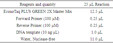
The ExoS, PlcH, ToXA and LasB genes were amplified by colony PCR using a specific set of primers (Table 2). The amplifications of these virulence genes were performed in a total of 25 μL reaction mixture containing 0.5 µL each primer, 12.5 μL EconoTaq PLUS GREEN 2X Master Mix, 1.0 μL of each primer (CinnaGen, Iran). 11μL Ultra-pure water was then added to make up a final volume to 25 µL (Table 1). The amplification was performed as follows: initial denaturation step at 94°C for two minutes (one cycle), followed by 30 cycles consisting of denaturation at 94°C for two minutes, annealing at 68°C for one minute, extension at 72°C for one minute and final extension at 72°C for seven minutes.
Table 1. Reaction set up for all resistance genes and virulence factors
     
Table 2. Primer sequence and PCR conditions used for detection of virulence genes in Pseudomonas aeruginosa isolates of various types of samples [73]
     

2.5. Biofilm Formation Assay

The biofilm production of P. aeruginosa isolates was analyzed for their ability to produce biofilm using 96 well tissue culture plate biofilm formation assay [21]. In this method, the P. aeruginosa isolated were cultured 18-24hrs. at 37°C in tryptic soy broth (TSB) containing 0.25% glucose. The cultures were diluted 1:100 in TSB medium. Sterile flat-bottomed 96-well tissue culture plates were inoculated with 125 µL of the bacterial suspension and incubated for 24 hours at 37°C without agitation. The 96 well tissue culture plate were cleaned three times with 300 µL distilled water, dried in an inverted position at room temperature and finally smeared with 125 µL of 0.1% crystal violet solution in water for about 15 minutes. After smearing, the wells were cleaned three times with distilled water. The wells were distained with 125 μL of 30% acetic acid in water. A new sterile flat-bottomed 96-well tissue culture plate was inoculated with 125 µL distaining solution in each well. The absorbance of the distaining solution was measured at 570 nm using spectrophotometer [53,60]. Each test was performed in triplicate. As control, uninoculated medium was used. Based on the spectrophotometer measurement of optical density of the samples (ODi) and on the average of the optical density of the negative control (ODc), the samples were analyzed as strong biofilm former (4xODc < ODi), moderate biofilm former (2xODc < ODi ≤ 4xODc), weak biofilm former (ODc < ODi ≤ 2xODc), or non-biofilm former (ODi < ODc) [61].

2.6. Statistical Analyses

To assess differences in Aerobic mesophilic bacterial count among the vegetable samples the serial dilutions (log10 of cfu/gm) and the standard plat count formula (number of colonies times dilution factor divided by volume) were performed [72]. Differences in overall clinically significant bacterial distribution among the vegetable types (cauliflower, lettuce, spinach and cabbage) were analyzed using average and percentage. Positive correlations in antibiotic sensitivity test between the bacterial isolates of the vegetable samples and clinical sources were assessed using a per mutational multivariate ANOVA test (PERMANOVA). ANOVA tests were also used to test for the resistance genes among the bacterial isolates. Significant differences in bacterial population were observed across vegetable types using the nonparametric Kruskal-Wallis test and between environmental samples and clinical sources using a t-test.

3. Results

3.1. Aerobic Mesophilic Count of Vegetable Samples

The analysis of aerobic mesophilic count for the vegetable samples in cfu/gm are provided in table 3 below. The total population count result indicated that all vegetable samples contain large bacteria load. In general, among the leafy vegetables that we analyzed the samples with the highest aerobic mesophilic counts were cauliflowers (4.9cfu/ml), spinach (4.5cfu/ml), and lettuce (4.7cfu/ml), while cabbage (4.0cfu/ml) was the lowest (table 3).
Table 3. Aerobic mesophilic count of vegetable samples
     

3.2. Plasmid Profile and Evolutionary Relationship of 16srRNA Sequenced Isolates

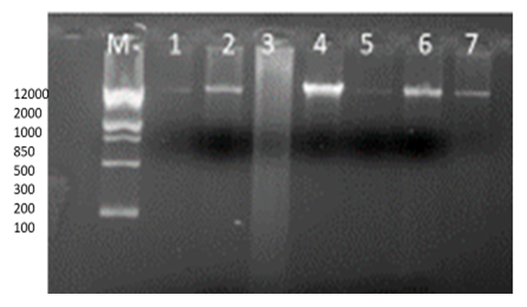
Plasmid profile was carried out on the 7of the 25 multiple antibiotic resistance Pseudomonas aeruginosa isolates. All the isolates 7(100%) were found to possess at least one plasmid bands. Plasmids were detected from three of the clinical isolates (1-3), the other four of the plasmids detected from vegetable isolates (5-7) as depicted in figure 1 below.
Figure 1. PCR analysis of Plasmids in Pseudomonas aeruginosa isolates
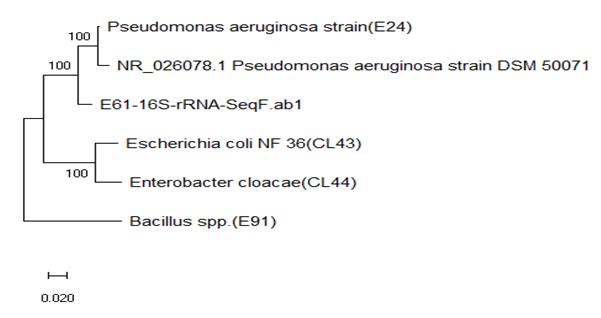
Vegetable isolate Clones (E24) Pseudomonas aeruginosa isolate clone appear to represent a phylogenetically coherent with the gene bank strain (figure 2). Sequences of clones E24(MN871898) and NR-026078.1 from a gene bank were 100% similar and shared 100% sequence similarity with the sequence E61(MN871946) P. aeruginosa isolate from vegetable sample (figure 2).
Figure 2. Evolutionary relationship of 16s rRNA sequenced Pseudomonas aeruginosa isolates

3.3. Antibiotic Susceptibility Result of P. aeruginosa Isolates

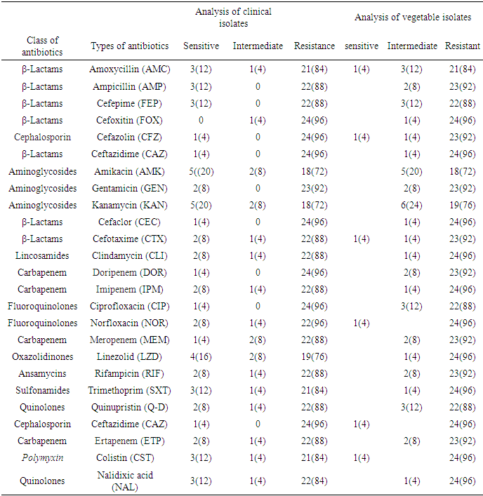
The antibiotic susceptibility results of the P. aeruginosa isolates are showed in (table 4) below. All the isolates were resistant to the 25 antibiotics tested. For clinical isolates which are tested for beta lactamase antibiotics Ceftazidime (CAZ), Cefaclor (CEC), Cefoxitin (FOX) and Ceftazidime (CAZ) were (96%) resistant, Ampicillin (AMP), Cefepime (FEP) and Cefotaxime (CTX) were (88%) resistant, and Amoxycillin (AMC) were (84%) resistance as mentioned in table 4. Also, the Pseudomonas isolates are resistant to other classes of antibiotics tested as indicated in table 4. For both clinical and vegetable isolates tested for 25 antibiotics 99% were resistant to more than three classes of antibiotics (figure 3). Most of the isolates were multiple antibiotic resistance to more than three classes of antibiotics. Also, isolates from vegetable sources and clinical samples were resistant to all common prescribed antibiotics in Gondar Ethiopia (table 4).
Table 4. Antibiotic Susceptibility patterns of P. aeruginosa isolates from vegetables and clinical samples
     
Figure 3. Antibiotic susceptibility pattern of clinical and vegetable Pseudomonas aeruginosa isolates the first three (S, I, R =clinical) second S, I, R=vegetables, ATCC2400 positive control

3.4. Analysis of Biofilm Formation in P. aeruginosa Isolates

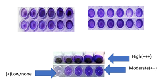
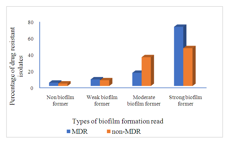
All isolates were further evaluated for biofilm production using the 96 well tissue culture plate. Ultimately, 88% (n=22) isolates produced biofilms, which were classified as strong, moderate, and weak based on their appearance. Among biofilm-producing isolates, 40% (n=10) produced strong, 32% (n=8) moderate, and 16% (n=4) weak biofilm patterns. Isolates that had not produced an observable amount of biofilm were classified as nonproducers and their strength was 12% (n=3), as demonstrated in table 5. The relationship between biofilm production and antibiotic resistance were analyzed, 76% of MDR isolates were strong biofilm formers and 46% of non MDR isolates were strong biofilm formers (figure 4).
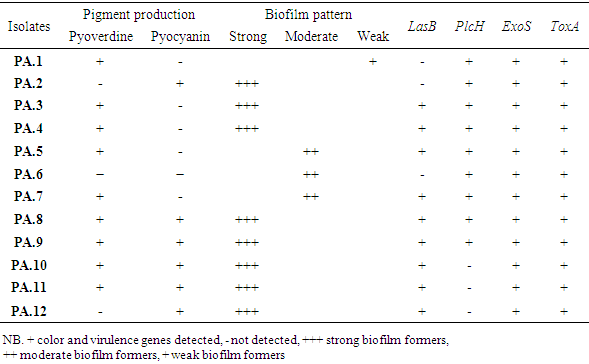
Table 5. Phenotypic characteristics and virulence genes profile antibiotic resistant Pseudomonas aeruginosa
     
Figure 4. Analysis of biofilm formation as high, moderate, and non-biofilm producers differentiated with crystal violet staining in tissue culture plates (TCP), OD570
In our study, biofilm producing P. aeruginosa showed (88%, 96%), (88%, 92%), (88%, 88%), (96%, 96%), (72%, 72), and (72%, 76%) resistance to Imipenem, Meropenem, cefepime, cefoxitin, amikacin and kanamycin, in clinical and vegetable isolates respectively. Resistance to aminoglycosides, beta lactamase and carbapenem resistant were comparatively higher among biofilm producing P. aeruginosa than nonproducer (Statistically significant; P < 0.001). MDR were comparatively higher among biofilm producing than nonproducer (statistically significant; P < 0.001). Relationship between MDR pattern of biofilm producing and nonproducing P. aeruginosa are shown in figure 5 below.
Figure 5. The relationship between biofilm formation and Multi- Drug Resistant Pseudomonas aeruginosa isolates

3.5. Analysis of Virulence Genes in P. aeruginosa Isolates

Beta-lactamase-producing Pseudomonas isolates (n=12) were further analyzed for phenotypic and virulence gene profiles. P. aeruginosa produced characteristic pigments in the form of pyoverdine or pyocyanin. A total of 96% (n=24) isolates produced these pigments. Among pigment-producing isolates, 76% (n=19) demonstrated pyoverdine and 24% (n=6) exhibited pyocyanin, as shown in table 5.
Virulence genes were investigated among twelve randomly selected beta-lactamase-producing isolates using PCR. Among them, 75% (n = 9) isolates were found to be LasB positive, while 100% (n = 12) were ExosA producers, 75% (9) were PlcH producers and 100% (12) were ToxA producers. Furthermore, all the virulence screened isolates coproduced more than two type of virulence factors (Figure 6 A, B, C, and D).
Figure 6. PCR analysis of virulence factors in P. aeruginosa: A) Tox A (352bp); B) LAS B (300bp); C) PlC H (307bP); and D) ExoS (504bP), (lane1-5 vegetable and 6-12 clinical) lane 8 ATCC 27853 + control
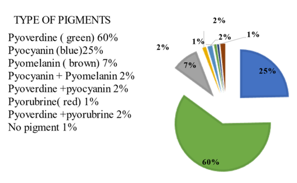
Among all the 24 Pseudomonas aeruginosa isolates 60% were pyoverdine fluorescein yellow green pigment producers, 25% were pyocyanin blue pigment producers and 18% others as depicted in figure 7 below.
Figure 7. Pigment production in P. aeruginosa isolates
3.5.1. Analysis of Resistance Gene Regions in Pseudomonas aeruginosa Isolates
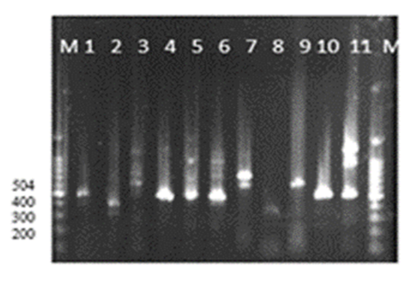
Colony PCR were performed in 12 selected P. aeruginosa isolates to detect OprL(504bp) resistance gene regions. Based on the PCR results out of the selected and screened P. aeruginosa isolates OprL(504bp) were detected in 6 of the vegetable and 5 of the clinical isolates Figure 8.
Figure 8. PCR result of Opr L(540bp) 1-6 vegetables and 7-11 clinical isolates

4. Discussions

Pseudomonas aeruginosa remains one among the foremost important opportunistic pathogen resistances to a variety of antimicrobial agents worldwide [49,63]. P. aeruginosa pathogens causes nosocomial infection by secreting various toxins in immunocompromised people [5,59]. The primary focus of this research was to determine the total population and distribution of the Pseudomonas aeruginosa pathogens in leafy vegetables, study their antibiotic susceptibility, resistance genes, biofilm formation, and other associated virulence factors comparing the result with the clinical isolates from Gondar University Hospital.
With regard to the bacterial load of the leafy vegetables, the mean aerobic mesophilic count of cauliflower, lettuce spinach and cabbage were 4.9x10¹⁰, 4.7x10¹⁰, 4.5 x10¹⁰, and 4.0x10⁹ cfu/g, respectively. These numbers are higher than previously reported data the mean aerobic mesophilic count of 1.68x10⁵ for cauliflower, 1.50x10⁵ for cabbage and 1.53x10⁵ for spinach [3,66]. The higher bacterial load in this study may be partly due to the use of contaminated irrigation water and organic fertilizers in the farms, coupled with the poor hygienic environment where open defecation is not uncommon. Together these factors contribute to the microbial contamination during preharvest, harvesting, and poor handling practices at the post-harvesting [23,44]. These poor sanitary conditions were clearly observed when the vegetables were been sourced from the various markets. The maximum aerobic plate count (9.8x10⁶ cfu/g) was from the vegetables obtained at Arada market while the lowest aerobic plate count was in the vegetables from Pizza market. Comparison of the mean aerobic mesophilic count of the vegetables based on the different markets shows that there was a statistically significant difference (P=0.006<0.05).
In an attempt to evaluate any causal relationship between the clinically relevant bacterial population from the vegetables and Gondar community Hospital, the P. aeruginosa isolates from both sources were compared on the bases of their phylogenetic, phenotypic characteristics, plasmid profiling, antibiotic susceptibility, resistance genes and key virulence factors. The 16SrRNA sequencing and the phylogenetic relationship of the randomly selected isolates indicated the clinical and vegetable isolates features a relationship as they grouped together. As studies suggested that antibiotic resistance genes in human bacterial pathogens originate from different bacterial genetic sources including plasmid [6]. The genes carried in plasmids provide bacteria with genetic advantages, like antibiotic resistance and besides their act as a carrier, these plasmids play a big role as a transferring agent to antibiotic resistant mobile genes [2,32]. During this study, the plasmid profile for all selected isolates confirmed that every isolate had a minimum of one plasmid.
Antibiotic susceptibility test analysis revealed the isolates were resistant to 99% the antibiotics used and the frequency of multiple antibiotic resistance was 98% in both vegetable and clinical isolates. In the past two to three decades, many reports confirmed an increasing multiple antibiotic resistance among P. aeruginosa isolated in several parts of the world [17,35,71]. Multiple antibiotic resistance in P. aeruginosa are mediated by several mechanisms, including the assembly of multidrug efflux systems, enzyme production, or outer membrane protein (porin) loss and target mutations [73]. Carbapenems are the effective antibiotic against multiple antibiotic resistance (MDR) isolates. However, the increasing frequency of carbapenem-resistant P. aeruginosa has recently become a worldwide challenge [42]. Our results showed high resistance to all or any carbapenem antibiotics (88%, 96%) imipenem, (88%, 92%) meropenem, (96%, 92%) doripenem and (88%, 92%) ertapenem resistant in clinical and vegetable isolates, respectively, which agrees to previous report, resistance to imipenem in Tehran was within the range of 16% - 90% [28,29,36].
Biofilm formation and virulence factors production were also further analyzed in extended spectrum beta lactamases (ESBL)-producing P. aeruginosa, and it had been established that a robust correlation exists between secretion of poisons and beta-lactamase producers [34]. Other factors contributing to the virulence of P. aeruginosa detected in this study are pigment production which have positive correlation with antibiotic resistance. In this study all P. aeruginosa isolates produced pigment. It had been revealed that pigment production was more significantly related to antibiotic resistance than the assembly of virulence factors like elastases and proteases in clinical isolates of P. aeruginosa [18]. In another study, pigment production was according to our results [73].
Biofilm formation and the correlation of multiple antibiotic resistance were analyzed and compared with previously reported cases of Pseudomonas isolates, and it had been noted that the prevalence of biofilm formation was above previous study results, which implied that biofilm formation features a strong correlation with the antibiotic resistance pattern [21]. Production of biofilm can boost the strength of ESBL-carrying bacteria more pathogenically compared with non-ESBL producers, as in our case, where all were biofilm producers [47]. Our data identified that 96% of the P. aeruginosa isolates produced biofilm. During a study by [26,61] in Iran, 96.9% of the isolates produced biofilm, which is in correlation with our results. Previously Reported studies of the connection between biofilm formation and antibiotic resistance/susceptibility revealed that the multiple antibiotic resistant isolates displayed significant biofilm formation as compared to susceptible isolates, probably due to the delayed penetration of antibiotics inside the bacterial cell. In this study, the analysis of the relationship between biofilm formation and multi drug resistant (MDR) confirmed that 72% of the multiple antibiotic resistant isolates displayed significant biofilm production compared to susceptible isolates, our results are according to previous reports [1,10]. This could be due to the delayed penetration of antibiotics target site in the bacterial cell.
In the current study a diverse antibiotic resistance and virulence determinant isolates were also detected among the ubiquitous Pseudomonas aeruginosa. The antibiotic resistant gene OprL (504bp) was present in 98% of the isolates. Similarly, the virulence factors ExoS and PlcH were detected in 100% of the P. aeruginosa isolates compared to ExoS (84.0%) and Plc H (71.0%) by [24,58]. However, the ToxA and LasB virulence factors were detected in 75% of the P. aeruginosa isolates. Distribution of virulence factor ExoS was higher compared with the info shown in another study previously. The ToxA prevalence was lower in our study than that during a previous study. Expression of virulence factors are often retarded using azithromycin, which inhibits autoinducers that are involved within the expression of those genes [68]. Overall, expression levels of both ExoS and ToxA were higher in our study as compared with those during a previous study [35]. When ExoS acts as a mitogen, it can promote activation of T lymphocytes resulting in inflammation, which is that the explanation for pathogenesis in CF patients [8]. An elevated incidence of ExoS was reported in drug resistance isolates during a study conducted in Iran [17]. The expression of those virulence factors is required to cause infection in patients, particularly in those having pulmonary infection [65]. Las B incidence was also higher in our isolates as compared with cases reported in another study, which involved the detection of virulence genes in pigment- and non-pigment producing P. aeruginosa [18]. ESBL-harboring bacteria have more LasB activity than non-ESBL-harboring bacteria, which highlights their strong force with the drug resistance pattern [36]. Other previous study reported that, the occurrence of LasB and PlcH virulence factors in clinical isolates of P. aeruginosa, however, the prevalence of both virulence factors was higher in our findings [26].

5. Conclusions

Antibiotic resistant bacterial pathogens represent a global threat to human health. The acquisition of antimicrobial resistance genes bacterial pathogens has reduced the treatment options for serious infections, increased the burden of disease, and increased death rates due to treatment failure and requires a coordinated global response for antimicrobial resistance surveillance. This looming health threat has restimulated interest in the development of new antimicrobial therapies, has demanded the need for better patient care, and has facilitated heightened governance over stewardship practices. In the current study beta-lactamase-harboring bacteria exhibit a robust antibiotic resistance pattern against different groups of antibiotics. Besides the assembly of resistance-related enzymes, production of various virulence genes and pigments could be the explanation for development of the resistance pattern in P. aeruginosa. Further studies are going to be helpful in revealing the correlation between the antibiotic resistance pattern and virulence factor expression.

ACKNOWLEDGEMENTS

I would like to express my sincere gratitude to Gondar university Microbiology department who assisted us in providing the resources for this project and Howard university biology department for the technical assistance.

Conflict of Interest

There is no conflict of interest with any party in this research work.

References

[1]  A. Abdi-Ali, M. Mohammadi-Mehr, Y. Agha Alaei, Bactericidal activity of various antibiotics against biofilm-producing Pseudomonas aeruginosa, International Journal of Antimicrobial Agents, Volume 27, Issue 3, 2006, Pages 196-200.
[2]  Akeredolu DO., Akinnibosun FI., Udochukwu U. (2017). Isolation and identification of plasmids of bacteria from petroleum products contaminated soil. Journal of Bacteriology & Mycology, Volume 5 Issue 6 – 2017.
[3]  Akoachere, J., Tatsinkou, B. F., & Nkengfack, J. M. (2018). Bacterial and parasitic contaminants of salad vegetables sold in markets in Fako Division, Cameroon and evaluation of hygiene and handling practices of vendors. BMC research notes, 11(1), 100. https://doi.org/10.1186/s13104-018-3175-2.
[4]  Altschuler M, Heddens DK, Diveley RR Jr, Kresheck GC (1994) Plasmid DNA isolation utilizing a novel nonionic detergent. Biotechniques 17: 434–436.
[5]  Balasubramanian, D., Schneper, L., Kumari, H., & Mathee, K. (2013). A dynamic and intricate regulatory network determines Pseudomonas aeruginosa virulence. Nucleic acids research, 41(1), 1–20. https://doi.org/10.1093/nar/gks1039.
[6]  Bennett P. M. (2008). Plasmid encoded antibiotic resistance: acquisition and transfer of antibiotic resistance genes in bacteria. British journal of pharmacology, 153 Suppl 1(Suppl 1), S347–S357. https://doi.org/10.1038/sj.bjp.0707607.
[7]  Birnboim, H. C., & Doly, J. (1979). A rapid alkaline extraction procedure for screening recombinant plasmid DNA. Nucleic acids research, 7(6), 1513–1523. https://doi.org/10.1093/nar/7.6.1513.
[8]  Bruno TF, Buser DE, Syme RM, Woods DE, Mody CH., (1998). Pseudomonas aeruginosa exoenzyme S is a mitogen but not a superantigen for human T lymphocytes. Infect Immun. 1998; 66:3072-3079.
[9]  C. Van Delden, B.H. Iglewski. Cell-to-cell signaling and Pseudomonas aeruginosa infections. Emerg Infect Dis, 4 (1998), pp. 551-560.
[10]  C.T. O'Loughlin, L.C. Miller, A. Siryaporn, K. Drescher, M.F. Semmelhack, B.L. Bassler, A quorum-sensing inhibitor blocks Pseudomonas aeruginosa virulence and biofilm formation Proc Natl Acad Sci U S A, 110 (2013), pp. 17981-17986.
[11]  Chowdhury EH, Akaike T (2005) Rapid isolation of high quality, multimeric plasmid DNA using zwitterionic detergent. J Biotechnol 119: 343–347.
[12]  Chowdhury K (1991) One step 'miniprep' method for the isolation of plasmid DNA. Nucleic Acids Res 19: 2792.
[13]  Currier TC, Nester EW (1976) Isolation of covalently closed circular DNA of high molecular weight from bacteria. Anal Biochem 76: 431–441.
[14]  D.E. Woods, S.J. Cryz, R.L. Friedman, B.H. Iglewski Contribution of toxin A and elastase to virulence of Pseudomonas aeruginosa in chronic lung infections of rats. Infect Immun, 36 (1982), pp. 1223-1228.
[15]  Donlan, R. M., & Costerton, J. W. (2002). Biofilms: survival mechanisms of clinically relevant microorganisms. Clinical microbiology reviews, 15(2), 167–193. https://doi.org/10.1128/CMR.15.2.167-193.2002.
[16]  E. Déziel, Y. Comeau, R. Villemur. Initiation of biofilm formation by Pseudomonas aeruginosa 57RP correlates with emergence of hyperpiliated and highly adherent phenotypic variants deficient in swimming, swarming, and twitching motilitiesJ Bacteriol, 183 (2001), pp. 1195-1204.
[17]  Fazeli N, Momtaz H., (2014). Virulence gene profiles of multidrug-resistant Pseudomonas aeruginosa isolated from Iranian hospital infections. Iran Red Crescent Med J. 2014; 16: e15722.
[18]  Finlayson EA, Brown PD., (2011). Comparison of antibiotic resistance and virulence factors in pigmented and non-pigmented Pseudomonas aeruginosa. West Indian Med J. 2011; 60:24-32.
[19]  Flores-Díaz, M., Monturiol-Gross, L., Naylor, C., Alape-Girón, A., & Flieger, A. (2016). Bacterial Sphingomyelinases and Phospholipases as Virulence Factors. Microbiology and molecular biology reviews: MMBR, 80(3), 597–628. https://doi.org/10.1128/MMBR.00082-15.
[20]  Fricks-Lima J, Hendrickson CM, Allgaier M, Zhuo H, Wiener-Kronish JP, Lynch SV, Yang K. (2011). Differences in biofilm formation and antimicrobial resistance of Pseudomonas aeruginosa isolated from airways of mechanically ventilated patients and cystic fibrosis patients. Int J Antimicrob Agents. 2011 Apr; 37(4): 309-15.
[21]  Ghanbarzadeh Corehtash Z, Khorshidi A, Firoozeh F, Akbari H, Mahmoudi Aznaveh A., (2015). Biofilm formation and virulence factors among Pseudomonas aeruginosa isolated from burn patients. Jundishapur J Microbiol; 8: e22345.
[22]  Guerry P, LeBlanc DJ, Falkow S (1973) General method for the isolation of plasmid deoxyribonucleic acid. J Bacteriol 116: 1064–1066.
[23]  Gultie, A., & Sahile, S. (2013). Microbial Spectrum of Fruit in Gondar town Markets, North Western Ethiopia. Journal of Microbiology Research, 3, 1-10.
[24]  Hassuna, N. A., Mandour, S. A., & Mohamed, E. S. (2020). Virulence Constitution of Multi-Drug-Resistant Pseudomonas aeruginosa in Upper Egypt. Infection and drug resistance, 13, 587–595. https://doi.org/10.2147/IDR.S233694.
[25]  Head NE, Yu H., (2004). Cross-sectional analysis of clinical and environmental isolates of Pseudomonas aeruginosa: biofilm formation, virulence, and genome diversity. Infect Immun; 72:133-144.
[26]  I. Mitov, T. Strateva, B. Markova. Prevalence of virulence genes among Bulgarian nosocomial and cystic fibrosis isolates of Pseudomonas aeruginosa, Braz J Microbiol, 41 (2010), pp. 588-595.
[27]  Iglewski BH. Pseudomonas. In: Baron S, editor. Medical Microbiology. 4th edition. Galveston (TX): University of Texas Medical Branch at Galveston; 1996. Chapter 27. Available from: https://www.ncbi.nlm.nih.gov/books/NBK8326/.
[28]  Ilori MON, Amund DL. Degradation of anthracene by bacteria isolated from oil polluted tropical soils. Z Naturforsch. 2000; 55(11–12): 890–897.
[29]  Itah AY, Essien JP., (2005). Growth profile and hydrocarbonoclastic potential of microorganisms isolated from tarballs in the bight of bonny. Nigeria. World J Microbial Biotechnol. 2005; 21(6–7): 1317–1322.
[30]  Jaffe, R. I., Lane, J. D., & Bates, C. W. (2001). Real-time identification of Pseudomonas aeruginosa direct from clinical samples using a rapid extraction method and polymerase chain reaction (PCR). Journal of clinical laboratory analysis, 15(3), 131–137. https://doi.org/10.1002/jcla.1016.
[31]  Jimenez, P. N., Koch, G., Thompson, J. A., Xavier, K. B., Cool, R. H., & Quax, W. J. (2012). The multiple signaling systems regulating virulence in Pseudomonas aeruginosa. Microbiology and molecular biology reviews: MMBR, 76(1), 46–65. https://doi.org/10.1128/MMBR.05007-11.
[32]  John, J. F., Jr, & Twitty, J. A. (1986). Plasmids as epidemiologic markers in nosocomial gram-negative bacilli: experience at a university and review of the literature. Reviews of infectious diseases, 8(5), 693–704. https://doi.org/10.1093/clinids/8.5.693.
[33]  Jurado-Martín I, Sainz-Mejías M, McClean S. Pseudomonas aeruginosa: An Audacious Pathogen with an Adaptable Arsenal of Virulence Factors. International Journal of Molecular Sciences. 2021; 22(6): 3128. https://doi.org/10.3390/ijms22063128.
[34]  Khalil MA, Ibrahim Sonbol F, Mohamed AF, Ali SS., (2015). Comparative study of virulence factors among ESbetaL-producing and nonproducing Pseudomonas aeruginosa clinical isolates. Turk J Med Sci; 45:60-69.
[35]  Khattab MA, Nour MS, El Sheshtawy NM. Genetic identification of Pseudomonas aeruginosa virulence genes among different isolates. J Microb Biochem Technol. 2015; 7:274-277.
[36]  L.R. Beuchat. Ecological factors influencing survival and growth of human pathogens on raw fruits and vegetables. Microbes and Infection, 4 (4) (2002), pp. 413-423
[37]  LaSarre, B., & Federle, M. J. (2013). Exploiting quorum sensing to confuse bacterial pathogens. Microbiology and molecular biology reviews: MMBR, 77(1), 73–111. https://doi.org/10.1128/MMBR.00046-12
[38]  Lezin, G., Kosaka, Y., Yost, H. J., Kuehn, M. R., & Brunelli, L. (2011). A one-step miniprep for the isolation of plasmid DNA and lambda phage particles. PloS one, 6(8), e23457. https://doi.org/10.1371/journal.pone.0023457.
[39]  Limoli, D. H., Jones, C. J., & Wozniak, D. J. (2015). Bacterial Extracellular Polysaccharides in Biofilm Formation and Function. Microbiology spectrum, 3(3), 10.1128/microbiolspec. MB-0011-2014. https://doi.org/10.1128/microbiolspec.MB-0011-2014.
[40]  Lister, P. D., Wolter, D. J., & Hanson, N. D. (2009). Antibacterial-resistant Pseudomonas aeruginosa: clinical impact and complex regulation of chromosomally encoded resistance mechanisms. Clinical microbiology reviews, 22(4), 582–610. https://doi.org/10.1128/CMR.00040-09.
[41]  M.A. Khattab, M.S. Nour, N.M. El Sheshtawy Genetic identification of Pseudomonas aeruginosa virulence genes among different isolates. J Microb Biochem Technol, 7 (2015), pp. 274-277.
[42]  Meletis G. (2016). Carbapenem resistance: overview of the problem and future perspectives. Therapeutic advances in infectious disease, 3(1), 15–21. https://doi.org/10.1177/2049936115621709.
[43]  Moazami Goudarzi S, Eftekhar F. Assessment of carbapenem susceptibility and multidrug resistance in Pseudomonas aeruginosa burn isolates in Tehran. Jundishapur J Microbiol. 2013; 6(2): 162–5.
[44]  Moges, F., Endris, M., Mulu, A., Tessema, B., Belyhun, Y., Shiferaw, Y., Huruy, K., Unakal, C., & Kassu, A. (2014). The growing challenges of antibacterial drug resistance in Ethiopia. Journal of global antimicrobial resistance, 2(3), 148–154. https://doi.org/10.1016/j.jgar.2014.02.004.
[45]  Moradali, M. F., Ghods, S., & Rehm, B. H. (2017). Pseudomonas aeruginosa Lifestyle: A Paradigm for Adaptation, Survival, and Persistence. Frontiers in cellular and infection microbiology, 7, 39. https://doi.org/10.3389/fcimb.2017.00039.
[46]  Nikbin VS, Abdi-Ali A, Feizabadi MM, Gharavi S. Pulsed field gel electrophoresis & plasmid profile of Pseudomonas aeruginosa at two hospitals in Tehran, Iran. Indian J Med Res. 2007; 126(2): 146–51.
[47]  Norouzi F, Mansouri S, Moradi M, Razavi M., (2010). Comparison of cell surface hydrophobicity and biofilm formation among ESBL-and non-ESBL-producing Pseudomonas aeruginosa clinical isolates. Afr J Microbiol Res; 4:1143-1147.of Foods and Beverages, Food Microbiology and Food Safety, DOI 10.1007/978-1-4419-0826-1-6.
[48]  P. Lanotte, S. Watt, L. Mereghetti, N. Dartiguelongue, A. Rastegar-Lari, A. Goudeau, et al. Genetic features of Pseudomonas aeruginosa isolates from cystic fibrosis patients compared with those of isolates from other origins. J Med Microbiol, 53 (2004), pp. 73-81.
[49]  P.R. Jacome, L.R. Alves, A.B. Cabral, A.C. Lopes, M.A., (2012). Maciel. Phenotypic and molecular characterization of antimicrobial resistance and virulence factors in Pseudomonas aeruginosa clinical isolates from Recife, State of Pernambuco, Brazil. Rev Soc Bras Med Trop, 45 (2012), pp. 707-712.
[50]  Pachori, P., Gothalwal, R., & Gandhi, P. (2019). Emergence of antibiotic resistance Pseudomonas aeruginosa in intensive care unit; a critical review. Genes & diseases, 6(2), 109–119. https://doi.org/10.1016/j.gendis.2019.04.001.
[51]  Paul B, Cloninger C, Felton M, Khachatoorian R, Metzenberg S (2008) A nonalkaline method for isolating sequencing-ready plasmids. Anal Biochem 377: 218–222.
[52]  Pearson, J. P., Feldman, M., Iglewski, B. H., & Prince, A. (2000). Pseudomonas aeruginosa cell-to-cell signaling is required for virulence in a model of acute pulmonary infection. Infection and immunity, 68(7), 4331–4334. https://doi.org/10.1128/IAI.68.7.4331-4334.2000.
[53]  Prasad, A.S.B., Shruptha, P., Prabhu, V. et al. Pseudomonas aeruginosa virulence proteins pseudolysin and protease IV impede cutaneous wound healing. Lab Invest 100, 1532–1550 (2020). https://doi.org/10.1038/s41374-020-00478-1.
[54]  Q. Wei, L.Z. Ma. Biofilm matrix and its regulation in Pseudomonas aeruginosa. Int J Mol Sci, 14 (2013), pp. 20983-21005.
[55]  R.M. Ostroff, A.I. Vasil, M.L. Vasil. Molecular comparison of a nonhemolytic and a hemolytic phospholipase C from Pseudomonas aeruginosa J Bacteriol, 172 (1990), pp. 5915-5923.
[56]  Rossolini GM, Mantengoli E, Docquier JD, Musmanno RA, Coratza G. Epidemiology of infections caused by multiresistant gram-negatives: ESBLs, MBLs, pan resistant strains. New Microbiol. 2007;30(3):332–9.
[57]  S. Ingle., K. Mahajan, B.S. Kumar, S. Singh, R.K. Agrawal, R., (2013). Verma Over-expression, and immunogenicity of outer membrane protein L (OprL) of Pseudomonas aeruginosa Proc Natl Acad Sci India Sec B Biol Sci (2015), pp. 1-7.
[58]  Sabharwal, N., Dhall, S., Chhibber, S., & Harjai, K. (2014). Molecular detection of virulence genes as markers in Pseudomonas aeruginosa isolated from urinary tract infections. International journal of molecular epidemiology and genetics, 5(3), 125–134.
[59]  Sadikot, R. T., Blackwell, T. S., Christman, J. W., & Prince, A. S. (2005). Pathogen-host interactions in Pseudomonas aeruginosa pneumonia. American journal of respiratory and critical care medicine, 171(11), 1209–1223. https://doi.org/10.1164/rccm.200408-1044SO.
[60]  Sanchez, C.J., Mende, K., Beckius, M.L. et al. Biofilm formation by clinical isolates and the implications in chronic infections. BMC Infect Dis 13, 47 (2013). https://doi.org/10.1186/1471-2334-13-47.
[61]  Saxena, S., Banerjee, G., Garg, R., & Singh, M. (2014). Comparative Study of Biofilm Formation in Pseudomonas aeruginosa Isolates from Patients of Lower Respiratory Tract Infection. Journal of clinical and diagnostic research: JCDR, 8(5), DC09–DC11. https://doi.org/10.7860/JCDR/2014/7808.4330.
[62]  Serghini MA, Ritzenthaler C, Pinck L (1989) A rapid and efficient 'miniprep' for isolation of plasmid DNA. Nucleic Acids Res 17: 3604.
[63]  Shaan L. Gellatly, Robert E.W. Hancock, Pseudomonas aeruginosa: new insights into pathogenesis and host defenses, Pathogens and Disease, Volume 67, Issue 3, April 2013, Pages 159–173, https://doi.org/10.1111/2049-632X.12033.
[64]  Shahcheraghi F, Feizabadi MM, Yamin V, Abiri R, Abedian Z. Serovar determination, drug resistance patterns and plasmid profiles of Pseudomonas aeruginosa isolated from burn patients at two hospitals of Tehran (IRAN). Burns. 2003; 29(6): 547–51.
[65]  Shaver, C. M., & Hauser, A. R. (2004). Relative contributions of Pseudomonas aeruginosa ExoU, ExoS, and ExoT to virulence in the lung. Infection and immunity, 72(12), 6969–6977. https://doi.org/10.1128/IAI.72.12.6969-6977.2004.
[66]  Singh, S., Singh, S. K., Chowdhury, I., & Singh, R. (2017). Understanding the Mechanism of Bacterial Biofilms Resistance to Antimicrobial Agents. The open microbiology journal, 11, 53–62. https://doi.org/10.2174/1874285801711010053.
[67]  Sousa, A. M., & Pereira, M. O. (2014). Pseudomonas aeruginosa Diversification during Infection Development in Cystic Fibrosis Lungs-A Review. Pathogens (Basel, Switzerland), 3(3), 680–703. https://doi.org/10.3390/pathogens3030680.
[68]  Tateda K, Comte R, Pechere JC, Kohler T, Yamaguchi K, Van Delden C., (2001). Azithromycin inhibits quorum sensing in Pseudomonas aeruginosa. Antimicrobial Agents Chemother; 45:1930-1933.
[69]  Tavajjohi Z, Moniri R, Khorshidi A. Detection and characterization of multidrug resistance and extended-spectrum-beta-lactamase-producing (ESBLS) Pseudomonas aeruginosa isolates in teaching hospital. African J Microbiol Res. 2011; 5(20): 3223–8.
[70]  Tsegaye Alemayehu, (2020). Prevalence of multidrug-resistant bacteria in Ethiopia: a systematic review and meta-analysis, Journal of Global Antimicrobial Resistance Volume 26, September 2021, Pages 133-139. https://doi.org/10.1016/j.jgar.2021.05.017.
[71]  W.H. Sperber, M.P. Doyle (eds.), Compendium of the Microbiological Spoilage 135.
[72]  Waheed Ullah, Muhammad Qasim, Hazir Rahman, Yan Jie, Noor Muhammad, Beta-lactamase-producing Pseudomonas aeruginosa: Phenotypic characteristics and molecular identification of virulence genes, Journal of the Chinese Medical Association, Volume 80, Issue 3, 2017, Pages 173-177, ISSN 1726-4901.
[73]  World Health Organization (WHO, 2007) www.who.int/whr/2007/whr07_en.pdf.
[74]  Zhang, S., Yang, G., Ye, Q., Wu, Q., Zhang, J., & Huang, Y. (2018). Phenotypic and Genotypic Characterization of Klebsiella pneumoniae Isolated from Retail Foods in China. Frontiers in microbiology, 9, 289. https://doi.org/10.3389/fmicb.2018.00289.
[75]  Zhao, X., Yu, Z., & Ding, T. (2020). Quorum-Sensing Regulation of Antimicrobial Resistance in Bacteria. Microorganisms, 8(3), 425. https://doi.org/10.3390/microorganisms8030425.