Journal of Microbiology Research

p-ISSN: 2166-5885    e-ISSN: 2166-5931

2021;  11(1): 21-32

doi:10.5923/j.microbiology.20211101.03

Received: Nov. 9, 2021; Accepted: Nov. 19, 2021; Published: Nov. 26, 2021


Characterization of Carbapenem and β-lactam Resistance in Klebsiella pneumoniae Associated with Leafy Vegetables and Clinical Isolates from Gondar, Ethiopia

Cherinet Yigrem1, Roman Fisseha2, Broderick Eribo3

1Department of Biology, Howard University, 400 Bryan St. NW Washington DC, USA

2Department of Microbiology, University of Gondar, Gondar, Ethiopia

3Department of Biology, Howard University, 415 College St. NW Washington DC, USA

Correspondence to: Cherinet Yigrem, Department of Biology, Howard University, 400 Bryan St. NW Washington DC, USA.

Email:

Copyright © 2021 The Author(s). Published by Scientific & Academic Publishing.

This work is licensed under the Creative Commons Attribution International License (CC BY).
http://creativecommons.org/licenses/by/4.0/

Abstract

Klebsiella pneumoniae is a gram negative, encapsulated, non-motile bacterium that is found in the environment and has been associated with pneumonia in the alcoholic and diabetic patient population. The bacterium typically colonizes human mucosal surfaces of the oropharynx and gastrointestinal tract. Klebsiella pneumoniae is an opportunistic pathogen of medical importance. In this study a total of 27 Klebsiella pneumoniae strains were identified using standard tests. The average aerobic mesophilic bacterial count was 3.85 cfu/g and the prevalence of Klebsiella pneumoniae was highest in cauliflower and lettuce 7(25.9%), 4(14.8%) respectively followed by spinach 2(7.4%), and cabbage (2(7.4%). The antimicrobial susceptibility for 25 antibiotics indicated >98% of the Klebsiella pneumonia strains were carbapenems and β-lactam resistant. All the strains were resistant to the carbapenem drugs, (95%) Imipenem (IMP), (100%) Meropenem (MEM) (95%) Doripenem (DOR) and (87%) Ertapenem (ETP) resistance. Nine randomly selected strains were PCR screened for the presence of carbapenems genes demonstrated 9(100%) harbor resistance determinants (NDM-1), 8(88.9%) harbor resistance determinants blaOXA48, 9(100%) harbor resistance determinants blaIMP and 9(100%) harbor resistance determinants blaVIM. Furthermore, representative K. pneumonia strains profiled for plasmid DNA demonstrated that all the isolates harbor at least one plasmid.

Keywords: β-lactams, Carbapenems, K. pneumoniae, Plasmid and resistance genes

Cite this paper: Cherinet Yigrem, Roman Fisseha, Broderick Eribo, Characterization of Carbapenem and β-lactam Resistance in Klebsiella pneumoniae Associated with Leafy Vegetables and Clinical Isolates from Gondar, Ethiopia, Journal of Microbiology Research, Vol. 11 No. 1, 2021, pp. 21-32. doi: 10.5923/j.microbiology.20211101.03.

1. Introduction

Klebsiella pneumoniae is an opportunistic bacterium found in various microbiological niches such as soil; the skin, intestines, and feces of mammals; and food. K. pneumoniae has been known to cause bacteremia, pneumonia, and urinary tract infection [45,46]; the gastrointestinal carriage of K. pneumoniae has been said to be a predisposing factor for liver abscess [47], and hypervirulent K. pneumoniae strains have emerged as a predominant cause of pyogenic liver abscess [48]. Studies have reported that most pathogens in liver abscess are susceptible to broad-spectrum antibiotics such as fluoroquinolones and third- and fourth generation cephalosporins [47,49,50], but over the years, antibiotic resistance in K. pneumoniae has been increasingly observed in both nosocomial and community settings. Studies of K. pneumoniae having developed an acquired resistance to last-line antibiotics (i.e., carbapenems) [51,52,53] have been most concerning. The interplay between mechanisms of antibiotic resistance and virulence in K. pneumoniae is not well established. It is generally accepted that antibiotic resistance comes with a fitness cost and decreased virulence, but recent studies have suggested otherwise the development of antibiotic resistance in K. pneumoniae has been reported to augment virulence [54], and increased virulence is said to naturally evolve in response to, or potentially be shared among, bacteria, leading to acquired resistance [55].
Although K. pneumoniae is more commonly associated with nosocomial infections, food has also been reported to be a possible transmission vector [56]. K. pneumoniae has been isolated from raw meat [57,59,61,63], raw vegetables [58,62], fruit juice [59], and ready-to-eat (RTE) food. Several studies on K. pneumoniae in food have also reported its worrying resistance to antibiotics, on several occasions citing foodborne K. pneumoniae as resistant to three or more antibiotic classes (multidrug resistance) [60,64,65]. The presence of antimicrobial-resistant and multidrug-resistant (MDR) K. pneumoniae strains in the food chain and its potential contribution to the resistome, particularly to the resistance of clinically relevant bacteria, should not be taken lightly.
Among the antibiotics, β–lactam is a large class of drugs that has different sub-classes including penicillin’s, cephalosporins, carbapenems, and monobactam. In Gram-negative bacteria, resistance to β–lactams are mediated by different strategies such as production of β–lactamases, efflux pumps, and alteration of penicillin binding proteins. The major mechanism involved in B lactam resistance is the production of β–lactamases [38,68]. Different β–lactamases have been discovered starting with broad spectrum β–lactamases now designated as extended spectrum β–lactamases (ESBLs), β–lactamases with reduced sensitivity to β–lactamase inhibitors, Amp C and β–lactamases that hydrolyze carbapenems. As β–lactamases are inducible enzymes, the presence of β–lactam antibiotics in food is considered a risk factor that generates the intrinsic resistance of the gram-negative bacteria [39]. Consequently, the presence of antimicrobial resistant K. pneumoniae in food is a major health problem.
On the other hand, carbapenems are broad spectrum antibiotics that are kept as the last treatment option for infections caused by multiple antibiotic resistant Klebsiella pneumoniae isolates [67]. However, uncontrolled administration of carbapenems leads to the development and the spread of carbapenem resistant isolates, which are usually resistant to other antibacterial agents such as fluoroquinolones [3,23], and these isolates have limited treatment options leading to significantly high morbidity and mortality rates [5]. Carbapenem hydrolyzing β-lactamases have been reported to be increasingly widespread. Ambler molecular class A (KPC), class B (VIM, IMP, NDM) and class D (OXA-48) types are the most often found among Klebsiella pneumoniae isolated during serious nosocomial infections [35]. Carbapenem resistant K. pneumoniae strains have been also recently reported in many countries in the world [14,15,16,17,18,35]. Carbapenem resistance in Klebsiella has been developed due to the acquisition of genes encoding carbapenemases (carbapenem hydrolyzing enzymes) [36]. Three classes of carbapenemases (A, B and D) are involved in the carbapenem resistance i) class A (K. pneumoniae carbapenemase) [6,7] ii) class B, metallo-β-lactamases (MBLs) such as Verona integron metallo-beta-lactamase (VIM), Imipenemase metallo-beta-lactamase (IMP) and New Delhi metallo-beta-lactamase (NDM) [5,8] and iii) class D, oxacillin hydrolyzing beta-lactamases (OXA) [9,10]. The carbapenem hydrolyzing genes are commonly encoded on mobile genetic elements and are accompanied by other antibiotic resistant genes resulting in co-transfer of the antimicrobial resistance genes and rapid spread of bacterial infections [37].
In this study, we conducted a molecular characterization of 27 multiple antibiotic resistant, K. pneumoniae strains. These included isolates from vegetable samples and clinical sources from Gondar, Ethiopia. The 16S rRNA sequencing and colony PCR has been used to investigate and compare their genetic relatedness, antimicrobial resistance, and the presence of virulence determinants. The outcome of this study also provided new insights into the pathogenesis and resistance of the previously under-recognized human pathogenic bacteria in Ethiopia.

2. Materials and Methods

2.1. Sample Collection

Vegetable samples of cauliflower, spinach, lettuces, and cabbage were collected from four different retail markets in Gondar, Ethiopia from June to August. The samples were placed in a cold box at a temperature approximately 4°C, tightly sealed with sterile plastic wrap, and transported to an accredited laboratory and subjected to microbiological analysis within 24hrs. Forty clinical isolates were collected from patients who were admitted to a hospital of Gondar university of medical science college. The clinical strains were collected from different specimens and no duplicate isolates from the same patient were included in this study.
On the other hand, in each place vegetable samples were randomly collected from two retail markets and two farmers' markets. The samples were placed in separate sterile plastic bags and then immediately transported to the laboratory in a cooler with ice packs (below 4°C) and processed within 4–6 hrs.
About 25 g of each food sample was enriched in 225 mL nutrient broth (Huankai Ltd., Guangzhou, China) for 24hrs at 37°C. Thereafter, the enrichment was streaked onto MacConkey agar (Huankai Ltd., Guangzhou, India), followed by incubation at 37°C for 24 h. From MacConkey agar, three pink, mucoid colonies were picked up and cultured onto nutrient agar at 37°C for 24 h, followed by biochemical identification using API 20 E (BioMe′rieux, Marcy I′Etoile, France). Finally, 27 K. pneumoniae isolates were recovered in this study. Among these isolates, 15 were from ready-to-eat leafy vegetables, 12 were in clinical source from Gondar University Hospital. Confirmed cultures were preserved in Luria-Bertani broth containing 20% glycerol and stored at −80°C for further study.

2.2. Antimicrobial Susceptibility Testing

All confirmed Klebsiella pneumonia isolates were tested for antibiotic susceptibility using the Kirby–Bauer method [75] which performed by standard disk diffusion on Mueller–Hinton agar incubated at 37°C for 24 h, following the guidelines of the Clinical and Laboratory Standards Institute (The Clinical and Laboratory Standards Institute, 2015). A total of 25 antibiotics (Oxoid, Basingstoke, UK) were classified into 11 different groups according to the WHO (Organization, 2007): amoxicillin (AMC, 30 μg), ampicillin (AMP, 10 μg), cefepime (FEP, 10 μg), Cefoxitin (FOX, 30 μg), Cefazolin (CFZ) (30 μg), ceftazidime (CAZ, 30 μg), amikacin (AK, 30 μg), gentamicin (CN, 10 μg), kanamycin (K, 30 μg), Cefaclor (CEC) (25 μg), Cefotaxime (CTX) (30 μg), clindamycin (DA, 2 μg), Doripenem (DOR) (30 μg), Imipenem (IPM) (30 μg), ciprofloxacin (CIP, 5 μg), Norfloxacin (NOR, 10 μg), Meropenem (MEM) (30 μg), Linezolid (LZD, 30 μg), rifampicin (RD, 5 μg), Trimethoprim/sulphamethoxazole (SXT, 25 μg), Quinupristin/dalfopristin (QD, 15 μg), Ceftazidime (CAZ (30 μg), Ertapenem (ETP) (30μg), Nalidixic acid (NAL) (30 μg) and Colistin (CST) (30 μg).

2.3. Plasmid Profile and 16s rRNA Sequencing of Bacteria Isolates

The isolates were subjected to plasmid profile analysis via the OMEGA/miniprep method. An overnight culture of each bacteria isolate was prepared in 5 mL of nutrient broth. The broth culture was properly mixed by vertexing, and 1.5 mL was then transferred into a prelabeled Eppendorf tube. The tubes were then centrifuged for 2 minutes at 13 000 revolutions per minute (rpm) to harvest the bacterial cells. The supernatant was gently decanted, leaving about 100 μL of the broth culture, which was then vortexed at high speed until the bacterial cell pellet became completely suspended. OMEGA solution (300 μL; 25mM Tris, 10mM EDTA, 0.1N NaOH, 0.5% SDS) was then added to lyse the bacterial cells. It was mixed by inversion 3 to 5 times until the solution became slimy, after which 150 μL of 3.0M sodium acetate (pH 5.2) was added and again vortexed for about 10 seconds. It was further centrifuged at 13 000 rpm for 5 minutes to pellet out cellular debris and chromosomal DNA. The supernatant was then transferred into another labeled 1.5-mL Eppendorf tube, and 900 μL of cold absolute ethanol was added. The solution was centrifuged at 13 000 rpm for 10 minutes. The supernatant was discarded, and the white pellet containing the plasmid DNA was rinsed twice with 1000 μL of 70% ethanol.
The pellet was then air-dried, and 40 μL of TE buffer (10mM Tris, 1mM Na2EDTA) was added to resuspend the pellet. Electrophoresis of the extracted plasmid DNA was carried out on 1.8% agarose gel and run for 3 hours at 150-200 V with HIND III digest of lambda phage DNA (Fermentas) as the molecular weight marker. It was visualized with ethidium bromide via the ultraviolet transilluminator. 13 representative isolates were sent to the GENWIZ for 16S rRNA genomic Sequencing [41,42,43,44].

2.4. Primers and PCR Confirmation of Klebsiella pneumoniae

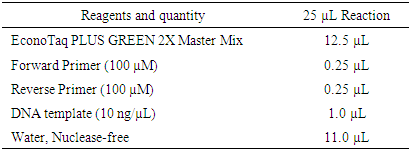
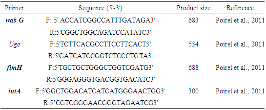
All confirmed K. pneumoniae isolates were grown overnight in lactose broth at 37°C. Genomic DNA was extracted using a commercial Universal DNA Extraction Kit (Sangon Biotech, Shanghai, China) according to the manufacturer's instructions. Confirmation of K. pneumoniae isolates was performed by PCR as previously described based on the reaction set up listed in table 1 [13]. The primer sequences and amplicon size are listed in table 2 and 3.
Table 1. Reaction set up for all resistance genes and virulence factors
     
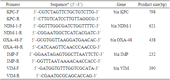
The characterization of bla-KPC, NDM-1, bla-VIM, bla-IMP, and blaOXA-48 β-lactamase and carbapenem’s genes was performed by PCR and sequencing in table 2 [24]. The presence of virulence factors was detected by PCR using the primers listed in table 3. All oligonucleotide primers were synthesized by Sangon Biotech. The PCR mixture (total volume 25 μL) contained 1 × DreamTaqTM Green PCR Master Mix (Fermentas, Waltham, MA, USA), 4 μL primer mixture, and 2 μL DNA template. PCR was conducted in a Bio-Rad PTC-200 Thermal Cycler (Bio-Rad, Hercules, CA, USA). The reference strain K. pneumoniae GIM 46117 (khe +) was used as a positive control. The amplified products were analyzed by electrophoresis on 1.5% agarose gels in 1 × TAE buffer (40 mM Tris–HCl, 1.18 mL acetic acid, 2 mM EDTA, pH 8.0), and the bands were visualized using an Image Quant 350 Capture system (GE Healthcare, Waukesha, WI, USA).
Table 2. Carbapenem’s gene regions in Klebsiella pneumonia [13]
     
Table 3. List of primers for virulence gene regions for Klebsiella pneumoniae
     

2.5. Statistical Analyses

To assess differences in Aerobic mesophilic bacterial count, total coliform count and fecal bacteria count among the vegetable samples, performed using the serial dilutions (log10 of cfu/gm) and the standard plat count formula (number of colonies times dilution factor divided by volume). Overall differences in the vegetable-borne clinically significant bacteria were analyzed using their means and standard deviation. Positive correlations in antibiotic sensitivity test between the bacterial isolates of the vegetable samples and clinical sources were assessed using a per mutational multivariate ANOVA. ANOVA and basic statistical tests(mean/SD) were also used to test for the resistance genes among the bacterial isolates. Significant differences in bacterial population were observed across vegetable types using the nonparametric Kruskal-Wallis test and between environmental samples and clinical sources using a t-test.

3. Result

3.1. Aerobic Mesophilic Count and Total Coliform Count of Vegetable Samples

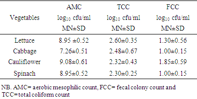
The vegetable types, aerobic mesophilic count, and total coliform counts of standard deviation and average in cfu/gm of samples are provided in (table 4) below. The total bacterial count result indicated that all vegetable samples contain large bacteria load. In general, among the leafy vegetables that we analyzed the samples with the highest average viable counts were cauliflowers (9.08cfu/ml), spinach (8.95cfu/ml), and lettuce (8.95cfu/ml), while cabbage (7.26cfu/ml) was the lowest (table 4). As the samples are collected randomly from three places there were no significant differences between the different markets. All samples were collected before their respective best before dates and even those with high counts were not spoiled based on their visual appearance.
The average total coliform count was high in cauliflower (2.37cfu/ml) on other hand the fecal colony count of bacteria was high in lettuce (2.60cfu/ml) and the lowest count was found in cabbage and spinach samples (table 4). Regarding to the market area the maximum mean viable count was found from Arada which was equal to 6.8x10⁶ cfu/ml. The lowest aerobic plate count was found around Piazza (1.5 x 10⁸ cfu/ml). The difference might be from the handling and sanitary differences in the market around Arada markets. The area was crowded with carrier animals and vehicles that emit dust particles; there were donkey and horse feces which are the source of different contaminant. Moreover, the vegetable handlers put vegetables on ground without using covering material and used the measuring balance for different commodities such as crops and cereal. These factors could increase the microbial load of leafy vegetables (table 4).
Table 4. The Aerobic mesophilic count from vegetable samples (log10 cfu/ml)
     

3.2. Antibiotic Susceptibility Testing

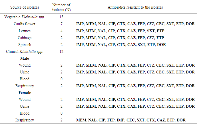
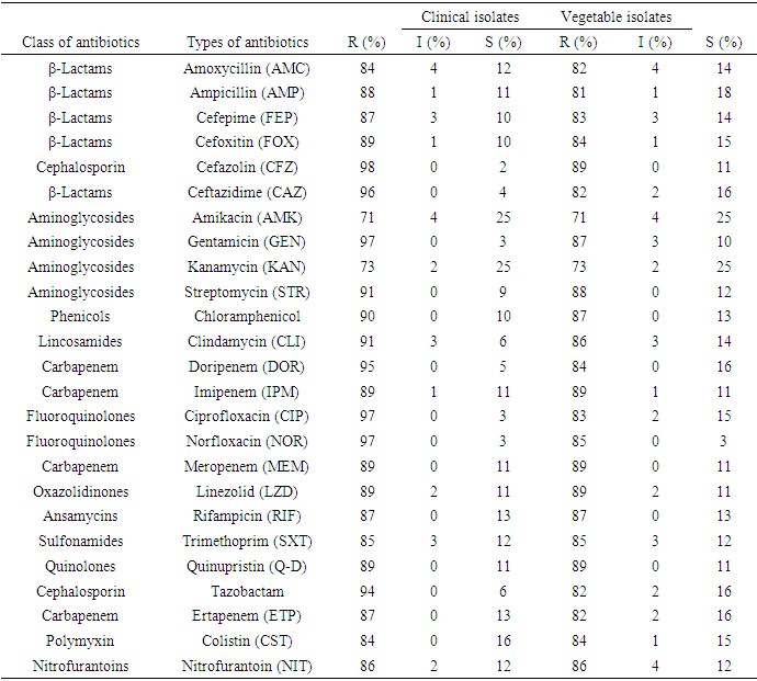
The antibiotic susceptibility results and multi drug resistance Klebsiella pneumonia isolates are showed in table 5 and 6. Overall, all the isolates were resistant to the 25 antibiotics tested. For clinical isolates which are tested for carbapenem antibiotics, (95%) were resistant to Doripenem (DOR), (89%) Imipenem (IPM), (89%) Meropenem (MEM) and (82%) Ertapenem (ETP) as mentioned in table 5. On the other hand, for vegetable isolates which are tested for carbapenem antibiotics (84%) were resistant to Doripenem (DOR), (89%) Imipenem (IPM), (89%) Meropenem (MEM) and (82%) Ertapenem (ETP) as mentioned in table 5. For both clinical and vegetable isolates tested for 25 antibiotics 99% were resistant to more than three classes of antibiotics. Most of the isolates were multiple antibiotic resistant to more than three classes of antibiotics. Also, isolates from vegetable sources and clinical samples were resistant to all common prescribed antibiotics in Gondar Ethiopia (table 5 and 6).
Table 5. Distribution of Antibiotics resistant among different K. pneumonia isolates and sample sources
     
Table 6. Antibiotics susceptibility Klebsiella pneumoniae strains for clinical and vegetable isolates
     

3.3. 16SrRNA Sequencing and Phylogenetic Relationships

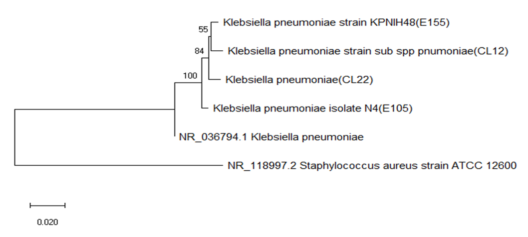
Molecular analysis of 16SrRNA sequenced vegetable and clinical isolates were aligned together. Aligned sequences were compared with the same regions of 16S rRNAs from gene bank and other bacterial groups. Phylogenetic trees were constructed by the least square’s method [76]. Five sequences from both sources were considered to group within the gene bank sequenced species (Fig. 1). The lines represented by clones CL12 (clinical isolate clone) and E155 (vegetable isolate clones) shared 55% similarity each other and shared 84% similarity in 16S rRNA sequence with sequence from the gene bank. The sequences of clones CL22 and E105 shared 100% similarity with sequence of species in gene bank. Vegetable isolate clones (E105) and clinical isolate clone (CL22) appear to represent a phylogenetically coherent group. Sequences of clones E105 and CL22 from a (vegetable and clinical clones respectively) were 100% similar and shared 84% sequence similarity with E155(vegetable) and CL12(clinical0 16S rRNA sequences.
Figure 1. Evolutionary relationship of 16s rRNA sequenced Klebsiella pneumonia isolates, (NR-036794.1 Klebsiella pneumonia from NCBI)

3.4. Plasmid Isolation and Profiling

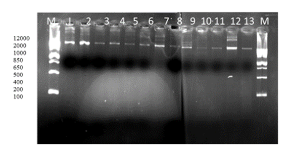
Plasmid profile was carried out on the 13 of the 32 multiple antibiotic resistance Klebsiella pneumonia isolates. All the isolates 13 (100%) were found to possess at least one plasmid bands. Plasmids were detected from eight of the clinical isolates (1-8), the other five of the plasmids detected from vegetable isolates (9-13) as depicted in figure 2 below.
Figure 2. Plasmid profile of Klebsiella pneumonia Lane 1-8 clinical isolates, L9-13 vegetable isolates, lane12 positive control

3.5. Analysis of the Carbapenems Resistant Gene Regions

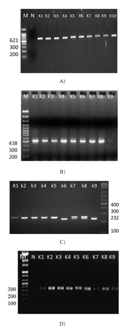
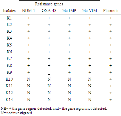
Two different multiplex PCR reaction mixtures were defined for four resistance genes (blaIMP(232bp), blaVIM(390bp), blaOXA-48(438bp), and blaNDM-1(621bp) were used to study nine K. pneumoniae strains. Among these isolates, oxacillinase (OXA-48) gene was determined in 8 out of 9 strains (90%); (NDM-1) 10 out of 10 strains (100%), bla IMP (232bp) 9 out of 9 strains (100%) and VIM bla (390 bp) 9 out of 9 strains (100%) (Figs. 3 A, B, C, D) and table 7 below.
Figure 3. A) PCR analysis of bla NDM-1 (621bp) resistance gene regions in Klebsiella pneumoniae; B) PCR analysis of bla OXA 48 (438bp) resistance gene regions in Klebsiella pneumoniae; C) PCR analysis of bla IMP (232bp) resistance gene regions in Klebsiella pneumoniae; D) PCR analysis of carbapenems resistant determinants of Klebsiella pneumoniae: VIM bla (390 bp) with clinical & vegetable strains; source of DNA: 1kb DNA molecular size marker; k2: positive control; k1: S3; k4: k5; k6: k7; k8: and k9 positive VIM bla390 bp
Table 7. Carbapenem's and β-lactamase resistance genes and virulence factor profile in Klebsiella pneumonia
     

3.6. Analysis of Virulence Gene Regions

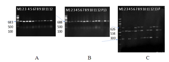
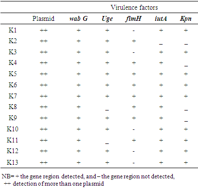
Different multiplex PCR reaction mixtures were defined for five virulence genes (uge, wabG, iutA, kpn and fimH) were used to study 13/27 K. pneumoniae strains. The five determined virulence gene regions, patterns of these strains, PCR positive for virulence genes, are given in fig 4(a-c) and table 8.
Figure 4. PCR amplification of K. pneumonia virulence gene regions, A) The WabG683bp and B) the fimH 688, C) iutA 300bp, uge534bp, and Kpn 626bp virulence regions of Klebsiella pneumoniae (1-8 vegetable and 9-13 clinical isolates)
Table 8. Distribution of virulence factors in Klebsiella pneumoniae isolates
     

4. Discussions

Multiple drug resistance (MDR) K. pneumoniae is an eminent threat to human health in an era when the discovery of new antibiotics lags behind the emergence and dissemination of antimicrobial resistance. Therefore, understanding the genetic and mechanistic basis for MDR becomes crucial, which may provide a hint on finding solutions to prevent its spread. The aim of this study was to determine the total population and distribution of multiple antibiotic resistance Klebsiella pneumonia associated with leafy vegetables and compare the antibiotic susceptibility patterns with clinical strains and determine the resistance genes in Gondar city districts. In the current study the bacterial load of the vegetables, the mean aerobic mesophilic count of cauliflower, lettuce, spinach, and cabbage were 9.08cfu/g, 8.95cfu/g, 8.95cfu/g, and 7.26 cfu/g, respectively. These numbers are higher than previous reports in Cameroon, the mean was AMC of 1.68MPN/g for cauliflower, 1.50MPN/g for cabbage and 1.53MPN/g for spinach [67]. The higher bacterial load in this study may be partly due to the use of contaminated irrigation water and organic fertilizers in the farms, coupled with the poor hygienic environment in the area. Together these factors contribute to the microbial contamination during preharvest, harvesting, and poor handling practices at the post-harvesting [68,69]. These poor sanitary conditions were clearly observed when the vegetables were been sourced from the various markets. The maximum aerobic plate count (9.08 cfu/g) was from the vegetables obtained at Arada market while the lowest aerobic plate count was in the vegetables from Pizza market. Additionally, the 16SrRNA sequencing and the phylogenetic relationship of the randomly selected isolates indicated the clinical and the vegetable isolates has a relationship as they grouped together. As studies suggested that antibiotic resistance genes in human bacterial pathogens originate from different bacterial genetic sources including plasmid [70]. The genes carried in plasmids provide bacteria with genetic advantages, such as antibiotic resistance. In this study, the plasmid profile for the thirteen selected isolates confirmed that each isolate harbor at least one plasmid [71].
On the other hand, antibiotics resistance has become a public health concern in developing countries including Ethiopia. Antibiotics resistance is dramatically accelerated when antibiotics are misused. This is critical, especially in developing countries where they are not only misused but are often underused due to financial constraints. Although large scale studies on antibiotics resistance in Ethiopia have not yet been conducted, the available reports indicate a trend towards increasing resistance rates among different bacteria [72]. Now a days, besides the common antibiotics carbapenem resistance is also widely spread all over the world and is becoming worrisome [24]. Hence, characterization of the carbapenem resistant isolates is the first step in the road map for controlling these isolates [23]. In this study, 99% of the Klebsiella pneumonia isolates were found to be resistant to beta lactams and carbapenems as well as all other various classes of antibiotics. Similarly, carbapenem-resistant Klebsiella pneumoniae isolates exhibited co-resistance to other multiple antibiotics [25,26]. The carbapenem-resistant isolates were carbapenemase positive and harbored the carbapenemase encoding genes; blaNDM-1, blaVIM-1, and blaOXA-48 particularly, the nine randomly selected isolates for multiplex PCR had multiple carbapenemases genes. Various studies detected carbapenemase genes in the carbapenem-resistant Klebsiella pneumoniae isolates especially bla VIM and bla NDM-1 however, carbapenemase bla IMP was not common in Enterobacteriaceae [24,27,28]. Klebsiella pneumoniae contained multiple metallo-β-lactamase genes have been previously detected in Germany [29], Italy [30], Colombia [31], and Sultanate of Oman [32]. OXA-48 was first identified from Klebsiella pneumoniae in Turkey [9] and spread of OXA-48 producing Klebsiella pneumoniae in the European countries and Mediterranean area has been observed [3]. NDM-1 (New Delhi metallo-β-lactamase), one of the most clinically significant carbapenemase producer, was first reported in New Delhi, India [8], followed by several case reports in United Kingdom, Pakistan and now worldwide [32]. Coproducing NDM-1 and OXA group carbapenem’s have been reported in different places [4] including Morocco [78], Oman [79] Singapore [16], the United States and Switzerland [80].
Several virulence factors were also detected in the K. pneumoniae isolates in the present study, wabG (92.3%), uge (84.6%), iutA (61.5%) and fimH (92.3%). These virulence factors encode the capsule, capsule lipoprotein, external membrane protein and enterobactin/ aerobactin production in both vegetable and clinical isolates. Similar findings were observed by [9,77] who found comparable virulence factors in K. pneumoniae.
It is known that virulence factors and antibiotic resistance are generally considered to play a significant role in bacterial pathogenesis [22]. Many studies have reported that virulence factors are associated with antibiotic resistance in pathogenic bacteria [73,74], however, the present study indicates that there is no significant correlation among virulence factors, carbapenem resistance and infection types. Recently, a few studies have indicated that quorum sensing affects these mechanisms [8,12]. Consequently, the study presented here demonstrated that virulence factors, antibiotic resistance and quorum sensing molecules should be considered in a collective manner in further studies on bacterial pathogenesis for developing effective treatments [21].
In this study, Klebsiella pneumoniae isolates were resistant to carbapenems and to several other antibiotics including beta lactams. Multiplex PCR confirmed the presence of carbapenems and multiple carbapenems in the isolates. Moreover, this study highlighted the prevalence of multiple copies of blaNDM-1 bla OXA48, bla IMP and blaVIM-1 in Klebsiella pneumoniae isolates with high-resistance levels. The situation is worrisome as the available treatment options are limited and the development of novel molecules to control this invasive infection is not easily accomplished. Hence, more restrictions on the administration of carbapenems should be applied in different hospitals to reduce the spread of the beta lactam and carbapenem resistant Klebsiella pneumoniae strains.

5. Conclusions

In conclusion, our results demonstrated the presence of beta lactam and carbapenem resistant K. pneumonia strains in leafy vegetables, which are related to the clinical strains. Leafy vegetables are considered “hot spots” of antibiotic resistant bacteria, resistance genes, and mobile genetic elements. The use of irrigation water might be one reason for the distribution of resistant bacteria and genetic elements through vegetables. However, still there remain several further knowledge gaps that must be filled to control and mitigate efficiently the emergence and spread of antibiotic resistance in the environment including vegetables. Antibiotic resistance is developing through complex interactions including de-novo mutation under clinical antibiotic pressure, the acquisition of mobile genes that evolve progressively in environmental bacteria, rapid demographic changes, human and animal population movement, as well as agricultural, landscape, climate, and environmental changes. Giving more attention to how anthropogenic activities are influencing the evolution of antimicrobial resistance and broad, multisectoral and interdisciplinary collaboration are key factors in addressing the rise of antibiotic resistance.

ACKNOWLEDGEMENTS

I would like to express my sincere gratitude to Dr. Garima Bansal and Dr. Adrian Allen who has been helping me throughout this study, to Dr. Gugssa Ayele who assisted us in providing the resources for this project and Dr. Abdullah M. and Dr. Bahailu Eshete who assisted with technical support. Also, would like to thank Gondar University Department of Microbiology and Howard University Biology department, applied microbiology laboratory.

Conflict of Interest

There is no conflict of interest within any party in this research work.

References

[1]  Huang Y.T., Hsueh P.R. (2008) Antimicrobial drug resistance in Taiwan. Int. J. Antimicrob. Agents; 32: S174–S178.
[2]  Pitout J.D. (2010) Infections with extended-spectrum beta-lactamase-producing Enterobacteriaceae: changing epidemiology and drug treatment choices. Drugs. 2010; 70: 313–333.
[3]  Nordmann P., Naas T., Poirel L. (2011) Global spread of carbapenemase-producing Enterobacteriaceae. Emerg. Infect. Dis. 17: 1791–1798.
[4]  Ørjan Samuelsen, Umaer Naseer, Nabil Karah, Paul Christoffer Lindemann, Anita Kanestrøm, Truls M. Leegaard, Arnfinn Sundsfjord, Identification of Enterobacteriaceae isolates with OXA-48 and coproduction of OXA-181 and NDM-1 in Norway, Journal of Antimicrobial Chemotherapy, Volume 68, Issue 7, July 2013, Pages 1682–1685, https://doi.org/10.1093/jac/dkt058.
[5]  Lee C-R., Lee J.H., Park K.S., Kim Y.B., Jeong B.C., Lee S.H. (2016) Global dissemination of carbapenemase-producing Klebsiella pneumoniae: epidemiology, genetic context, treatment options, and detection methods. Front. Microbiol. 7: 895.
[6]  Pournaras S., Kristo I., Vrioni G., Ikonomidis A., Poulou A., Petropoulou D., et al. (2010) Characteristics of meropenem heteroresistance in Klebsiella pneumoniae carbapenemase (KPC)-producing clinical isolates of K. pneumoniae. J. Clin. Microbiol. 48: 2601–2604.
[7]  Yigit H., Queenan A.M., Anderson G.J., Domenech-Sanchez A., Biddle J.W., Steward C.D., et al. (2001) Novel carbapenem-hydrolyzing beta-lactamase, KPC-1, from a carbapenem-resistant strain of Klebsiella pneumoniae. Antimicrob. Agents Chemother. 45: 1151–1161.
[8]  Yong D., Toleman M.A., Giske C.G., Cho H.S., Sundman K., Lee K., et al. (2009) Characterization of a new metallo-beta-lactamase gene, bla(NDM-1), and a novel erythromycin esterase gene carried on a unique genetic structure in Klebsiella pneumoniae sequence type 14 from India. Antimicrob. Agents Chemother. 53:5046–5054.
[9]  Palacios, M., Miner, T. A., Frederick, D. R., Sepulveda, V. E., Quinn, J. D., Walker, K. A., & Miller, V. L. (2018). Identification of Two Regulators of Virulence That Are Conserved in Klebsiella pneumoniae Classical and Hypervirulent Strains. mBio, 9(4), e01443-18. https://doi.org/10.1128/mBio.01443-18.
[10]  Walther-Rasmussen J., Høiby N. (2006) OXA-type carbapenemases. J. Antimicrob. Chemother. 2006; 57(3): 373–383.
[11]  Kaczmarek F.M., Dib-Hajj F., Shang W., Gootz T.D. (2006) High-level carbapenem resistance in a Klebsiella pneumoniae clinical isolate is due to the combination of blaACT-1 β-lactamase production, porin OmpK35/36 insertional inactivation, and down-regulation of the phosphate transport porin PhoE. Antimicrobes. Agents Chemother. 50: 3396–3406.
[12]  Wang X.D., Cai J.C., Zhou H.W., Zhang R., Chen G.X. (2009) Reduced susceptibility to carbapenems in Klebsiella pneumoniae clinical isolates associated with plasmid-mediated beta-lactamase production and OmpK36 porin deficiency. J. Med. Microbiol. 58: 1196–1202.
[13]  Zhang, S., Yang, G., Ye, Q., Wu, Q., Zhang, J., & Huang, Y. (2018). Phenotypic and Genotypic Characterization of Klebsiella pneumoniae Isolated from Retail Foods in China. Frontiers in microbiology, 9, 289. https://doi.org/10.3389/fmicb.2018.00289.
[14]  Pfeifer Y, Schlatterer K, Engelmann E, Schiller RA, Frangenberg HR, Stiewe D, Holfelder M, Witte W, Nordmann P, Poirel L (2012) Emergence of OXA-48-type carbapenemase-producing Enterobac-teriaceae in German hospitals. Antimicrobe Agents Chemother56: 2125–2128. http://dx.doi.org/10.1128/AAC.05315-11.
[15]  Azap Ö, Otlu B, Yeşilkaya A, Yakupoğulları Y (2013) Detection of OXA-48 like carbapenemase-producing Klebsiella pneumoniae in a tertiary care center in Turkey: molecular characterization and epi-demiology. Balkan Med J30: 259–260. http://dx.doi.org/10.5152/balkanmedj.2013.7499.
[16]  Balm MND, La MV, Krishnan P, Jureen R, Lin RTP and Teo JWP (2013) Emergence of Klebsiella pneumoniae coproducing NDM-type and OXA-181 carbapenemases.Clin Microbiol Infec19: E421–E423. http://dx.doi.org/10.1111/1469-0691.12247.
[17]  Doi Y, O’Hara JA, Lando JF, Querry AM, Townsend BM, Pasculle AW, Muto CA (2014). Co-Production of NDM-1 and OXA-232 by Klebsiella pneumoniae. Emerg Infec Dis20:163–164. http://dx.doi.org/10.3201/eid2001.130904.
[18]  Shibl A, Al-Agamy M, Memish Z, Senok A, Khader SA, Assiri A (2013) The emergence of OXA-48- and NDM-1-positive Klebsiella pneumoniae in Riyadh, Saudi Arabia. Int J InfectDis17: e1130–e1133. http://dx.doi.org/10.1016/j.ijid.2013.06.016.
[19]  M. Saı¨dani, et.al., (2012) Emergence of carbapenem-resistant OXA-48 carbapenems-producing Enterobacteriaceae in Tunisia, Journal of Medical Microbiology, 61, 1746–1749.
[20]  Sunday B. et.al., (2016) Microbes in Irrigation Water and Fresh Vegetables: Potential Pathogenic Bacteria Assessment and Implications for Food Safety, https://doi.org/10.1177/1535676016652231
[21]  Esra D. et.al. (2015) Klebsiella pneumoniae: Characteristics of carbapenem resistance and virulence factors. Vol. 62, No 4/2015. http://dx.doi.org/10.18388/abp.2015_1148 pp. 867-874.
[22]  Beceiro, A., Tomás, M., & Bou, G. (2013). Antimicrobial resistance and virulence: a successful or deleterious association in the bacterial world? Clinical microbiology reviews, 26(2), 185–230. https://doi.org/10.1128/CMR.00059-12.
[23]  Gupta N., Limbago B.M., Patel J.B., Kallen A.J. (2011) Carbapenem-resistant Enterobacteriaceae: epidemiology and prevention. Clin. Infect. Dis. 53:60–67.
[24]  Poirel L., Héritier C., Tolün V., Nordmann P. (2004) Emergence of oxacillinase-mediated resistance to imipenem in Klebsiella pneumoniae. Antimicrob. Agents Chemother. 48: 15–22.
[25]  Shi W., Li K., Ji Y., Jiang Q., Wang Y., Shi M., et al. (2013) Carbapenem and cefoxitin resistance of Klebsiella pneumoniae strains associated with porin OmpK36 loss and DHA-1 β-lactamase production. Braz. J. Microbiol. 44: 435–442.
[26]  Steinmann J., Kaase M., Gatermann S., Popp W., Steinmann E., Damman M., et al. (2011) Outbreak due to a Klebsiella pneumoniae strain harbouring KPC-2 and VIM-1 in a German university hospital, July 2010 to January 2011. Euro Surveill. 16:19944.
[27]  Sahin K., Tekin A., Ozdas S., Akin D., Yapislar H., Dilek A.R., et al. (2015) Evaluation of carbapenem resistance using phenotypic and genotypic techniques in Enterobacteriaceae isolates. Ann. Clin. Microbiol. Antimicrob. 14:44.
[28]  Nordmann P., Poirel L. (2014) The difficult-to-control spread of carbapenemase producers among Enterobacteriaceae worldwide. Clin. Microbiol. Infect. 20:821–830.
[29]  Steinmann J., Kaase M., Gatermann S., Popp W., Steinmann E., Damman M., et al. (2011). Outbreak due to a Klebsiella pneumoniae strain harbouring KPC-2 and VIM-1 in a German university hospital, July 2010 to January 2011. Euro Surveill. 16:19944.
[30]  Perilli M., Bottoni C., Grimaldi A., Segatore B., Celenza G., Mariani M., et al. (2013) Carbapenem-resistant Klebsiella pneumoniae harbouring blaKPC-3 and blaVIM-2 from central Italy. Diagn. Microbiol. Infect. Dis. 75:218–221.
[31]  Rojas L.J., Mojica M.F., Blanco V.M., Correa A., Montealegre M.C., De La Cadena E., et al. Emergence of Klebsiella pneumoniae coharboring KPC and VIM carbapenemases in Colombia. Antimicrob. Agents Chemother. 2013; 57:1101–1102.
[32]  Brink, A. J., Coetzee, J., Corcoran, C., Clay, C. G., Hari-Makkan, D., Jacobson, R. K., Richards, G. A., Feldman, C., Nutt, L., van Greune, J., Deetlefs, J. D., Swart, K., Devenish, L., Poirel, L., & Nordmann, P. (2013). Emergence of OXA-48 and OXA-181 carbapenemases among Enterobacteriaceae in South Africa and evidence of in vivo selection of colistin resistance as a consequence of selective decontamination of the gastrointestinal tract. Journal of clinical microbiology, 51(1), 369–372. https://doi.org/10.1128/JCM.02234-12.
[33]  Effah CY, Sun T, Liu S, Wu Y. Klebsiella pneumoniae: an increasing threat to public health. Ann Clin Microbiol Antimicrob. 2020 Jan 9; 19(1): 1. doi: 10.1186/s12941-019-0343-8. PMID: 31918737; PMCID: PMC7050612.
[34]  Richard D. et.al., 1994. A Phylogenetic Tree of 16S rRNA Sequences from Sulfate Reducing Bacteria in a Sandy Marine Sediment. Applied and environmental Microbiology, Sept. 1994, p. 3437-3439, Vol. 60, No.
[35]  Hammoudi Halat D, Ayoub Moubareck C. The Current Burden of Carbapenemases: Review of Significant Properties and Dissemination among Gram-Negative Bacteria. Antibiotics (Basel). 2020 Apr 16; 9(4): 186. doi: 10.3390/antibiotics9040186. PMID: 32316342; PMCID: PMC7235769.
[36]  Codjoe, F. S., & Donkor, E. S. (2017). Carbapenem Resistance: A Review. Medical sciences (Basel, Switzerland), 6(1), 1. https://doi.org/10.3390/medsci6010001.
[37]  Queenan AM, Bush K. Carbapenemases: the versatile beta-lactamases. Clin Microbiol Rev. 2007 Jul; 20(3): 440-58, table of contents. doi: 10.1128/CMR.00001-07. PMID: 17630334; PMCID: PMC1932750.
[38]  Chavez, M. V., Caicedo, L. D., & Castillo, J. E. (2019). Occurrence of β-Lactamase-Producing Gram-Negative Bacterial Isolates in Water Sources in Cali City, Colombia. International journal of microbiology, 2019, 1375060. https://doi.org/10.1155/2019/1375060.
[39]  Bush K. Past and Present Perspectives on β-Lactamases. Antimicrob Agents Chemother. 2018 Sep 24; 62(10): 01076-18. doi: 10.1128/AAC.01076-18. PMID: 30061284; PMCID: PMC6153792.
[40]  Sheu, C. C., Chang, Y. T., Lin, S. Y., Chen, Y. H., & Hsueh, P. R. (2019). Infections Caused by Carbapenem-Resistant Enterobacteriaceae: An Update on Therapeutic Options. Frontiers in microbiology, 10, 80. https://doi.org/10.3389/fmicb.2019.00080.
[41]  Akinde1, S. B., Abiodun A. Sunday, A. A., Folasade M. Adeyemi, F. M., Iyabobola B. Fakayode1, I. B, Odunola O. Oluwajide, O.O., Adetoun A. Adebunmi1, A. A., Julius K. Oloke, J. K., and Clement O. Adebooye. C.O. (2016). Microbes in Irrigation Water and Fresh Vegetables: Potential Pathogenic Bacteria Assessment and Implications for Food Safety, Applied Biosafety: Journal of ABSA International 2016, Vol. 21(2) 89-97.
[42]  Venkatesan, M. M., Goldberg, M. B., Rose, D. J., Grotbeck, E. J., Burland, V., & Blattner, F. R. (2001). Complete DNA sequence and analysis of the large virulence plasmid of Shigella flexneri. Infection and immunity, 69(5), 3271–3285. https://doi.org/10.1128/IAI.69.5.3271-3285.2001.
[43]  Clarridge J. E., 3rd (2004). Impact of 16S rRNA gene sequence analysis for identification of bacteria on clinical microbiology and infectious diseases. Clinical microbiology reviews, 17(4), 840–862. https://doi.org/10.1128/CMR.17.4.840-862.2004.
[44]  Janda, J. M., & Abbott, S. L. (2007). 16S rRNA gene sequencing for bacterial identification in the diagnostic laboratory: pluses, perils, and pitfalls. Journal of clinical microbiology, 45(9), 2761–2764. https://doi.org/10.1128/JCM.01228-07.
[45]  Karlowsky, J. A., Jones M. E., Thornsberry C., Friedland I. R., and Sahm D. F. (2003) Trends in antimicrobial susceptibilities among Enterobacteriaceae isolated from hospitalized patients in the United States from 1998 to 2001. Antimicrob. Agents Chemother. 47: 1672– 1680.
[46]  Siu, L. K, Fung C. P., Chang F. Y., Lee N., Yeh K. M., Koh T. H., and Ip M. (2011) Molecular typing and virulence analysis of serotype K1 Klebsiella pneumoniae strains isolated from liver abscess patients and stool samples from noninfectious subjects in Hong Kong, Singapore, and Taiwan. J. Clin. Microbiol. 49 (11): 3761– 3765.
[47]  Fung, C. P., Lin Y. T., Lin J. C., Chen T. L., Yeh K. M., Chang F. Y., Chuang H. C., Wu H. S., Tseng C. P., and Siu L. K. (2012) Klebsiella pneumoniae in gastrointestinal tract and pyogenic liver abscess. Emerg. Infect. Dis. 18 (8): 1322– 1325.
[48]  Lübbert, C., Wiegand J., and Karlas T. (2014) Therapy of liver abscess. Gastrointest. Med. Surg. 30: 334– 341.
[49]  Rahimian, J., Wilson T., Oram V., and Holzman R. S. (2004) Pyogenic liver abscess: recent trends in etiology and mortality. Clin. Infect. Dis. 39: 1654– 1659.
[50]  Siu, L. K, Yeh K. M., Lin J. C., Fung C. P., and Chang F. Y. (2012) Klebsiella pneumoniae liver abscess: a new invasive syndrome. Lancet Infect. Dis. 12: 881– 887.
[51]  Arnold, R. S., Thom K. A., Sharma S., Philips M., Johnson J. K., and Morgan D. J. (2011) Emergence of Klebsiella pneumoniae carbapenemase (KPC)–producing bacteria. South Med. J. 104 (1): 40– 45.
[52]  Paterson, D. L., and Bonomo R. A. (2005) Extended-spectrum B-lactamases: a clinical update. Clin. Microbiol. Rev. 18 (4): 657– 686.
[53]  Pitout, J. D. D., Nordmann P., and Poirel L. (2015) Carbapenemase-producing Klebsiella pneumoniae a key pathogen set for global nosocomial dominance. Antimicrob. Agents Chemother. 59 (10): 5873– 5884.
[54]  Kidd, T. J., Mills G., Sa-Pessoa J., Dumigan A., Frank C. G., Insua J. L., Ingram R., Hobley L., and Bengoechea J. A. (2017) A Klebsiella pneumoniae antibiotic resistance mechanism that subdues host defences and promotes virulence. EMBO Mol. Med. 9 (4): 430– 447.
[55]  Shroeder, M., Brooks B. D., and Brooks A. E. (2017) The complex relationship between virulence and antibiotic resistance. Genes 8 (39): 1– 23.
[56]  Calbo, E., Freixas N., Xercavins M., Riera M., Nicolás C., Monistrol O., Solé Mdel M., Sala M. R., Vila J., and Garau J. (2011) Foodborne nosocomial outbreak of SHV1 and CTX-M-15–producing Klebsiella pneumoniae: epidemiology and control. Clin. Infect. Dis. 52 (6): 743– 749.
[57]  Davis, G. S., Waits K., Nordstrom L., Weaver B., Aziz M., Gauld L., Grande H., Bigler R., Horwinski J., Porter S., Stegger M., Johnson J. R., Liu C. M., and Price L. B. (2015) Intermingled Klebsiella pneumoniae populations between retail meats and human urinary tract infections. Clin. Infect. Dis. 61 (6): 892– 899.
[58]  Falomir, M. P., Rico H., and Gozalbo D. (2013) Enterobacter and Klebsiella species isolated from fresh vegetables marketed in Valencia (Spain) and their clinically relevant resistances to chemotherapeutic agents. Foodborne Pathog. Dis. 10: 1002– 1007.
[59]  Ghenghesh, K. S., Belhaj K., El-Amin W. B., El-Nefathi S. E., and Zalmum A. (2004) Microbiological quality of fruit juices sold in Tripoli-Libya. Food Control 16 (10): 855– 858.
[60]  Guo, Y., Zhou H., Qin L., Pang Z., Qin T., Ren H., Pan Z., and Zhou J. (2016) Frequency, antimicrobial resistance, and genetic diversity of Klebsiella pneumoniae in food samples. PLoS ONE 11 (4): e0153561.
[61]  Kim, S. H., Wei C. I., Tzou Y. M., and An H. (2005) Multidrug-resistant Klebsiella pneumoniae isolated from farm environments and retail products in Oklahoma. J. Food Prot. 68 (10): 2022– 2029.
[62]  Puspanadan, S., Afsah-Hejri L., Loo Y. Y., Nillian E., Kuan C. H., Goh S. G., Chang W. S., Lye Y. L., John Y. H. T., Rukayadi Y., Yoshitsugu N., Nishibuchi M., and Son R. (2012) Detection of Klebsiella pneumoniae in raw vegetables using most probable number-polymerase chain reaction (MPN-PCR). Int. Food Res. J. 19 (4): 1757– 1762.
[63]  Wu, H., Liu B. G., Liu J. H., Pan Y. S., Yuan L., and Hu G. Z. (2012) Phenotypic and molecular characterization of CTX-M-14 extended-spectrum β-lactamase and plasmid-mediated ACT-like AmpC β-lactamase produced by Klebsiella pneumoniae isolates from chickens in Henan Province, China. Genet. Mol. Res. 11 (3): 3357– 3364.
[64]  Haryani, Y., Noorzaleha A. S., Fatimah A. B., Noorjahan B. A., Patrick G. B., Shamsinar A. T., Laila R. A. S., and Son R. (2007) Incidence of Klebsiella pneumoniae in street foods sold in Malaysia and their characterization by antibiotic resistance, plasmid profiling, and RAPD-PCR analysis. Food Control 18: 847– 853.
[65]  Sabota, J. M., Hoppes W. L., Ziegler J. R., DuPont H., Mathewson J., and Rutecki G. W. (1998) A new variant of food poisoning: enteroinvasive Klebsiella pneumoniae and Escherichia coli sepsis from a contaminated hamburger. Am. J. Gastroenterol. 93: 118– 119.
[66]  Tooke, C. L., Hinchliffe, P., Bragginton, E. C., Colenso, C. K., Hirvonen, V., Takebayashi, Y., & Spencer, J. (2019) β-Lactamases and β-Lactamase Inhibitors in the 21st Century. Journal of molecular biology, 431(18), 3472–3500. https://doi.org/10.1016/j.jmb.2019.04.002.
[67]  Akoachere, F. T., Bertrand, F., and Joseph, M. N. (2018). Bacterial and parasitic contaminants of salad vegetables sold in markets in Fako Division, Cameroon and evaluation of hygiene and handling practices of vendors. BMC Research Notes, 11, 100. https://doi.org/10.1186/s13104-018-3175-2.
[68]  Asmaru, G., Samuel S. (2013). Microbial Spectrum of Fruit in Gondar town Markets, Northwestern Ethiopia, Journal of Microbiology Research, 3(1): 1-10. doi: 10.5923/j.microbiology.20130301.01.
[69]  Bruce, S. K., Schick, D. G., Tanaka, L., Jimenez, E. M., & Montgomerie, J. Z. (1981). Selective medium for isolation of Klebsiella pneumoniae. Journal of clinical microbiology, 13(6), 1114–1116. https://doi.org/10.1128/jcm.13.6.1114-1116.1981.
[70]  Bennett P. M. (2008). Plasmid encoded antibiotic resistance: acquisition and transfer of antibiotic resistance genes in bacteria. British journal of pharmacology, 153 Suppl 1 (Suppl 1), S347–S357. https://doi.org/10.1038/sj.bjp.0707607.
[71]  Bruce Alberts, Alexander Johnson, Julian Lewis, Martin Raff, Keith Roberts, and Peter Walter (2002) Molecular Biology of the Cell, 4th edition
[72]  Moges, F., Endris, M., Mulu, A., Tessema, B., Belyhun, Y., Shiferaw, Y., Huruy, K., Unakal, C., & Kassu, A. (2014). The growing challenges of antibacterial drug resistance in Ethiopia. Journal of global antimicrobial resistance, 2(3), 148–154. https://doi.org/10.1016/j.jgar.2014.02.004.
[73]  El Fertas-Aissani, R., Messai, Y., Alouache, S., & Bakour, R. (2013). Virulence profiles and antibiotic susceptibility patterns of Klebsiella pneumoniae strains isolated from different clinical specimens. Pathologie-biologie, 61(5), 209–216. https://doi.org/10.1016/j.patbio.2012.10.004.
[74]  Mishra, M., Panda, S., Barik, S., Sarkar, A., Singh, D. V., & Mohapatra, H. (2020). Antibiotic Resistance Profile, Outer Membrane Proteins, Virulence Factors and Genome Sequence Analysis Reveal Clinical Isolates of Enterobacter Are Potential Pathogens Compared to Environmental Isolates. Frontiers in cellular and infection microbiology, 10, 54. https://doi.org/10.3389/fcimb.2020.00054.
[75]  A. W. Bauer, M.D., W. M. M. Kirby, M.D., J. C. Sherris, M.D., M. Turck, M.D., Antibiotic Susceptibility Testing by a Standardized Single Disk Method, American Journal of Clinical Pathology, Volume 45, Issue 4_ts, April 1966, Pages 493–496, https://doi.org/10.1093/ajcp/45.4_ts.493.
[76]  Joseph Felsenstein, An Alternating Least Squares Approach to Inferring Phylogenies from Pairwise Distances, Systematic Biology, Volume 46, Issue 1, March 1997, Pages 101–111, https://doi.org/10.1093/sysbio/46.1.101.
[77]  Kuş, H., Arslan, U., Türk Dağı, H., & Fındık, D. (2017). Hastane enfeksiyonu etkeni Klebsiella pneumoniae izolatlarında çeşitli virülans faktörlerinin araştırılması [Investigation of various virulence factors of Klebsiella pneumoniae strains isolated from nosocomial infections]. Mikrobiyoloji bulteni, 51(4), 329–339. https://doi.org/10.5578/mb.59716.
[78]  Barguigua, A., El Otmani, F., Lakbakbi El Yaagoubi, F., Talmi, M., Zerouali, K., & Timinouni, M. (2013). First report of a Klebsiella pneumoniae strain coproducing NDM-1, VIM-1 and OXA-48 carbapenemases isolated in Morocco. APMIS: acta pathologica, microbiologica, et immunologica Scandinavica, 121(7), 675–677. https://doi.org/10.1111/apm.12034.
[79]  Contreras, D. A., Fitzwater, S. P., Nanayakkara, D. D., Schaenman, J., Aldrovandi, G. M., Garner, O. B., & Yang, S. (2020). Coinfections of Two Strains of NDM-1- and OXA-232-Coproducing Klebsiella pneumoniae in a Kidney Transplant Patient. Antimicrobial agents and chemotherapy, 64(4), e00948-19. https://doi.org/10.1128/AAC.00948-19.
[80]  P. Nordmann and L. Poirel1, (2014) The difficult-to-control spread of carbapenem’s producers among Enterobacteriaceae worldwide, 10.1111/1469-0691.12719.