Journal of Microbiology Research

p-ISSN: 2166-5885    e-ISSN: 2166-5931

2016;  6(4): 75-81

doi:10.5923/j.microbiology.20160604.02


Distribution of Microbial Population Associated with Penaeus monodon Larvae in Marine Nursery Ponds in Mtwapa Creek, Kenya

Mutai Edwin Kipyegon 1, Mutai Raymond 2

1Department of Biological Sciences, School of Science, University of Eldoret, Kenya

2Department of Biotechnology, School of Agriculture and Biotechnology, University of Eldoret, Kenya

Correspondence to: Mutai Edwin Kipyegon , Department of Biological Sciences, School of Science, University of Eldoret, Kenya.

Email:

Copyright © 2016 Scientific & Academic Publishing. All Rights Reserved.

This work is licensed under the Creative Commons Attribution International License (CC BY).
http://creativecommons.org/licenses/by/4.0/

Abstract

One of the mainchallenges of prawnculture is poor survival of the larvae, which makes offspring production difficult, unreliable and expensive. The immune system of the larvae is immature and infections with pathogenic bacteria are the major cause of larval mortality. Since the larvae require life feed such as microalgae, rotifers and Artemia that need to be present in high densities, levels of dissolved and particulate nutrients are high. Due to this; saprophytic and opportunistic pathogenic bacteria thrive in cultures of marine larvae and cause infections. The present study aims to evaluate the occurrence and distribution of microbial diversity of bacterial population present in prawn larvae samples cultured in nursery ponds in Mtwapa Creek, Kenya. Microbial species were characterized based on morphological and biochemical tests. Total number of bacteria ranged from 21.7 × 10⁵ cfu to 32 × 105 cfu. Microorganisms presumably belong to genus Vibrio, Pseudomonas, Aeromonas, Alcaligenes, Bacillus, Staphylococcus, Hafnia and Fusarium. The absence of V. harveyi pathogen indicated that the fusant serve as the main source on increasing of resistance to diseases and thus reducing the mortality of prawn larvae.

Keywords: Microbes, Pathogenic bacteria, Mtwapa, Penaeus monodon

Cite this paper: Mutai Edwin Kipyegon , Mutai Raymond , Distribution of Microbial Population Associated with Penaeus monodon Larvae in Marine Nursery Ponds in Mtwapa Creek, Kenya, Journal of Microbiology Research, Vol. 6 No. 4, 2016, pp. 75-81. doi: 10.5923/j.microbiology.20160604.02.

1. Introduction

Inorganic and organic contaminants entering coastal waters may be concentrated by edible marine organisms to varying degrees from either water, their food or sediments [1]. Understanding the transfer of contaminants through the food web is critical to predict the exposure of humans to contaminants either through subsistence or commercial consumption of seafood and the possible health consequences of such exposure. In addition, such information is crucial in making accurate risk assessment for seafood safety purposes. Infectious microbial diseases and parasites are not only a major obstacle to closing lifecycles and breeding, but also inflict tremendous economic losses on the aquaculture industry. As an example, it isestimated that more than 3 billion US$ per year are lost as an effect of infectious diseases in prawn culture alone [2]. The most prevalent diseases in aquaculture are caused by bacteria (54.9%), followed by viruses (22.6%), parasites (19.4%) and fungi (3.1%) [3]. The most prevalent causative agents of bacterial infections in marine environment belong to the family Vibrionaceae of the γ proteobacteria [4]. The World’s most prominent and deleterious pathogen is Vibrio (Listonella) anguillarum. It has the ability to persist in nutrient-free sea water for more than one year and can increase a thousand fold in coastal sea water as an effect of carbohydrate-rich wastewater discharge, which may be a reason for its global abundance [5]. Vibrio harveyi has the largest impact on Crustacean and fish culture with multi drug resistant V. harveyi being a major problem in prawn culture. Outbreaks of photobacterium damselae spp. Piscicida, a member of the Vibrionaceae family, have been reported from marine aquacultures [6]. V. vulnificus and V. parahaemolyticus are of special concern, since they do not only commonly account for mortalities in aquaculture, but some strains can also cause disease in humans [7]. Pathogenic bacteria in sea water are most abundant in sediments [8] but are also seen in increased concentrations in the surface film, as compared with the water column [9]. As a result, shellfish and other benthic fish, such as Flounder, show elevated levels of these bacteria, which can also cause disease in fish, as well as human hosts [8, 10]. Many bacterial species of enteric origin can be isolated from harbours which are located around sites of human habitation, including Bacillus cereus, Staphylococcus aureus, Vibrio parahaemolyticus, Salmonella spp, Eschirichia coli, Shigellaspp, Listeria monocytogenes and Klepsiella spp. These bacterial species are commonly isolated from waters which contain fecal materials [8, 11]. The global importance of food safety is not fully appreciated by many public health authorities despite the constant increase in the prevalence of food borne illnesses. The surveillance for food borne illness has been stressed because of centralization of food production and increased International trade and tourist, the responsibility for food safety has expanded from individuals to industries and government, and thus these changes have created potentials for epidemiological outbreaks of food borne diseases. Aquatic ecosystem although harbors a sizable population of microbes [20] are often considered as an index of water quality. These ubiquitous microorganisms do find various surfaces or organs of aquatic organisms for colonization. The present study provides an account of distribution of microbial population associated with penaeus monodon larvae in marine nursery ponds in mtwapa creek, kenya.

2. Materials and Methods

Study area and study site
The Kenyan Coast is situated immediately south of the equator; it covers a distance of about 500 km while the actual length of the seafront is about 600 km. The coastline forms part of the western border of the Indian Ocean and has an almost continuous fringing coral reef. Other features of the Kenyan coast include mangrove forests and estuaries as well as a number of islands to the south, which protect several embayments and harbours [21]. Approximately three million people inhabit the Kenyan coastal areas, at a density of 300-400 persons/km2. The marine environment provides this population with employment and food in the form of shell and finfish. Fish contributes over 70% of the protein consumed by the coastal inhabitants [13]. Artisanal fishery lands 95% of the total marine catch, contributing 6% to the coastal economy, and this is the main source of livelihood for more than 60,000 households [12]. Mariculture in the Kenyan coast at the moment is still at its infancy stage. It is thus important to understand the likely ecological changes that mariculture may introduce and their remedies so that the farmers and policy makers can be guided accordingly.
Facility design
The study was carried out in six nursery pond units with 14m by 6m dimensions constructed at Kwetu Training Centre, Mtwapa Creek (Figure 1). The pond culture system consists of the water column and surface sediment in the ponds. The water from the creek moves through the mangrove ecosystem before getting into the ponds. The ponds were constructed in such a way that there is regulated inflow and out flow of water which is controlled by a sluice gate at the main channel entry. Water enters the ponds when the tide level rises above 3.4m but when the tide is below 3.4m no water enters the ponds. This means that the ponds are subjected to periods of no water exchange alternating with periods where there is water exchange during the high spring tide. The length of these periods varies with behavior of tides but on average water exchange takes place between 10-14 days.
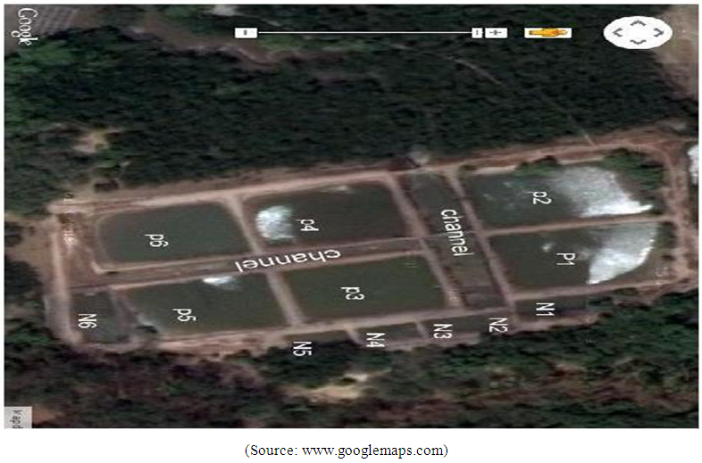
Figure 1. Map showing the study site in Mtwapa Creek, Kenya
Sample collection
P. monodon larvae were obtained from culture ponds. The stage of prawn was chosen at the larval stage 10-12 days (L.10-L.12), with complete gill development. They were sampled during the period from July 2014- June 2015. The primary criteria for the selection of prawns for use in this study were that they are culturable and edible. Samples were aseptically transferred in iceboxes and transported to the laboratory where they were frozen until analysis. 2000 samples were analyzed during the study period.
Quantitative analysis of bacteria
Culture and identification of bacterial types was performed using slightly modified methods described by Buller [14]. The samples of P. monodon were collected and homogenized and further used for bacterial isolation [15]. To estimate bacterial numbers, the inoculated plates were incubated at 25°C - 32°C for two days and duplicates were prepared for each dilution. Following incubation, the total number of colony forming unit (CFU) was determined and representative colonies were subcultured for identification. Bacterial numbers were calculated as the average of each set of duplicates and expressed as CFU/ml of the homogenate. Bacteria were isolated by a random collection of colonies from the agar plates. The colonies were purified by repeatedly sub culturing them on agar.
Bacterial identification
The isolated bacterial species were identified based on the morphological and biochemical characteristics of the individual colony and recorded. The individual colony of bacteria was transferred to NA and NB. The isolates were subjected to different morphological and biochemical test include Gram staining, motility test, gelatin liquefaction, casein hydrolysis test, catalase test, nitrate test and carbohydrate fermentation test, growth on salinity test and colony pigment appearance [16].

3. Results and Discussion

Bacterial Identification
Larval stages of the life cycle is the most vulnerable to prawn disease. The present study deals with the distribution of bacteria which are suspected to be the major reason in causing mortality of P. monodon larvae in Nurseries. The total numbers of isolated bacteria were 21.7 × 10⁶ (approx. 5.4 × 10⁴/g of larval macerate) from 2000 samples of prawn larvae as shown in table 1 and figure 2. This bacterial number was very important to note since one of the first criteria of microbiological test for evaluating prawnlarvae quality is that the maximum total bacteria count should be 1.0 ×10³ CFU/g of larval macerate in agar, of which more than 90% of the colonies should be yellow [17]. The study shows that the occurrence of total bacteria in P. monodon larvae exceeded the allowed maximum number and therefore the mortality of larvae was mainly due to bacteria. Furthermore, most of the colony on NA plate had a yellow colour, instead of white and pale which could have been possibly due to presence of suspected several genus of Vibrio and its related genera consisting of Aeromonas Hafnia, Pseudomonas and Alcaligenes. Research has shown that Vibrio strain are pathogenic and can cause Vibriosis, a serious infectious disease in maricultured organisms [18]. Several Vibrios associated with shrimp larvae, juvenile and adult stages consist of V. alginilyticus, V. parahaemolyticus, Photobacterium damselae, and V. mimicus [19].
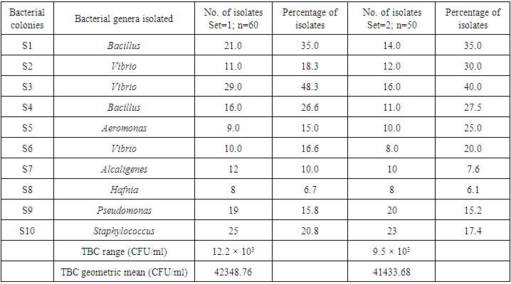
Table 1. The number and percentage of bacterial isolates from Penaeus monodon larvae (set=1; n=1000 and set=2; n=1000)
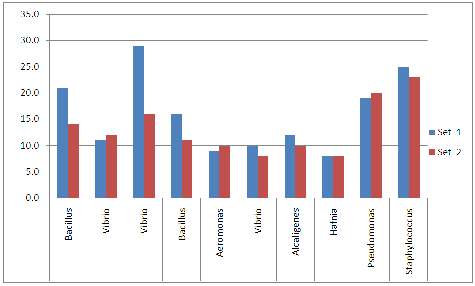
Figure 2. The number and percentage of bacterial isolates from Penaeus monodon larvae (set=1; n=1000 and set=2; n=1000)
Identification based on the morphological and biochemical characteristics compared with Bergey’s manual of determinative bacteriology is tabulated on Table 2. Based on the comparison, the bacteria were confirmed as members of genus Bacillus, Vibrio, Aeromonas, Alcaligenes, Hafnia, Pseudomonas, and Staphylococcus. Other researchers have supported this finding that disease in prawn larvae is caused by bacteria of the genus Aeromonas, Pseudomonas, and Plaubacterium [20]. The distribution of bacteria also shows widespread distribution of the common Gram negative bacteria in the outer area of prawn larvae comparing with inner area of hepatopancreas. This result indicated that Gram negative bacteria mainly came from coastal, fresh or seawater.
Table 2. Characteristics of the genus of selected bacteria
The isolation of Pseudomonas sp. from the prawn larvae samples is of high importance because this bacterium plays a considerable role as potential pathogenic bacteria for human and as an indicator of food quality as spoilage organism. This is in accordance with previous studies by Jeyasekaran et al. [21] and Koutsoumanis and Nychas [22] who identified pseudomonas as a good spoilage index. Although Pseudomonas sp. is not recognized as the cause of food borne illnesses they are closely associated to food deterioration [23]. According to Tripathy et al. [24] Pseudomonas sp. are frequently associated to fish and have been isolated from skin, gills and intestine. Their load is explained by the population density in water. In aquaculture, P. aeruginosa and P. fluorescens have been identified as opportunistic pathogenic species [25]. Aeromonas sp. has been recognized as potential food borne pathogens for more than 20 years. Aeromonas salmonicida can be causative agents not only of human enteritis [26], but also of a fatal septicaemia and is the causative agent of the fish disease called furunculosis [27]. Aeromonas is one of the major causes of bacterial infections affecting tilapia [28]. Alcaligenes is commonly found in the environment [29]. Alcaligenes sp. had been isolated from water as well as in the mussel samples [30]. Alcaligenes faecalis which produce disease in crustaceans such as lobster, being isolated from the hemolymph and inducing a softening of the shells [31]. Pseudomonas is the most common genera in crustaceans, marine fish and bivalves [32]. The group of bacteria related to the genus Pseudomonas is very broad and includes species pathogenic for humans and plants commonly found in fresh altered water. In the case of marine organisms species of the genus Pseudomonas have been isolated and identified from the microbiota of farmed fish such as rainbow trout (Oncorhynchus mykiss), perch (Perca fluviatilis) and rohu (Labeo rohita) [33]. Some species of the genus Aeromonas are considered to some to possibly cause gastro-enteritis in humans and these may also be present naturally in the marine or, more especially, the estuarine environment. Although many such organisms pose significant health risks for immuno-compromised individuals or other susceptible groups, several species such as Pseudomonas and Aeromonas spp. commonly form part of the natural flora of seafood. These observations and ensuing inferences of this study are useful for managing effluent out fall in to coastal ecosystem. However, we must rely on men to take social awareness and learn to care for the ocean, minimizing the contamination into the sea. Every effort leading to reduction in pollution indicator bacteria and microbes of human health concern has to be promoted and implemented.
Prawn larvae quality
Despite the above result, second evaluation for good quality of prawn larvae was dependent on the presence of V. harveyi (bioluminescent bacteria), which can be detected in agar. In marine invertebrates, notably larval penaeid prawn. V. harveyi is a major constraint on production, particularly in Asia [34]. The result shows that from ten selected bacterial isolates on luminescent test medium, luminescent bacteria like V. harveyii from the samples did not exist. It was suggested that V. harveyi is a marine Gram-negative luminous organism with a growth requirement for sodium chloride [35]. Further observation using 3.0-15.0% of NaCl (Table 2) did not show luminescence area as an indicator of V. harveyi existence. Luminous bacterial disease in Indonesia occur during the rainy season which decrease salinities (10-15 ppt) and bases pH resulting in significantly enhanced penaeid prawn larvae mortalities [36]. The result exhibited negative incidence of luminescent bacteria during larval stage in the nurseries which could be an indicator that the environment still have good quality. These results have an implication that the early larval stages and the research environment did not give possibility to development of luminuous bacterial disease. The sea water was providing some advantageous effect in diminishing the load of V. harveyi in the nurseries [37]. This supported the earlier work that we did in the same site on impact of water and sediment quality on temporal variations of bacterial densities and diversities where we found out that the estimates for bacterial groups (normally isolated from marine environments) genera richness in both sediment and water samples were greater than those indicated by Torsvike et al. [38]. This could have been due to the fact that the impact of organic loading from aquaculture to the pond culture system is not as great as that seen in other regions where production is more intensive. There was a positive correlation between the negative existence of V. harveyi in the death of larvae of P. monodon. This non-pathogenic probiotic bacterium has a high specificity to the cultured prawns host and provides a healthy balance of indigenous organisms in the host’s intestines. This is an indication that these microorganisms have the potency of supporting prawn larvae survival [39]. Furthermore, some other bacteria consisting of several strains of B. subtilis, B. cereus, V. pelagius, V. mediterranei, A. media, Pseudomonas and Thalasso bacterutilis also can be used as probiotics or biological controls against Vibrio.

4. Conclusions

Total number of bacteria ranged from 21.7 × 10⁵cfu to 32 × 105cfu. Microorganisms presumably belong to genus Vibrio, Pseudomonas, Aeromonas, Alcaligenes, Bacillus, Staphylococcus, Hafnia and Fusarium. The absence of V. harveyi pathogen indicated that the fusant serve as the main source on increasing of resistance to diseases and thus reducing the mortality of prawn larvae. The study hasalso indicated the possibility of microorganisms on prawn larvae as probiotic which remain to be explored.

ACKNOWLEDGMENTS

We are grateful to the National Council of Science and Technology (NCST) for funding the project and Kwetu Training Centre for hosting the project. We thank Mr. Brendan Muli for technical assistance in the field during the study.

References

[1]  Fowler, S.W., 1982. Biological transfer and transport processes. In: Pollutant transfer and transport in the sea. G. Kullenberg (Ed.) Vol.2, Chapter 1. CRC Press, Inc., Boca Raton, pp. 1-6.
[2]  Stentiford, G.D., Neil, D.M., Peeler, E.J., Shields, J.D., Small, H.J., Flegel, T.W., Vlak, J.M., Jones, B., Morado, F., Moss, S., Lotz, J., Bartholomay, L., Behringer, D.C., Hauton, C., Lightner, D.V., 2012.
[3]  Kibenge, F.S.B., Godoy, M.G., Fast, M., Workenhe, S., Kibenge, M.J.T., 2012. Countermeasures against viral diseases of farmed fish. Antiviral Research 95, 257‐281.
[4]  Austin, B., 2010. Vibrios as causal agents of zoonoses. Veterinary Microbiology 140, 310‐317.
[5]  Hoff, K.A., 1989. Survival of Vibrio anguillarum and Vibrio salmonicida at different salinities. Appl. Environ. Microbiol. 55, 1775‐1786.
[6]  Zhou, L.Y., Wang, X.H., Liu, Q., Wang, Q.Y., Zhao, Y., Zhang, Y.X., 2010. A novel multivalent vaccine based on secretary antigendelivery induces protective immunity against Vibrio anguillarum and Aeromonas hydrophila. J. Biotechnol. 146, 25‐30.
[7]  Kumar, S.R., Parameswaran, V., Ahmed, V.P.I., Musthaq, S.S., Hameed, A.S.S., 2007. Protective efficiency of DNA vaccination in Asian seabass (Lates calcarifer) against Vibrio anguillarum. Fish & Shellfish Immunology 23, 316‐326.
[8]  Badley, A., B. Philips, D.J.M. Haldane and Dalton, M.T. 1990. Pathogenic marine Vibrio species in selected Nova Scotian recreational coastal waters. Can. J. Public Heal. 81: 263-267.
[9]  Mikkelsen, H., Lund, V., Larsen, R., Seppola, M., 2011. Vibriosis vaccines based on various serosubgroups of Vibrio anguillarum O2 induce specific protection in Atlantic cod (Gadus morhua L.) juveniles. Fish & Shellfish Immunology 30, 330‐339.
[10]  Plusquellec, A., M. Beucher, C. Le Lay, Y. Le Gal and Cleret, J.J. 1991. Quantitative and qualitative bacteriology of the marine water surface micro layer in a sewage polluted area. Mar.Environ. Res. 31: 227-239.
[11]  Martinez-Manzanarez, E., M.A. Morinigo, D. Castro, M.C. Balebona, J.M. Sanchez and Borrego, J.J. 1992. Influences of the faecal pollution of marine sediments on the microbial content of shell fish. Mar.Pollu. Bull. 24(7): 342-349.
[12]  Austin, B., 1982. Taxonomy of bacteria isolated from a coastal marine fishrearing unit. J.Mar.Biol.Assoc. 63:583592.
[13]  Government of Kenya: (2011) Integrated Coastal Zone Management Action for Kenya, 2011-2015. Towards an integrated Kenya’s coastal and marine resources. NEMA, Nairobi. 90.
[14]  Richmond, D.M. (1997) (Ed.) A guide to the sea shores of eastern African and the western Indian Ocean islands. Sida Department for Research Cooperation/ SAREC, 448.
[15]  Kumar M, Pratap Singh M. Mixotrophic cultivation of microalgae for the production of biofuel. Patent No. WO 2014083534 A1. Indian Oil Corporation Limited. 2014.
[16]  Buchanan RE, Gibbons NE. Bergey's manual of determinative bacteriology, 8-Ed, New York: Springer; 1985.
[17]  Krieg NR, Staley JT, Brown DR, Hedlund BP, Paster BJ, Ward NL, Ludwig W, Whitman WB. Ed. Bergey's manual of systematic bacteriology, 2nd edn, vol. 4. The Bacteroidetes, Spirochaetes, Tenericutes (Mollicutes), Acidobacteria, Fibrobacteres, Fusobacteria, Dictyoglomi, Gemmati monadetes, Lentisphaerae, Verrucomicrobia, Chlamydiae, and Planctomycetes. New York: Springer. 2010.
[18]  Vandenberghe J, Verdonck L, Robles-arozarena R, Rivera G, Bolland A, Balladares M, Gomez-gil B, Calderon J, Sorgeloos PP, Swings J. Vibrios associated with Litopenaeus vannamei post larvae, post larvae, brood stock, and hatchery probionts. Appl Environ Microbiol. 1999; 65(6): 2592–2597.
[19]  Lightner DV, Redman RM. Shrimp Diseases and Current Diagnostic Methods. Aquaculture.1998. 164(1-4) : 201 – 220.
[20]  Taslihan A, Sunaryoto. Pest and disease management in P. monodon culture. Proceedings of the shrimp culture industry workshop FAO. Background papers Brackish water Aquaculture Development Centre, Jepara, Central Java.1984.
[21]  Jeyasekaran, G., P. Ganesan, R. Anandaraj, J.R. Shakilaand Sukumar, D. 2006. Quantitative and 2005. Prevalence and Indian white shrimp (Penaeusindicus) stored in dry ice. J. Food Microbiol. 23(6): 526-533.
[22]  Koutsoumanis, K., and Nychas, G.J. 2000. Application of systemic experimental procedure to develop amicrobial model for rapid fish shelf life predictions. Int. J. Food Microbiol. 60(2-3): 171-184.
[23]  Tryfinopoulo, P,. E. Tsakalidou and Nuchas, G.J.E. 2002. Characterization of Pseudomonas spp. Associated with spoilage of gilt-heat bream stored under various conditions. Appl.Environ. Microbiol. 68(1): 65-72.
[24]  Tripathy, S., 2007. Characterisation of Pseudomonas aeruginosa isolated from freshwater culture systems. Microbiol. Res. 162(4): 391-396.
[25]  Altinok et al., 2006.
[26]  Sukroongreung, S., C. Nilakul and Tantimavanich, S. 1983. Distribution of IMVC biogroups of Aeromonashydrophila and Aeromonassobria isolated from human, fish and water. Southeast Asian J. Trop. Med. Public Health. 14: 330 335.
[27]  Isonhood J.H., and Drake M. 2002. Aeromonas species in foods. J. Food. Protect. 65: 575 582.
[28]  Li, Y., and Cai, S.H. 2011. Identification and pathogenicity of Aeromonassobria on tail-rot disease in juvenile tilapia Oreochromisniloticus. Curr. Microbiol. 62: 623-627.
[29]  Wang, GX., F.Y. Li, J. Cui, Y. Wang, Y.T. Liu, J. Han and Lei, Y. 2011. Immunostimulatory activities of a decapeptide derived from Alcaligenesfaecalis FY-3 to crucian carp. Scand. J. Immunol. Epub ahead of print.
[30]  Cavallo, R.A., M. I. Acquaviva and Stabili, L. 2009. Culturable heterotrophic bacteria in seawater and Mytilusgalloprovincialis from a Mediterranean area (Northern Ionian Sea Italy). Environ Monit Assess. 149: 465 475.
[31]  Buller N.B., 2004. Bacteria from Fish and other Aquatic Animals: A Practical Identification Manual. CAB International, Oxfordshire, UK.pp. 361.
[32]  Alexopoulos, A., S. Plessas, C. Voidarou, H. Noussias, E. Stavropoulou, I. Mantzourani, A. Tzora, I. Skoufos and Bezirtzoglou E. 2011. Microbial ecology of fish species on growing in Greek sea farms and their watery environment. Anaerobe. Epub ahead of print.
[33]  Salgado-Miranda, C., E. Palomares, M. Jurado, A. Marin, F. Vega and Soriano-Vargas E. 2010. Isolation and Distribution of Bacterial Flora in Farmed Rainbow Trout from Mexico. J. Aquat. Animal. Health. 2010. 22: 244-247.
[34]  Austin B and Zang XH. Vibrio harveyi: a significant pathogen of marine vertebrates and invertebrates. Lett in Appl Microbiol 2006; 43(2): 119-124.
[35]  Chrisolite B, Thiyagarajan S, Alavandi SV, Abhilash EC, Kalaimani N, Vijayan KK, Santiago TC. 2008. Distribution of luminescent Vibrio harveyi and their bacteriophages in a commercial shrimp hatchery in South India. Aquaculture. 2008: 275: 13–19.
[36]  Prayitno SB, Latchford JW. Experimental infections of crustaceans with luminous bacteria related to Photobacterium and Vibrio. Effect of salinity and pH on infectiosity. Aquaculture. 1995; 132: 105–112.
[37]  Noguerola I, Blanch AR. Identification of Vibrio spp. with a set of dichotomous keys. J of ApplMicrobiol 2008; 105: 175–185.
[38]  Torsvike V, Sorheim R and Goksoyr J “Total bacterial diversity in soil and sediment communities - a review”. J Ind Microbiol 17 (1996): 170–178.
[39]  Luis-Villaseñor IE, Campa-Córdova AI, Huerta-Aldaz N, Luna-González A, Mazón-Suástegui JM, Flores-Higuera F. Effect of beneficial bacteria on larval culture of Pacific whiteleg shrimp, Litopenaeusvannamei. African J of Microbiol Res DOI: 2013. 10.5897/AJMR12.1360. ISSN 1996-0808. http://www.academicjournals.org/AJMR 72013; 27: 3471-3478.