Journal of Microbiology Research

p-ISSN: 2166-5885    e-ISSN: 2166-5931

2015;  5(3): 109-117

doi:10.5923/j.microbiology.20150503.05

Phenotypic and Genotypic Characterization of a Marine Bacterium Isolated from Sundarbans, Bangladesh

Ashish Kumar Sarker1, Md. Anwarul Haque1, Mohammad Sayful Islam2, Md. Uzzal Haque2, Netish Kumar Kundo2, Abu Syed Md Anisuzzaman2, 3, Urmi Saha4, Md. Anwar Ul Islam2

1Department of Pharmacy, Pabna University of Science and Technology, Pabna, Bangladesh

2Department of Pharmacy, University of Rajshahi, Rajshahi, Bangladesh

3Department of Hematology & Medical Oncology, Winship Cancer Institute, Emory University, Atlanta, GA, USA

4Eastern Medical College and Hospital, Comilla, Bangladesh

Correspondence to: Md. Anwar Ul Islam, Department of Pharmacy, University of Rajshahi, Rajshahi, Bangladesh.

Email:

Copyright © 2015 Scientific & Academic Publishing. All Rights Reserved.

Abstract

The mangrove ecosystem is a largely unexplored source for bacteria with the potentiality to produce biologically active secondary metabolites. Consequently, we set our research study to isolate, characterize and screen a bacterium producing bioactive compounds from marine soil of Sundarbans, Bangladesh. A total number of 39 marine bacterial colonies were isolated and purified. Representative bioactive isolate, ANAM-5 was characterized using phenotypic and genotypic procedures since it exhibited highest antibacterial activity against a series of test organisms. The 16S rDNA sequence of the strain ANAM-5 showed close similarity with only one bacterium Planctomyces brasiliensis, complete genome (99.64%). But the cultural, morphological, physiological and biochemical characteristics of the strain and P. brasiliensis were completely different. So, it may not be logical to assign the strain ANAM-5 to the genus Planctomyces. Thus, to confirm the taxonomic position of the strain and to have a correct solution for this taxonomic problem, further studies are needed in respect to the DNA relatedness studies, fatty acid profiling, DNA-DNA hybridization, matrix-assisted laser desorption/ionization time-of-flight (MALDI-TOF), metabolic finger profiling using BIOLOG, ribotyping, small subunit (SSU) sequences, cell wall composition and other characterizations.

Keywords: Phenotypic, Genotypic, Marine Bacteria, Sundarbans

Cite this paper: Ashish Kumar Sarker, Md. Anwarul Haque, Mohammad Sayful Islam, Md. Uzzal Haque, Netish Kumar Kundo, Abu Syed Md Anisuzzaman, Urmi Saha, Md. Anwar Ul Islam, Phenotypic and Genotypic Characterization of a Marine Bacterium Isolated from Sundarbans, Bangladesh, Journal of Microbiology Research, Vol. 5 No. 3, 2015, pp. 109-117. doi: 10.5923/j.microbiology.20150503.05.

1. Introduction

Antibiotic resistance is now recognized as a global health problem. Although many pre-existing antibiotics have been modified to yield new derivatives, bacteria have the potential to mutate known resistance mechanisms to combat these molecules [1, 2]. The situation is exacerbated by the fact that no novel antibiotics have been discovered for last 20 years. For this, constant increasing of bacterial resistance, it is now desperately need to search for new sources of antibiotics.
Nature is always a boundless source for novel microorganisms producing bioactive compounds having antagonistic activity against pathogenic organisms. Among the bioactive metabolites producers of commercially important drugs, bacteria have proven to be a prolific source with a surprisingly small group of taxa accounting for the vast majority of compounds discovered till date [3]. Among bacteria, actinomycetes particularly the genus Streptomyces is of special interest because it able to produce a wide range of bioactive secondary metabolites, such as antibiotics, antitumor agents, immunosuppressive agents, antifungal, neuritogenic, anticancer, antialgal, antimalarial, anti-inflammatory and enzymes [4].
But, presently, the chance of isolating a novel actinomycete strain from a terrestrial habitat, which would produce new biologically active metabolites, has reduced [5]. So, now much attention has been focused on screening of microorganism from diverse environments such as desert biomes and marine ecosystems. Marine environment harboring a vast variety of organisms differing in their physiology and adaptation capacity. The immense diversity of this habitat along with its underexploitation is the fundamental reason for attracting researchers towards it for discovering novel metabolite producers [5]. It is estimated that less than 1% of potentially useful chemicals from marine environment has been screened so far, with microbial products representing approximately 1% of the total number [6]. As marine environmental conditions are extremely different from terrestrial ones, it is summarized that marine bacteria have different characteristics from those of terrestrial counterparts and therefore might produce different types of bioactive compounds [7].
Marine bacteria are efficient producers of new secondary metabolites that show a range of biological activities including antibacterial, antifungal, anticancer, insecticidal and enzyme inhibition. Bioactive compounds from marine bacteria possess distinct chemical structures that may form the basis for synthesis of new drugs that could be used to combat resistant pathogens [5]. So, the exploration of microbial secondary metabolites from marine environment has led to the discovery of hundreds of biologically active compounds. Marine bacteria occur on the marine soil, sediments, water and also other biomass (mangrove) and substrates.
In order to explore the diversity, antimicrobial activity of marine bacteria from mangrove environments, in the present investigation, an effort was made to screen marine soils from Sundarbans, Bangladesh which is a large unscreened and diverse ecosystem for the isolation of potent antibiotic producing marine bacteria. This research study revealed that the diversity of the Sundarbans as a rich source of new and potential marine bacteria species producing bioactive secondary molecules.

2. Materials and Methods

2.1. Isolation and Selection of Marine Bacteria

From different locations of Sundarbans, Bangladesh (08-16 August, 2010), totally nine marine soil samples were collected carefully from the various depths of the earth, ranging from layers just beneath the upper surface to 1.5 meters depth. By using a spread plate technique [8], isolation of the marine bacteria from these soil samples were done. A total of 39 strains [ANAM-1 to ANAM-48] were isolated and purified as pure culture from these soil samples. All of these purified isolates were preserved on yeast-extract-glucose-agar slants at 4°C. Then by using streak plating technique [9] on yeast-extract-glucose-agar medium, all of these pure isolates were preliminary screened for antibacterial activity. The isolates ANAM-5, a brownish gray colored microorganism was chosen for further characterization since it showed highest antibacterial activity in preliminary antibacterial screening, visibility of the metabolite as well as excellent growth properties.

2.2. Morphological Characterization of the Selected Marine Bacterium

Morphology is one of the major phenotypic characteristics to identify and distinguish bacteria species. By using the method described by Shirling and Gottlieb [10], morphological characteristics of the strain ANAM-5 was determined. Microscopic characterization was done by cover slip culture method. In this method, three to four sterile covers slip were inserted aseptically into the sterile solidified agar medium in a petri dish at an angle of 45° to 60° by using a sterile forceps and the strain was inoculated by streak plating method, at the free space on the plate. The hyphae of the strain grew and spread on the cover slip and produced spores during the incubation period (at 30.5°C for 8 days). After this incubation period, the cover slips were then taken out smoothly by using sterile forceps, mounted on slides and observed under microscope, using lactophenol cotton blue [11]. Colonies were identified on the basis of their colony morphology and color [10]. The characteristics of the spore-bearing hyphae and spore chains were determined by direct microscopic examination of the culture surface on opining dishes. To establish the presence or absence of chains of spores 200x – 700 x magnifications were used.

2.3. Cultural Characterization of the Selected Marine Bacterium

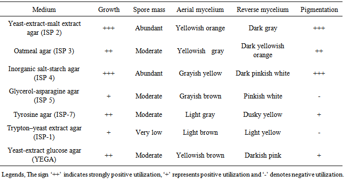
The cultural characteristic of the strain was also determined according to the method described by Shirling and Gottlieb [10]. Cultural characteristics are best made on a variety of standard cultivation media such as International Streptomyces Project (ISP) 2 (yeast-extract-malt extract agar); ISP 3 (oatmeal agar); ISP 4 (inorganic salts-starch agar); and ISP 5 (glycerol-asparagine agar), ISP 1 (Trypton-yeast extract agar), ISP 7 (Tyrosine agar) and yeast-extract -glucose agar (YEGA). A National Bureau of Standards Color Chart was used to determine the color of the substrate mycelia, aerial mycelia, and spore mass and pigment production [12].

2.4. Biochemical and Physiological Characterization of the Selected Marine Bacterium

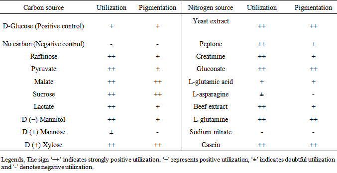
The biochemical and physiological characteristics of the strain were determined as described by Shirling and Gottlieb [10]. The growth of the strain ANAM-5 was determined on different carbon sources like D-glucose, sucrose, raffinose, D(+)-Xylose, D(−)- Mannitol, fumarate, lactate, malate and pyruate. Here, basal mineral salts agar (ISP medium 9) and different carbon sources were used for the medium of the test. The optimization of nitrogen sources such as yeast extract, peptone, L-glutamic acid, L-glutamine, gluconate, casein, beef extract, creatinine, sodium nitrate and L-asparagine was carried out by adding nitrogen source (0.2%) to the basal medium containing glucose (3%). The pH of the medium was adjusted using hydrochloric acid (1M) and sodium hydroxide (1M). For the optimization of temperature, after sterilizing and inoculating with spores the flasks were incubated at different temperatures (25-45°C).

2.5. Phylogenetic Characterization of the Selected Marine Bacterium

Phylogenetic studies were also performed. Thus, for the preparation of genomic DNA, the isolated colony from the agar plate was dispersed in 500μl of saline-EDTA buffer (NaCl 150 Mm; EDTA 10 Mm; ph 8.2) and incubated for 1h at 37°C. Then, 10 μl of lysozyme solution (5mg/ml), 5 μl of proteinase K solution (15mg/ml) and 10 μl SDS solution (25%) were added and incubated for 30 minutes at 55°C. Lysate was extracted, purified and dissolved in nano pure, sterile water (60 μl). The 16S rDNA was amplified by the PCR in a reaction mixture containing KOD FX buffer with 200 Μm Dntp, 100 ng genomic DNA and 0.5 μg forward (5’-AGAGTTTGATCCTGGCTCAG-3’) and 0.5 μg reverse (5’-GGTTACCTTGTTACGACTT-3’) primers. Thermal cycle was performed with a model 22331 eppendrof (Germany). The samples were subjected to an initial denaturing step consisting of 2 minutes 98°C, after which 2U of Taq polymerase was added to each sample at 90°C. The thermal profile used was 30 cycles consisting of 1 min annealing at 52°C, 2 min extension at 72°C and 1 min denaturation at 94°C. A final extension step consisting of 4 min at 65°C. PCR amplificants were detected by agarose gel electrophoresis and visualized in Dolphin WaveTech. The band was excised and isolated using TAE buffer. Purified by phenol chloroform extraction followed by ethanol precipitation and dissolved in water. The resultant PCR solution was used in sequencing reaction with Pre-Mix and primer. The reaction mixture was applied to thermal cycles in 30 cycles consisting of 10Sec at 98°C, 30Sec at 47°C and 2 min at 68°C. DNA was purified and by using Prep-A-Gene Kit (Bio-Rad). The liquid was evaporated at reduced temperature and using vacuum pressure. The dried DNA was dissolved in 20μl of polyacrylamide sequencing gel and sequenced by Applied Biosystems automated DNA sequencer. The resulting sequences were analyzed with DDBJ/EMBL/GenBank database using analysis softwares- BLAST and BIBI (Bioinformatics Bacterial Identification Tool) [13].

3. Results and Discussion

Marine environment has been proven as an outstanding and attractive resource for innovating new and potent bioactives compounds producing microorganisms. Marine microbes are particularly attractive because they have higher potency required for bioactive compounds to be effective in the marine environment, due to the diluting effect of sea water [14]. This present study was designed with an aim to discover a new marine bacterium strain from marine environment which is capable of producing antibacterial secondary metabolites. Considering this, the isolate ANAM-5, a brownish gray colored microorganism was chosen for further characterization due to the highest preliminary antibacterial activity (Figure 1), visibility of the metabolite as well as excellent growth properties of the strain.
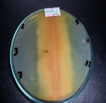
Figure 1. Antibacterial activity of ANAM-5 isolates. The perpendicular lines marked with numerical digits are the test organism- 2. Streptococcus agalactiae, 3. Bacillus cereus, 7. Pseudomonas aeruginosa, 9. Escherichia coli, 11. Shigella dysenteriae and 12. Shigella sonnei
The isolate ANAM-5 grew moderately at first day and after 2nd day white colonies were appeared, gradually it turned into thick and velvety cream colonies on to 3rd days. After the 3rd day ANAM-5 was started to release soluble yellow pigment through the medium. It became brownish gray (Figure 2) after the 4th day and gave deep gray colonies on the 5th day. As the time passed, the aerial mycelium became more powdery as well as deep yellowish due to sporulation of the microorganism.
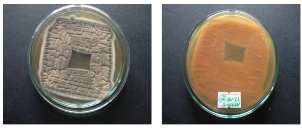
Figure 2. Gray and brown colonies of ANAM-5
Morphology had played a major role in distinguishing bacteria and in the characterization of bacteria species [15, 16] and, in fact, it was the only characteristic used in many early descriptions in the first few editions of Bergey's Manual. Under the microscope (Figure 3), the marine bacteria ANAM-5 produce well developed substrate mycelium and aerial hyphae which are differentiated into long and extended filamentous chains with cylindrical, smooth spores occurring in chains of more than 5. Spores are observed after six days. The isolate ANAM-5 exhibited morphology of typical Streptomyces; the colonies were slow growing, aerobic, glabrous or chalky, folded, and with aerial and substrate mycelia [13]. In addition, this isolate possessed an earthy odor and fitted to the description of genus Streptomyces in Bergey’s Manual of Systemic Bacteriology.
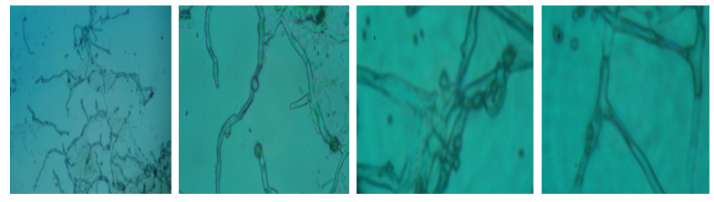
Figure 3. Microscopic view of the ANAM-5 isolates at different magnification
Cultural characteristics of the strain ANAM-5 in different cultural media (Table 1) showed morphology and properties of typical Streptomyces. The growth and pigmentation of the strain ANAM-5 was very fast in both ISP 2 and ISP 4 medium compared with both ISP 1 and ISP 5 medium where the growth of the strain was very slow. So, in ISP 2 and ISP 4 medium, the spore mass was profuse in the medium and pigmentation was very high. The growth of the strain was moderate in ISP 3, ISP 7 and YEGA medium.
Table 1. Cultural characteristics of strain ANAM-5
     
The production of antibiotic by various Streptomyces species was greatly influenced by suitable carbon and nitrogen sources [17]. Utilization of carbon and nitrogen sources by the strain ANAM-5 is presented in Table 2. The growth as well as production of yellow pigment was good by the strain ANAM-5 in the presence of malate, sucrose and D (+) Xylose as carbon sources in the medium. Utilization and metabolite production by the strain in the presence of D(+)-Mannose was doubtful. In the presence of D-glucose as a sole carbon source in the medium, growth and pigmentation were moderate. We also observed that, in the presence of raffinose, pyruvate, lactate and D(−)-Mannitol in the medium as carbon sources, the growth of the strain was high but the frequency of pigmentation was moderate. In the absence of any carbon source (Negative control) the strain could not grow. Therefore, it may be concluded that, proper carbon source is essential for optimum growth of the strain and for optimum production of pigment in the medium. In case of Streptomyces species, with regards to carbon and nitrogen sources, species specific variation may occur in cell growth and secondary metabolites production [18]. In this present study, we observed that the frequency of growth and production of secondary metabolites by the strain ANAM-5 was varied with the medium with different carbon sources. So, by studying the utilization of carbon sources by the strain we may conclude that the strain ANAM-5 showed the characteristics of typical Streptomyces.
Table 2. Utilization of carbon and nitrogen sources by the strain ANAM-5
     
The growth of the strain as well as metabolite production was very good in the medium supplemented with yeast extract, gluconate, L-glutamine and casein as nitrogen sources. In the presence of peptone, creatinine, L-glutamic acid and beef extract as nitrogen sources, the growth was high but metabolites production of the strain were moderate. The growth and production of pigment were moderate for the strain in the medium containing L-glutamic acid. The growth of the strain was doubtful and metabolite production was negative in the medium containing l-asparagine. In the presence of sodium nitrate as nitrogen source in the medium, growth and metabolite production was negative. So, it may be concluded that, the strain used different inorganic and organic nitrogen sources differently and exhibited differences in metabolites production. For, various Streptomyces species, different types of carbon and nitrogen sources were found to have significant effect on growth and secondary metabolites production [19]. Considering this, the strain ANAM-5 showed similar characteristics with Streptomyces species. The growth of the strain was very high and production of high levels of antibiotic when culture medium incubated at 29°C and pH 6.1. Extreme pH and temperature were unfavorable for antibiotic production for the strain. So, the morphological, cultural, biochemical and physiological properties of the strain ANAM-5 exhibited consistency with its assignment to the genus Streptomyces.
For confirmation and identification of the strain ANAM-5 at the species level, incongruities were observed. The 16S rDNA sequence of strain ANAM-5 was determined and compared to those deposited in GenBank using the BLAST and BIBI. Species identification was based on maximum score, identity and coverage values. The length of the sequence was 1131 bp. It was observed that the sequence was actually similar to only one sequence (Table 3).
Table 3. Sequence producing significant alignment with the 16S rDNA sequence of ANAM-5
     
According to BLAST, The 16S rDNA sequence of strain ANAM-5 exhibited close similarity (99.64%) with Planctomyces brasiliensis, complete genome. P. brasiliensis is a member of the order Planctomycetales was first isolated in 1982 in water from a salt pit, Lagoa Vermelha, Brazil. According to [20], >99% and >97% sequence similarities were used as the cut-offs for species and genus identification, respectively, depending on the bacterial genus under investigation. On the other hand, Janda and Abbott in their recommended guidelines suggested that a minimum of >99%, and ideally >99.5%, sequence similarity be used as the criteria for species identification [21]. They also proposed that other properties, e.g. phenotype, should be considered in final species identification. So, before assignment of a strain in a definite genus and species based on 16S rDNA sequencing there must be a comparative study based on phenotypic characteristics between the respective strain and most closely related bacteria.
Since, only the sequence of the 16S rRNA gene may not be sufficient for the description of a new bacterial species, phenotypic and genotypic characters of newly described bacterial species should be compared with those of reference neighboring species and genera in order to identify precise phenotypic and genotypic characters that will discriminate the newly described bacterial species. Some basic and distinctive phenotypic characters should be included in the description of new toxins. These include conditions for optimum growth, the Gram staining value, colony morphology, motility, spore forming capacity and median measurements as obtained by electron microscopic examination. Biochemical profiles of the isolate, including the results of oxidase and catalase testing, antibiotic susceptibility profile and the ability to metabolize major carbohydrates, should also be useful for description of a toxin [20]. Morphological features, such as capsules, flagella, cell size and shape are also used for the identification and classification of bacterial species [22]. When identifying bacteria, much attention is paid to how they grow on the media in order to identify their cultural characteristics, since different species can produce very different colonies [23]. Each colony has characteristics that may be unique to it and this may be useful in the preliminary identification of a bacterial species.
For comparison based on phenotypic study, we examined Planctomyces brasiliensis and the strain ANAM-5, but we found important differences. P. brasiliensis differs from other bacterial taxa by several distinctive features such as internal cell compartmentalization, multiplication by forming buds directly from the spherical, ovoid or pear-shaped mother cell and a cell wall consisting of a proteinaceous layer rather than a peptidoglycan layer [24]. It requires NaCl for growth and has a broad tolerance to salt (0.1 to 1.7 mol Na+/l). The cells are spherical to ovoid with a diameter of 0.7 to 1.8 μm which bear crateriform structures (Figure 4) and, in some cases, nonprosthecate appendages [25]. The colonies have a dry, rough surface and a yellow to ochre pigmentation but no sporulation. It has a distinct cell cycle, with sessile mother cells forming stalks that attach to surfaces or to other stalks and motile daughter cells that bud from the mother cell. Mother cells are spherical to ovoid with stalks composed of twisted fibrils. The diameter of the mother cell is 1.1 to 1.5 um. Multiplication occurs by budding on the distal cell pole, yielding daughter cells, which are mono-trichously and popularly flagellated. Both the mother cell and bud have crateriform structures scattered over the whole of the cell surface [25]. Cells have rigid stalk fibers and can form multicellular rosettes. Although it lacks peptidoglycan it has a cell envelope, which consists almost entirely of protein which is rich in proline and cysteine and is stabilized to a high degree by disulfide bonds.
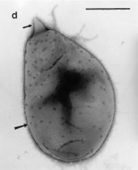
Figure 4. P. brasiliensis having crateriform structures and the polar unicorn prostheca-like projection [26]
On the other hand, the strain ANAM-5 was aerobic, produce well developed substrate mycelium and aerial hyphae which are differentiated into long and extended filamentous chains with cylindrical, smooth spores occurring in chains of more than 5, colonies glabrous or chalky, folded, and with aerial and substrate mycelia and possessed an earthy odor. The strain ANAM-5 was gram positive in gram staining technique and produce spore. So, it can be concluded that the strain has peptidoglycan in their cell structure but P. brasiliensis have no peptidoglycan layer in their cell structure.
P. brasiliensis grows chemoorganotrophically on the following carbon sources: D-cellobiose, D(+)-glucose, D(+)-galactose, maltose, melibiose, rhamnose, ribose, trehalose, N-acetyl Glucosamine and glucuronate but it can not grows in the medium containing D(−) fructose, D-fructose, D(−) lyxose, α-D-melezitose, raffinose, L(−)-sorbose, D(+)-xylose, methanol, ethanol, glycerol, D(−)-mannitol, D(−)-sorbitol, acetate, fumarate, lactate, malate, pyruvate and succinate as carbon sources [25]. On the other hand, the growth and metabolite production of ANAM-5 was moderate to high in the presence of malate, sucrose, D(+)-Xylose, D-glulose, raffinose, pyruvate, lactate and D(−)-Mannitol in the medium as carbon sources (Table 2). But in the presence of D(+)-Mannose in the medium, the utilization and metabolite production by the strain was doubtful. On the basis of utilization of carbon sources, it may be concluded that the strain ANAM-5 and P. brasiliensis are discriminative. P. brasiliensis can utilize ammonia, nitrate and N-acetyl glucosamine as a nitrogen source, but it does not utilize L-glutamic acid, L-glutamine, L-serine amygdalin, gluconate or creatinine and does not require vitamins for growth [25]. But, the growth and metabolite production of ANAM-5 was moderate to high in the presence of L-glutamic acid, L-glutamine, gluconate and creatinine as nitrogen sources in the medium (Table 2). In the presence of sodium nitrate as nitrogen source in the medium, there was no growth and metabolite production by the strain was observed. Based on utilization of nitrogen sources, it may be concluded that the strain ANAM-5 and P. brasiliensis exhibited differences.
Although 16S rRNA sequencing has been by far the most common, reliable and convenient method of bacterial species identification [27], this technique has some major limitations. The presence of mosaicism considered as one of the major limiting factors for 16S sequence based microbial identification. PCR amplification bias and cloning bias can also result in an imprecise representation of the microbial diversity. Similarly, the presence of abundant partial sequences of 16S genes in the databases results in ambiguous classification [22].
The major difficulties and controversies in interpreting 16S rRNA gene sequence data relate to the assignment of bacterial species based on similarity search results, as no threshold values are available as for DNA–DNA hybridization [28]. Another major limitation to its wider use is the difficulty associated with interpretation of 16S rRNA gene sequence results. The use of 16S rRNA gene sequencing for bacterial identification depends on a significant interspecies difference and a small intraspecies difference in 16S rRNA gene sequences. Therefore, one of the major limitations is that when two different bacterial species share almost the same 16S rRNA gene sequence; this technique alone would not be useful for distinguishing them confidently [29]. Identification of bacterial species based on sequence analysis of the 16S rRNA gene relies on matching the obtained sequence with the existing sequence. Matching with a sequence that was incorrectly identified leads to incorrect identification [30]. So, 16S gene sequencing lacks taxonomic resolution at the species level for some closely related species [21], subspecies, or recently diverged species [31]. For this reason, Escherichia coli and E. fergusonii species, as well as the subspecies Bartonella vinsonii subsp. arupensis and B. vinsonii subsp. vinsonii, are indistinguishable, when using 16S sequence similarity comparisons [32].
The presence of multiple copies of the rRNA operon and intragenomic heterogeneity of the 16S genes is considered as one of the limiting factor for the use of this gene for species identification. Several recent reports have suggested the existence of divergent 16S sequences within a single organism [22]. The reported intragenomic variability of 16S was as high as 6.4% in the case of the Thermobispora bispora genome [33]. Thus, the sequence heterogeneity within a genome can affects vigorusly in the phylogenetic resolution of a particular species based on the16S sequences. Horizontal gene transfer and recombination events have also been reported in ribosomal genes [34], which distort phylogenetic signals and thereby affect the phylogenetic classification. Horizontal transfers of 16S gene segments and the presence of mosaic like structures have been reported in Rhizobium, Aeromonas, Bradyrhizobium, Streptococcus and actinomycetes [35, 36]. Therefore, species identification using 16S gene based probes or homology analysis of partial 16S sequences may lead to misidentification, because it may represent only a part of the mosaic like structure.
However, there are ‘blind spots’ within some major genera, in which 16S rDNA sequences are not sufficiently discriminating for the identification of certain species [37, 38]. 16S rDNA gene sequencing is highly useful in regards to bacterial classification; it has low phylogenetic power at the species level and poor discriminatory power for some genera [39, 40]. In some cases, different species of bacteria with considerably different physiologies were reported to contain nearly identical 16S genes [41]. For example, the 16S sequences of certain species of Rhizobium such as R. galegae were found to be more similar to Agrobacterium sequences than other Rhizobium species. On the basis of these observations, it has been proposed that the genus Rhizobium may be corrected as a species within the genus Agrobacterium [42]. However, this proposal was not accepted because of the ecological and genomic differences that exist between these two genera [43]. In such circumstances, species identification solely based on the 16S sequences may lead to misidentifications.
Considering these limitations, in order to increase the accuracy of 16S rDNA sequencing for identification of bacteria demands for the use of alternative methods to confirm findings. More importantly, it is necessary to interpret the results of 16S rDNA sequencing with preliminary phenotypical, biochemical and physiological test results for the confirmation upto species level. Among the alternative methods fatty acid profiling, matrix-assisted laser desorption/ionization time-of-flight (MALDI-TOF), metabolic finger profiling using BIOLOG, ribotyping, together with the computational tools employed for querying the databases that are associated with these identification tools and high-throughput genomic sequencing can use in bacterial identification [30]. Whole genomic DNA relatedness are considered to provide the absolute resolution in bacterial taxonomy. It is generally accepted that all taxonomic information about a bacterium is incorporated in the complete nucleotide sequence of its genome [44]. Sequencing of several conserved genes (multilocus sequence analysis; MLSA) within the bacterial genome has been proposed to improve the discriminatory power [45]. The MLSA approach represents the bacterial genome to some extent and has higher discriminatory power than single locus analysis. Similarly, genomic fingerprinting method such as Rep-PCR (ERIC-, REP-, BOX-PCR), RAPD and AFLP provide more discriminatory power. However, whole genomic DNA–DNA hybridization (DDH) is considered as the ‘‘gold standard’’ for bacterial species identification. Organisms that show more than 70% DDH values and less than 5% difference in their melting temperature (DTm) are considered to belong to the same species [45, 46].
Therefore, in order to prevent misinterpretation of 16S rRNA gene sequence results, they should always be interpreted in the context of basic phenotypic test results [29]. Moreover, key phenotypic tests can help to distinguish between ‘the closest match’ by 16S rRNA gene sequencing and medically important bacterial species with similar 16S rRNA gene sequences, should be performed for differentiation of these possibilities [47-50]. Particular attention has to be paid if there is discrepancy between 16S rRNA gene sequence and phenotypic test results. For example, 16S rRNA gene sequencing able to identify the various isolates as Tsukamurella to the genus level, but all Tsukamurella species possess >99% nucleotide identities with each other. Phenotypic tests have to be performed to distinguish among the various Tsukamurella species [48]. New bacterial species are reported based on the polyphasic approach and this polyphasic approach utilizes phenotypic, genotypic and phylogenetic data of the microorganisms [51-53]. The results obtained by analyzing various phenotypic characteristics and phylogenetic studies based on 16S rDNA sequencing demonstrated that though the strain ANAM-5 is most closely related to the organism Planctomyces brasiliensis in phylogenetic development as well as sequence similarity, but the various phenotypic characteristics of ANAM-5 like morphological, cultural, physiological and biochemical were completely different from P. brasiliensis. So, the consignment of the strain ANAM-5 to the genus Planctomyces may not be logical. Thus, further studies should be carried out in respect of the DNA relatedness studies, fatty acid profiling, DNA-DNA hybridization, small subunit (SSU) sequences, cell wall composition, matrix-assisted laser desorption/ionization time-of-flight (MALDI-TOF), metabolic finger profiling using BIOLOG, ribotyping and other characteristics to confirm the taxonomic position of the strain ANAM-5.

4. Conclusions

The marine environment represents a relatively untapped source of biologically active compounds that can be applied in pharmaceutical and cosmeceutical industries. Our findings explore the resource of a mangrove forest, the Sundarbans for finding antibacterial metabolites producing marine bacterium. The strain ANAM-5 differs morphologically, culturally, physiologically and biochemically from P. brasiliensis. Strain ANAM-5 is phylogenetically most similar to the genus Planctomyces, but the phylogenetic relationship is not sufficient to permit classification of strain ANAM-5 in this genus and for the description of a new bacterial species. So, the strain ANAM-5 may not be assigned to the genus Planctomyces. Thus to confirm the taxonomic position and to provide absolute resolution to these taxonomic problem of the strain ANAM-5, more studies should be carried out in respect of the of the DNA relatedness studies, fatty acid profiling, DNA-DNA hybridization, small subunit (SSU) sequences, cell wall composition, matrix-assisted laser desorption/ionization time-of-flight (MALDI-TOF), metabolic finger profiling using BIOLOG, ribotyping and other characterizations.

ACKNOWLEDGEMENTS

This study was supported by Pharmaceutical Microbiology Laboratory, Department of Pharmacy, University of Rajshahi, Bangladesh. The authors are also grateful to Dr. Abu Sayed Md. Anisuzzaman, Associate Professor, Department of Pharmacy, University of Rajshahi for providing 16S rDNA gene sequencing data to carry out of this research work.

References

[1]  Knowles, D. J. C., 1997, New strategies for antibacterial drug design., Tre. Micro., 5(10), 379-383.
[2]  Levy, S. B., 1998, The challenge of antibiotic resistance., Sci. Am., 278(3), 32-39.
[3]  Bull, A. T., 2004, Microbial diversity and biosprospecting, ASM Press.
[4]  Ravikumar, S., Inbaneson, S. J., Uthiraselvam, M., Priya, S. R., Ramu, A., and Banerjee, M. B., 2011, Diversity of endophytic actinomycetes from Karangkadu mangrove ecosystem and its antibacterial potential against bacterial pathogens., J. Pharm. Res., 4(1), 294-296.
[5]  Solanki, R., Khanna, M., and Lal, R., 2008, Bioactive compounds from marine actinomycetes., Indian J. Microbiol., 48, 410–431.
[6]  Neidleman, S. L., and Laskin, A. L., 2000, Adv. AppL. Micro., ElsE., 47, 88-90.
[7]  Imada, C., Koseki, N., Kamata, M., Kobayashi, T., and Hamada-Sato, N., 2007, Isolation and characterization of antibacterial substances produced by marine actinomycetes in the presence of seawater., Actinomyce., 21, 27-31.
[8]  Bernard, B., 2004, Access excellence @ the national health museum, Isolation of Antibiotic strains from soils.
[9]  Alcamo, E., and Pomerville, P. C., 2004, Alcamo’s Laboratory Fundamental of microbiology.
[10]  Shirling, S. B., and Gottlieb, D., 1966, Methods of characterization of Streptomyces species., Inter. J. Sys. Bact., 16(3), 313-340.
[11]  Korzybski, T., Kowszyk, G., and Kurilowicz, W., 1967, Antibiotics, Origin, nature and properties, Translated by parayski. E. Per Pre, Oxf., 47, 450-725.
[12]  Zhao, H., Parry, R. L., Fllis, D. L., Griffith, G. W., and Goodacre, R., 2006, The rapid differentiation of Streptomyces isolates using fourier transform infrared spectroscopy., Vibrat. Spec., 40, 213-218.
[13]  Clarridge, J. E., 2004, Impact of 16S rRNA Gene Sequence Analysis for Identification of bacteria on clinical Microbiology and infectious diseases., Clin. Microbi. Rev., 17(4), 840-862.
[14]  Valli, S., Sugasini, S. S., Aysha, O. S., Nirmala, P., Kumar, P. V., and Reena, A., 2012, Antimicrobial potential of Actinomycetes species isolated from marine environment., Asi. Pac. J. Trop. Biom., 45, 469-474.
[15]  Williams, S. T., Goodfellow, M., Alderson, G., Wellington, E. M. H., Sneath, P. H., and Sackin, M., 1983, Numerical classification of Streptomyces and related genera., J. Gen. Appl. Microbio., 129, 1743–1813.
[16]  Anderson, A.S., and Wellington, E. M. H., 2001, The taxonomy of Streptomyces and related genera., Int. J. Syst. Evol. Microbiol., 51, 797–814.
[17]  Hobes, G., Frazer, C. M., Flett, F., and Oliver, S. G., 1990, Pigmented antibiotic production by S. coelicolor A3(2), kinetics and the influence of nutrients., J. Gen. Microbi., 136, 2291-2296.
[18]  Jonsbu, E. M., Intyre, M., and Nielsen, J., 2002, The influence of carbon source and morphology on nystatin production by Streptomyces noursei., J. Biotec., 95, 133-144.
[19]  Mansour, F. A., El-Shirbiny, S. A., and El-Metwaly, N. A., 1996, Demethyl tetracycline biosynthesis by Streptomyces aureofaciens Sub-species viridulans as influenced by medium composition., Egyp. J. Microbi., 31, 221-235.
[20]  Drancourt, M., Bollet, C., Carlioz, A., Martelin, R., Gayral, J. P., and Raoult, D., 2000, 16S ribosomal DNA sequence analysis of a large collection of environmental and clinical unidentifiable bacterial isolates., J. Clin. Microbi., 38, 3623-3630.
[21]  Janda, J. M., and Abbot, S. L., 2007, 16S rRNA gene sequencing for bacterial identification in the diagnostic laboratory: pluses, perils, and pitfalls., J. Clin. Microbiol., 45(9), 2761-2764.
[22]  Rajendhran, J., and Gunasekaran, P., 2011, Microbial phylogeny and diversity: Small subunit ribosomal RNA sequence analysis and beyond, Microbiol. Res., 166, 99-110.
[23]  Christopher, K., Bruno, E., 2003, Identification of bacterial species. In Tested studies for laboratory teaching. Ed., M.A. O’Donnell. Proceedings of the 24th Workshop/Conference of the Association for Biology Laboratory Education., 24, 103-130.
[24]  Scheuner, C., Tindall, B. J., Lu, M., Nolan, M., Lapidus, A., Cheng, J. F., Goodwin, L., Pitluck, S., Huntemann, M., Pati, A., Chen, A., Palaniappan, K., Jeffries, C.D., Hauser, L., Land, M., Mwirichia, R., Rohde, M., Abt, B., Markowitz, V., Hugenholtz, P., Mgöker, M., Kyrpides, N.C., and Klenk, H.P., 2014, Complete genome sequence of Planctomyces brasiliensis type strain (DSM 5305T), phylogenomic analysis and reclassification of Planctomycetes including the descriptions of Gimesia gen. nov., Planctopirus gen. nov. and Rubinisphaera gen. nov. and emended descriptions of the order Planctomycetales and the family Planctomycetaceae., Stand. Gen. Sci., 9(10), 1245-1257.
[25]  Schlesner, H., 1989, Planctomyces brasiliensis sp. Nov., a halotolerant bacterium from a salt pit., Syst. Appl. Microbiol., 12, 159–61.
[26]  Fuerst., John, A., Gwilliam, H. G., Lindsay, M., Lichanska, A., Belcher, C., Vickers, J. E., and Hugenholtz, P., 1997, Isolation and molecular identification of planctomycete bacteria from postlarvae of the giant tiger prawn, Penaeus monodon., App. Environ. Microbiol., 63(1), 254-262.
[27]  Song, Y., McTeague, M., and Finegold, S. M., 2003, 16S ribosomal DNA sequence-based analysis of clinically significant gram-positive anaerobic cocci., J. Clin. Microbiol., 41(4), 1363-1369.
[28]  Petti, C. A., 2007, Detection and identification of microorganisms by gene amplification and sequencing., Clin. Infect. Dis., 44, 1108–1114.
[29]  Woo, P. C. Y., Teng, J. L. L., Wu, J. K. L., Leung, F. P. S., Tse, H., Lau, S. K. P., and Yuen, K. Y., 2009, Guidelines for interpretation of 16S rRNA gene sequence-based results for identification of medically important aerobic Gram-positive bacteria., J. Medi. Microbiol., 58, 1030–1036.
[30]  Phumudzo, T., Nnzeru, R., Ntushelo, K., and Mudau, F., 2013, Bacterial species identification getting easier., Afri. J. Biotech., 12(41), 5975-5982.
[31]  Fox, G. E., Wisotzkey, J. D., and Jurtshuk, P. J., 1992, How close is close: 16S rRNA sequence identity may not be sufficient to guarantee species identity., Int. J. Syst. Bacteriol., 42, 166-170.
[32]  Adekambi, T., Colson, P., and Drancourt, M., 2003, RpoB-based identification of nonpigmented and late-pigmenting rapidly growing mycobacteria., J. Clin. Microbiol., 41, 5699-5708.
[33]  Wang, Y., Zhang, Z., and Ramanan, N., 1997, The actinomycete Thermobispora bispora contains two distinct types of transcriptionally active16S rRNA genes., J. Bacteriol., 179, 3270–6.
[34]  Miller, S. R., Augustine, S., Blankenship, R. E., and Selker, J., 2005, Discovery of a free-living chlorophyll producing cyanobacterium with a hybrid proteobacterial/cyanobacterial small-subunit rRNA gene., Proc. Natl. Acad. Sci., 102, 850-855.
[35]  Eardly, B. D., Wang, F. S., and Berkum, P. V., 1996, Corresponding 16S rRNA gene segments in Rhizobiaceae and Aeromonas yield discordant phylogenies., Plant. Soil., 186, 69–74.
[36]  Schouls, L. M., Schot, C. S., and Jacobs, J. A., 2003, Horizontal transfer of segments of the 16S rRNA genes between species of the Streptococcus anginosus group., J. Bacteriol., 185, 7241–6.
[37]  Kwok, A. Y., and Chow, A. W., 2003, Phylogenetic study of Staphylococcus and Macrococcus species based on partial hsp60 gene sequences., Int. J. Syst. Evol. Microbi., 53, 87–92.
[38]  Kwok, A. Y., Su, S. C., and Reynolds, R. P., 1999, Species identification and phylogenetic relationships based on partial HSP60 gene sequences within the genus Staphylococcus., Int. J. Syst. Bact., 49(3), 1181–1192.
[39]  Gupta, R., Lanter, J. M., and Woese, C. R., 1983, Sequence of the 16S ribosomal RNA from Halobacterium volcanii, an Archaebacterium., Sci., 221, 656–659.
[40]  Woo, P. C., Chong, K. T., and Yuen, K., 2001, Identification of Arcobacter cryaerophilus isolated from a traffic accident victim with bacteremia by 16S ribosomal RNA gene sequencing., Diag. Microbi. Infect. Dis., 40, 125–127.
[41]  Palys, T., Nakamura, L. K., and Cohan, F. M., 1997, Discovery and classification of ecological diversity in the bacterial world: the role of DNA sequence data., Int. J. Syst. Evol. Microbiol., 47, 1145–56.
[42]  Young, J. M., Kuykendall, L. D., Martinez-Romero, E., Kerr, A., and Sawada, H., 2001, A revision of Rhizobium Frank 1889, with anemended description of the genus, and the inclusion of all species of Agrobacterium Conn, 1942 and Allorhizobium undicola de Lajudie etal., 1998 as new combinations: Rhizobium radiobacter, R. rhizogenes, R. rubi, R. undicola and R. vitis., Int. J. Syst. Evol. Microbiol., 51, 89–103.
[43]  Farrand, S. K., Berkum, P. V., and Ger, P., 2003, Agrobacterium is a definable genus of the family Rhizobiaceae., Int. J. Syst. Evol. Microbiol., 53, 681–7.
[44]  Goris, J., Konstantinidis, K. T., and Klappenbach, J. A., 2007, DNA–DNA hybridization values and their relationship to whole-genome sequence similarities., Int. J. Syst. Evol. Microbiol., 57, 81-91.
[45]  Gevers, D., Cohan, F. M., and Lawrence, J. G., 2005, Re-evaluating prokaryotic species., Nat. Rev. Microbiol., 3, 733–9.
[46]  Wayne, L. G., Brenner, D. J., and Colwell, R. R., 1987, Report of the ad hoc committee on reconciliation of approaches to bacterial systematics., Int. J. Syst. Bacteriol., 37, 463–4.
[47]  Woo, P. C., Leung, A. S., Leung, K. W., and Yuen, K. Y., 2001, Identification of slide-coagulase positive, tube-coagulase negative Staphylococcus aureus by 16S ribosomal RNA gene sequencing., Mol. Pathol., 54, 244–247.
[48]  Woo, P. C., Ngan, A. H., Lau, S. K., and Yuen, K. Y., 2003d, Tsukamurella conjunctivitis: a novel clinical syndrome. J. Clin. Microbiol., 41. 3368–3371.
[49]  Lau, S. K., Woo, P. C., Woo, G. K., and Yuen, K. Y., 2002, Catheterrelated Microbacterium bacteremia identified by 16S rRNA gene sequencing., J. Clin. Microbiol., 40, 2681–2685.
[50]  Cloud, J. L., Hoggan, K., Belousov, E., Cohen, S., Brown-Elliott, B. A., Mann, L., Wilson, R., Aldous, W., Wallace, R. J., and Woods, G. L., 2005, Use of the MGB Eclipse system and Smart Cycler PCR for differentiation of Mycobacterium chelonae and M. abscessus., J. Clin. Microbiol., 43, 4205–4207.
[51]  Colwell, R. R., 1970, Polyphasic taxonomy of the genus Vibrio: numerical taxonomyof Vibrio cholerae, Vibrio parahaemolyticus, and related Vibrio species., J. Bacteriol., 104, 410–33.
[52]  Vandamme, P., Pot, B., Gillis, M., de Vos, P., Kersters, K., and Swings, J., 1996, Polyphasic taxonomy, a consensus approach to bacterial systematics., Microbiol. Rev., 60, 407–38.
[53]  Ghosh, A., Bhardwaj, M., Satyanarayana, T., Mayilraj, S., and Jain, R. K., 2007, Bacillus lehensis sp. nov. an alkali tolerant bacterium isolated from soil., Int. J. Syst. Evol. Microbiol., 57, 238–42.