Journal of Microbiology Research

p-ISSN: 2166-5885    e-ISSN: 2166-5931

2015;  5(1): 1-10

doi:10.5923/j.microbiology.20150501.01

Isolation and Identification of Egyptian Ras Cheese (Romy) Contaminating Fungi during Ripening Period

Husain M. El-Fadaly1, Sherif M. El-Kadi1, Mohamed N. Hamad2, Abdelhady A. Habib1

1Agric. Microbiology Dept., Fac. of Agric., Damietta University, Damietta, Egypt

2Dairy Dept., Fac. of Agric., Damietta University, Damietta, Egypt

Correspondence to: Sherif M. El-Kadi, Agric. Microbiology Dept., Fac. of Agric., Damietta University, Damietta, Egypt.

Email:

Copyright © 2015 Scientific & Academic Publishing. All Rights Reserved.

Abstract

The fungal counts on Ras cheese samples obtained from ripening rooms of different factories from were determined. The lowest total fungal count was in Akel's factory samples, but El-Eman's factory was the highest count being 0.8×10⁵ and 1.6×10⁵ colony forming unit/gram (cfu/g), respectively. A total of 66 fungal isolates were examined in this study. The classification position of obtained fungal isolates were classified in three families (Endomycetaceae, Mucoraceae and Trichocomaceae), 6 genus and 13 species as following Geotrichum candidum, Aspergillus ochraceus, A. alliaceus, A. oryzae, A. niger, A. nidulans, Emericella nidulans, A. flavus, A. glaucus, A. flavipes, Penicillium sp., Mucor sp. and Rhizopus stolonifer. Most of fungal strains were found in El-Ashmawy's factory and Abdo Gohar's factory being 7 strains, but Akel's factory and El-Eman's factory were lower being 5 species and the last were El-Safa's factory and El-Faiomy's factory being 4 strains belonging to the genus Aspergillus being A. ochraceus; A. oryzae; A. niger and A. glaucus. A. oryzae was observed in all factories except Abdo Gohar and El-Eman's factories being 39.39% while the other strains were attributed according to their percentages. A. glaucus was in the second order being16.67% that presented also in all factories except El-Ashmawy's factory. A. ochraceus was in the third order being 15.15% that observed in all factories. A. niger which the fourth order being 6.66% that presented in El-Ashmawy, Abdo Gohar and El-Faiomy factories. Moreover, the distribution of other fungi were only in two factories, the percentage of Geotrichum candidum was 4.55% that presented in El-Ashmawy and Abdo Gohar factories. But, Aspergillus flavus was 4.55% and presented in Abdo Gohar and Akel factories. Penicillium sp. was 3.03% in El-Ashmawy and El-Eman factories. Finaly, Rhizopus sp. was 3.03% in El-Safa and El-Eman factories.

Keywords: Fungal isolation, Fungal identification, Egyptian Ras cheese (Romy), Ripening rooms

Cite this paper: Husain M. El-Fadaly, Sherif M. El-Kadi, Mohamed N. Hamad, Abdelhady A. Habib, Isolation and Identification of Egyptian Ras Cheese (Romy) Contaminating Fungi during Ripening Period, Journal of Microbiology Research, Vol. 5 No. 1, 2015, pp. 1-10. doi: 10.5923/j.microbiology.20150501.01.

1. Introduction

Ras cheese (Romy) is the main traditional hard cheese in Egypt, it is manufactured in a high proportion under artisan conditions from raw cow’s or mixture of cow’s and buffalo’s milk without using starter cultures and marketing when it has a queried sharp flavor closed to kefalotyic cheese after 3 to 6 months [1, 2].
Cheese ripening is a complex and dynamic biochemical process that includes protein breakdown, fat hydrolysis and lactose metabolism. These processes are catalyzed by agents such as residual coagulant, indigenous milk enzymes, starter or nonstarter microflora and secondary organisms. The secondary organisms include moulds and presence of moulds on the surface of mould-ripened cheese gives them a different appearance and flavor from other cheeses. The moulds have more complex enzymatic systems than bacteria and their enzymes contribute in cheese maturing of the cheese, i.e., to proteolysis and lipolysis which are more extensive in these cheeses [3].
Fungi are significant spoilage microorganisms of foodstuffs during the storage, rendering them unfit for human consumption by retarding their nutritive value and sometimes by producing mycotoxins. Fungal growth on cheese is a common problem for the cheese manufacture during ripening and curing as well as for the retailer and consumer during refrigeration storage. Species of Penicillium and Aspergillus are common contaminants of cheese [4]. By the searching in the medical references, it was observed that, most of this fungi had the ability to human and animal pathogenicity or produced toxins [5-12].
The growth of toxigenic fungi during ripening of Ras cheese must be considered as a problem of safety for human consumption. During the ripening of Ras cheese, non-toxigenic strains of fungi should be avoid, moreover, fungi growth on the cheese surface causes economic losses and quality problems. Our isolated fungi will tested for their mycotoxin production, selected and will used as an inoculum in an attempt to control Ras cheese contamination during storage period.
This work aims to isolation and identification of fungi growing on the Egyptian Ras cheese during ripening period.

2. Materials and Methods

2.1. Cheese Samples

Samples of Egyptian Ras cheese were collected from ripening rooms of different factories (Fa) during (June of 2013), namely El-Ashmawy (AL-Sharqia governorate) (Fa1), Abdo Gohar (Kafr EL Sheikh governorate) (Fa2), El-Safa (Dakahlia governorate) (Fa3), El-Faiomy (Damittae governorate) (Fa4), Akel (Damittae governorate) (Fa5) and El-Eman (Kafr EL Sheikh governorate) (Fa6).

2.2. Cultivation Media

Potato dextrose agar (PDA) medium [13]; Rose Bengal yeast extract sucrose agar (RYS) medium [14] and Czapek yeast extract agar with 20% sucrose (CY20S) medium [14] were used for isolation and identification of fungi from examined Ras chees.

2.3. Fungal Count and Isolation

Scraping of Ras cheese wheel surface was done to get the contaminate fungal load then mixed well and 1.0 gm was taken for isolation. One gram of the above sample was suspended in 9.0 ml sterilized tap water then serial dilutions from 10-1 to 10-5 were done using a vortex (VM-300 power: 220 VAC, 50Hz, 0.16A/Made in Taiwan-Associated with Cannic, inc U.S.A.) to homogenate the obtained solution. One ml of each last three dilutions (10-3, 10-4 and 10-5) was put into petri dish then melted PDA medium was poured and mixed well then left to solidify. These petri dishes were then incubated in a digital incubator (Switc, MPM Instruments s.r.l., Bernareggio/Made in Italy) at 25°C for 5 days. Then single colonies of fungi developed on PDA medium were counted and picked-up [15]. Colonies were transferred to PDA plates for sub culturing to obtain pure fungal isolates [16]. The fungal isolates were maintained on PDA slants at 5°C till use. Before use, the fungal isolates were subcultured on new slants of PDA and incubated at 25°C for 5 days.

2.4. Identification of Obtained Fungal isolates

Identification was based on the visual observation of fungal isolates grown on PDA plates. Fungal isolates were identified by morphological characteristics of colonies in different cultivation media such as PDA, RYS and CY20S. In addition, the vegetative and reproduction strictness observed using a light microscope (Olympus CX31 Binocular Halogen Microscope made in Japan) with a magnification power 400x, were also considered. The following taxonomic keys were used [4, 11, 17-24].

3. Results and Discussion

3.1. Fungal Counts, Isolation and Identification

Fungal counts of Ras cheese samples are presented in Table 1. Akel's factory (Fa5) samples had the lowest total fungal count but El-Eman's factory (Fa6) had the highest count being 0.8×10⁵ and 1.6×10⁵ (cfu/g), respectively. This result is in line with those Florez and Mayo [25] who studied Cabrales cheese the most famous of the traditional, Spanish, blue veined cheeses and they found that, Cabrales cheese had been subject microbial characterization. Colonies of all morphologies from this culturing study were chosen (48 yeasts and 35 moulds). Mould and yeasts isolates were classified by morphotypic and biochemical criteria. Also, Hayaloglu and Kirbag, [3] studied the fungal growing on the surface of mould-ripened cheeses variety produced in the middle and eastern region of Turkey and they found that, a total of 24 different species of mould were isolated and identified in Kuflu cheese samples. These species made up 36.7% of the total number of moulds. The genus most frequently isolated was Penicillium spp. Which represented 70.25% (111 isolates of Penicillium spp.) of total isolates. Other genera isolated were Alternaria, Acremonium, Aspergillus, Cladosporium, Geotrichum, Mucor, Rhizopus and Trichoderma. Similar results was obtained by Santi et al., [26] who, isolated and identified filamentous fungi from traditional Italian cheese (Fossa cheese) and its ripening environment. After ageing for 60 days at a dairy, it was ripened for an additional three months in a pit. In the fully ripened cheese, moulds ranged from 3 to 3.4 log cfu/g. Pit environmental fungi ranged from 530 to 750 cfu/m3 (air) and from 130 to 340 cfu/cm2 (surfaces).
Table 1. Fungal counts of Ras cheese samples
Sample examinedFungal count (cfu x 105 /g Ras cheese)
El-Ashmawy (Fa1)1.2
Abdo Gohar (Fa2)0.9
El-Safa (Fa3)1.3
El-Faiomy (Fa4)0.9
Akel (Fa5)0.8
El-Eman (Fa6)1.6
A total of 66 fungal isolates obtained were examined in this study. Thirty different isolates were isolated from all samples. Among all samples, El-Eman's factory (Fa6) was the highest count of fungal isolate being 17 isolates, but Akel's factory (Fa5) was the lowest count being 8 fungal isolates. Fungal isolate number 9 was observed 26 times and it was observed in the most samples, but fungal isolate numbers 3, 6, 7, 11 and 13 were observed only one time.

3.2. Characterization and Identification of Fungal Isolates

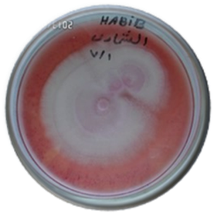
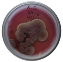
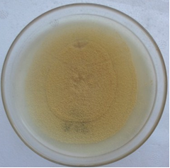
The cultural characteristics of the fungal isolates showed that, colonies were white, dry, powdery to cottony colonies on PDA medium (Fig. 1). When disturbed on the surface, the colony becomes yeast-like or slimy. The microscopic features showed that, arthroconidia and coarse true hyphae were observed. Blastoconidia, conidiophores and pseudohyphae were absent. They are either rectangular in shape or rounded at the ends resembling the barrel shape. From these characteristics, isolate No. 1, 2 and 35 were identified as Geotrichum candidum following the protocol of Buchta and Otcenasek [19]. Similar result was obtained by Hayaloglu and Kirbag, [3] who studied the semihard texture, rectangular shape Kuflu cheese which was a mould-ripened variety produced in the middle and eastern region of Turkey. This cheese is made from non-pasteurized, skimmed (or partially-skimmed) sheep's or goat's milk or their mixture. Penicillium sp, and Geotrichum candidum were the most frequently isolates. Also, a total of 24 fungi were isolated from Kuflu cheese being Aspergillus fumigatus, A. flavus, Rhizopus nigricans, Acremonium alternatum, Alternaria alternate, A. niger, Cladosporium cladosporioides, C. herbarium, Mucor himealis, Rhizopus nigricans and Trichoderma harzianum. Moreover, Cathrine and Skaar, [27]; Kure et al., [28] and Florez et al., [29] isolated and identified Geotrichum candidum from Norwegian semi-hard cheeses, Jarlsberg cheese and Spanish blue-veined Cabrales cheese.
Figure 1. Geotrichum candidum on RYS medium
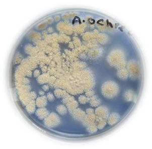
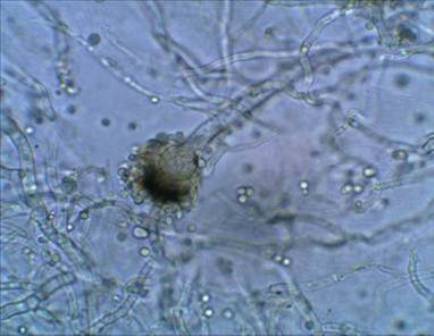
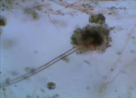
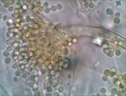
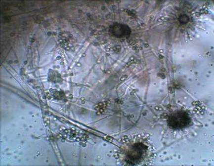
The colonies of isolates Nos. 3, 4, 14, 28, 31, 38, 49, 57, 65 and 66 grew rapidly on PDA medium (Fig. 2) while the conidial heads are typically arranged in zones. The color of the colony is yellow. This isolate formed pinkish to purple color on PDA medium, irregular, pebble like, the conidiophores appear as powdery mass. Microscopically, the appearances of these conidiophores are granular with pale yellow-brown walls that attach abruptly to a "globose to subglobose vesicle" (Fig. 3). From these characteristics, these isolates were identified as Aspergillus ochraceus following Chung and Bennett [21].
Few studies on the presence of Aspergillus ochraceus in cheese have been published [30]. Particularly worthy of mention are those by Spanish [31], Turkish [32] and Italian researchers [33]. In Egypt, Abdel-All et al., [34] isolated and identified twenty five of different fungi from different Gouda cheese samples and they found that, A. parasiticus, A. flavus, A. niger, A. versicolor, A. terrus, A. ochraceus, A. candidus, A. nidulans, P. requforti and P. chresogenum were the most isolated fungi. Also, Pattono et al., [30] reported that, Ochratoxin A is produced by different species of the genus Aspergillus such as A. ochraceus, A. melleus, A. sulphureus, A. niger, A. lanosus, A. alliaceus, A. carbonarius and A. awamori and different species of the genus Penicillium such as P. verrucosum, P. chrysogenum and P. nordicum.
Figure 2. Aspergillus ochraceus on PDA medium
Figure 3. Aspergillus ochraceus under light microscope (400x)
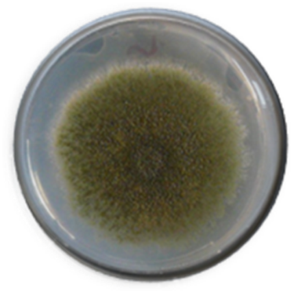
The characteristics of isolate No. 30 showed to be green, on PDA medium (Fig. 4). The isolate grew predominantly as sterile hyphae interspersed with large ellipsoidal to cylindrical dark brown ascomata produced directly from the vegetative hyphae. Conidial heads indicative of the genus Aspergillus. Conidiophores were smooth with globose vesicles that gave rise to radiating, biseriate conidial heads producing smooth-walled, globose conidia. From these characteristics and following the protocol of Balajee et al., [11] isolate No. 30 was identified as Aspergillus alliaceus (recently Petromyces alliaceus).
Figure 4. Aspergillus alliaceus on RYS medium
They found that, with the combined phenotypic and genotypic evidence, the unknown fungal isolate was identified as P. alliaceus. The teleomorphic species P. alliaceus is one of only three members assigned to the genus Petromyces. Although Petromyces spp. have been isolated from surfaces of plants and soil, the P. alliaceus is rarely recovered from an invasive fungal infection and has been previously isolated from a case of chronic otorrhea (drainage from the auditory canal). Petromyces alliaceus produces a variety of secondary metabolites, including ochratoxin A and B. Although the anamorphous of the teleomorphic genus Petromyces was originally assigned to the Aspergillus section Circumdati, recent chemotaxonomic and genotypic evidence places this genus in Aspergillus section Flavi, whose members often produce sclerotia [11]. The fungal contamination of Gouda cheese was studied by Berlkten and Kivanc [35] and they isolated and identified 21 isolates as Aspergillus alliaceus, Geotrichum candidum and Penicillium sp.
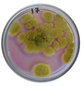
Characterization of the isolates showed that, colonies were yellow green on Rose Bengal medium (Fig. 5), conidiophores colorless, long, coarsely roughened. conidial heads typically radiate, conidia globose to subglobose. From these characteristics following the schemes of Chung and Bennett [21], isolate Nos. 13, 15, 16, 17, 21, 22, 29, 33, 34, 37, 39, 40, 43, 45, 50, 52, 54, 58, 59, 60, 61, 62, 63, 67, 69 and 70 were identified as Aspergillus oryzae.
Figure 5. Aspergillus oryzae on RYS medium
The fungal contamination of cheese at the stage of consumption in Saudi Arabia (including Egyptian cheese) was studied by Nasser, [36] who found that, 13 species of fungi were isolated from 12 cheese samples. The total fungal counts in cheese samples were relatively low and ranged from 95 and 125 cfu/g. Members of Penicillium and Aspergillus (A. flavus, A. niger and A. oryzae) were the most prevalent fungi on cheese samples. Ando et al., [37] isolated A. oryzae from Iranian commercial cheese (Caspian cheese). Also, A. oryzae was isolated from spoiled cheese by Sharma et al., [38] and used it for producing alkaline protease.
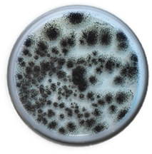
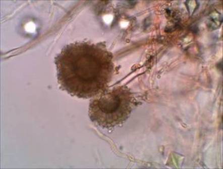
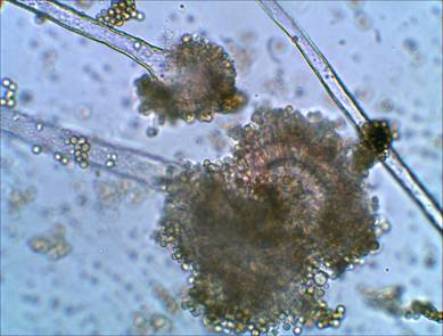
Mycelia of colonies of isolates Nos. 7, 27, 28 and 46 on PDA medium were white, conidial heads dark brown, greenish black (Fig. 6), brownish black to black reverse colorless, conidial heads globose, radiate or splitting into several irregular or well-defined columns of conidial chains, conidiophores hyaline to brown and smooth-walled (Fig. 7). Vesicles globose to subglobose hyaline to dark brown. From these characteristics, these isolates were identified as Aspergillus niger according to the protocol of Chung and Bennett [21]. Many authors isolated A. niger from cheese such as; Hassan and El-Deeb, [39] (Ras cheese); El-Deeb et al., [40] (Ras cheese); Nasser, [36] (white cheese); Hayaloglu and Kirbag, [3] (Kuflu cheese); Abdel-All et al., [34] (Ras cheese); Anwar and Sabah [41] (Qastqawan cheese) and Santi et al., [26] (Fossa cheese).
Figure 6. Aspergillus niger on PDA medium
Figure 7. Aspergillus niger under light microscope (400x)
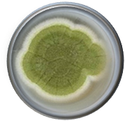
Some isolates showed pale green or dark yellow green colonies PDA on (Fig. 8), Conidiophores rarely exceed long, colourless, smooth, columnar, conidial heads looscly columanar vesicles very small, conidia globose or subglobose, smooth, ascospores present (Fig. 9). From these characteristics, isolate No. 26 was identified as Aspergillus nidulans [21].
Similar results were obtained by Barrios et al., [42] who reported that, contamination by the genus Aspergillus was studied in 52 samples of commercial cheeses made with different types of milk (cow's milk, ewe's milk, goat's milk and mixture of them) produced in southern Spain. The frequency of appearance of various species of the genus Aspergillus such as A. glaucus, A. niger, A. nidulans, A. sulphureus, A. terreus, and A. flavus, in the different types of cheese was determined. Also, Abdel-All et al., [34]; Santi et al., [26] and Ando et al., [37] isolated A. nidulans from Gouda, Fossa cheese and Caspian cheese, respectively.
Figure 8. Aspergillus nidulans on PDA medium
Figure 9. Aspergillus nidulans under light microscope (400x)
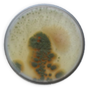
Figure 10. Emericella nidulans on PDA medium
Figure 11. Emericella nidulans under light microscope (400x)
Cleistothecia and conidial structures of isolate No. 8 duplicating those of nidulans, ascosp orange red on PDA medium (Fig. 10), lenticular, convex wall smooth, equatorial ridges absent (Fig. 11), but ascospres showing the usual bivalve construction. From these characteristics, isolate No. 8 was identified as Emericella nidulans [21].
Isolates Nos. 18, 23 and 24 showed conidial heads pale to intense yellow green when young, colonies not shifting to brown in age on PDA medium (Fig. 12); conidia definitely echinulate predominane; conidial heads radiate or very loosely columnar, colonies shifting to brownish in age; conidia smooth to roughened; conidiophores arising primarily from the substrate (Fig. 13). From these characteristics, these isolates were identified as Aspergillus flavus [21]. Many authors isolated A. flavus from cheese such as; Hassan and El-Deeb, [39] (Ras cheese); El-Deeb et al., [40] (Ras cheese); Nasser, [36] (white cheese); Hayaloglu and Kirbag, [3]; (Kuflu cheese); Abdel-All et al., [34] (Ras cheese); Gandomi et al., [4] (white cheese); Anwar and Sabah [41] (Qastqawan cheese) and Santi et al., [26] (Fossa cheese).
Figure 12. Aspergillus flavus on PDA medium
Figure 13. Aspergillus flavus under light microscope (400x)
Colonies of isolates Nos. 5, 6, 9, 10, 15, 19, 41, 44, 47, 48 and 53 on PDA medium were grayish turquoise to deep green (Fig. 14). Reverse was pale yellow to pale brown hyphae are septate and hyaline with a cleistothecia. Conidial heads were radiate to loosely columnar. Conidiophores were smooth walled and uncolored to pale brown. Vesicles were globose to subglobose and uniseriate. Conidia were globose to subglobose (Fig. 15). From these characteristics, these isolates were identified as Aspergillus glaucus [20]. Similar results were obtained by Barrios et al., [42] who isolated A. glaucus from commercial cheeses produced in southern Spain. Also, Santi et al., [26] isolated and identified Aspergillus glaucus, from traditional Italian cheese (Fossa cheese).
Figure 14. Aspergillus glaucus on RYS medium
Figure 15. Aspergillus glaucus under light microscope (400x)
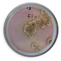
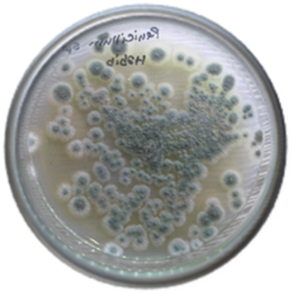
Figure 16. Penicillium sp. on PDA medium
Figure 17. Penicillium sp. under light microscope (400x)
The colonies of isolate Nos. 11 and 68 were rapid grown, flat, filamentous, and velvety, woolly, or cottony in texture on PDA medium (Fig. 16). The colonies were initially white and become blue green, gray green, olive gray, yellow or pinkish in time. visualized as globose to elongated sausage-shaped cells that multiply by fission (Fig. 17). From these characteristics, isolates No. 11 and 68 was identified and designated as Penicillium sp. [24].
Many authors isolated Penicillium from cheese such as; Hassan and El-Deeb [39] (Ras cheese); El-Deeb et al., [40] (Ras cheese); Cathrine and Skaar, [27] (Norwegian semi-hard cheeses); Kure et al., [28] (Jarlsberg cheeses); Nasser, [36] (white cheese); Florez et al., [29]; Hayaloglu and Kirbag, [3] (Kuflu cheese); Abdel-All et al., [34] (Ras cheese); Gandomi et al., [4] (white cheese), Anwar and Sabah [41] (Qastqawan cheese) and Santi et al., [26] (Fossa cheese). Penicillium roqueforti and P. commune were also isolated from spoilage Cheddar cheese by Taniwaki et al., [43]. Moreover, Ando et al., [37] isolated P. crysogenum and P. citrinum from Iranian commercial cheese (Caspian cheese).
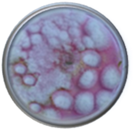
Colonies of isolate No. 12 grow rapidly at 25°C and quickly covered the surface of the medium. Its fluffy appearance with a height of several cm resembles cotton candy. From the front, the color is white initially and becomes grayish brown in time on Rose Bengal medium (Fig. 18). From the reverse, it was white. Nonseptate or sparsely septate, broad hyphae, sporangiophores, sporangia, and spores were visualized. Intercalary or terminal arthrospores (oidia) located through or at the end of the hyphae and few chlamydospores maybe also produced by some species. Apophysis, rhizoid and stolon were absent. Sporangiophores are short, erect, taper towards their apices and may form short sympodial branches. Columella were hyaline or dematiaceous and were hardly visible if the sporangium has not been ruptured. Smaller sporangia may lack columella. Sporangia were round, gray to black in color, and filled with sporangiospores. From these characteristics, isolate No. 12 was identified as Mucor sp. [23].
Figure 18. Mucor sp. on RYS medium
The growth of Mucor plumbeus and mycotoxin production on cheese under modified atmospheres was studied by Taniwaki et al., [43]. Also, Mucor racemosus and Mucor sp. were isolated and identified from Qashqawan Cheese and traditional Italian cheese (Fossa cheese) by Anwar and Sabah [41] and Santi et al., [26], respectively. Ando et al., [37] isolated Mucor hiemalis, M. javanicus and M. roxianus from Iranian commercial cheese (Caspian cheese). Also, Cheong et al., [44] reported that, Moulds are the most common cheese spoilage organisms which can lead to economic loss as well as raising public health such as Penicillium solitum, Aspergillus versicolor and Cladosporium herbarum, Mucor circinelloides and Geotrichum candidum. On the other hand, Lynch et al., [45] published that, the most frequently isolated fungi from cheese were Alternaria, Aspergillus, Cladosporium, Eurotium, Fusarium, Mucor, Penicillium and Phoma.
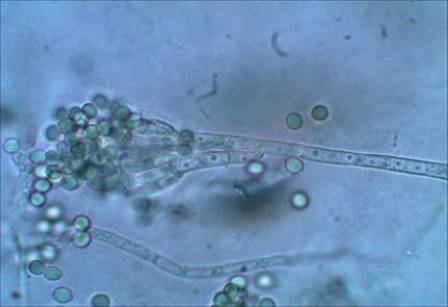
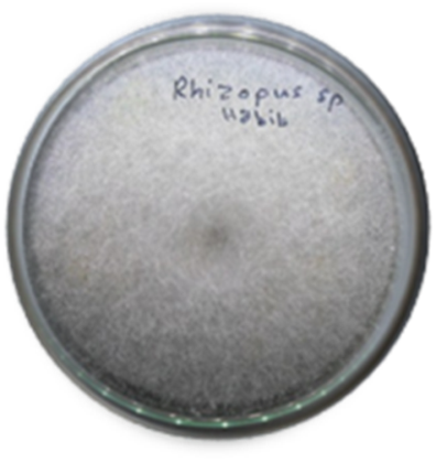
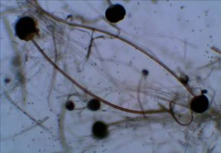
Colonies of isolate Nos. 42 and 64 grew very rapidly, fill the Petri dish, and mature in 4 days. The texture is typically cotton-candy like on PDA medium (Fig. 19). From the above data, the color of the colony was white initially and turns grey to yellowish brown in time. The reverse was white to pale. Nonseptate or sparsely septate broad hyphae, sporangiophores, rhizoids (root-like hyphae), sporangia, and sporangiospores were visualized. Sporangiophores were brown in color and usually unbranched (Fig. 20). They can be solitary or form clusters. Rhizoids were located at the point where the stolons and sporangiophores meet. Sporangiospores are unicellular, round to ovoid in shape, hyaline to brown in color, and smooth or striated in texture. From these characteristics, isolates Nos. 42 and 64 were identified as Rhizopus stolonifer [8]. Also, Rhizopus sp. was isolated and identified by Santi et al., [26] from traditional Italian cheese (Fossa cheese). In addition, Ando et al., [37] isolated Rhizopus retlexus, R. stolonifer and R. javanicus from Iranian commercial cheese (Caspian cheese).
Figure 19. Rhizopus stolonifer on PDA medium
Figure 20. Rhizopus stolonifer under light microscope (400x)
Conidiophores of isolate No. 56 were smooth, in yellow to light brown shades or colourless on PDA medium (Fig. 21). Conidial heads radiate when young, becoming broadly to irregularly columnar in age; persistently with or changing to avellaneous, pllebuff or greyish buff. Vesicles subglobose to ovate or elongate. Metulae present, covering the entire surface of the vesicle, or only the upper part. Conidia globosse to subglobose, colourless, smooth. Ascomata known in tow species; ascospores hyaline to pale yellow. Conidiophores definitely pigmented in yellow to light brown shades, conidial heads were white to very pale buff. From these characteristics, isolate No. 56 was identified as Aspergillus flavipes [21]. Alternaria, A. flavus, A. niger, A. glaucus, A. flavipes, A. terreus, Cladosporium sp., Fusarium solani, Mucor sp., Penicillium sp., Phoma sp. and Rhizopus sp. were isolated and identified by Santi et al., [26] from traditional Italian cheese (Fossa cheese).
Figure 21. Aspergillus flavipes on PDA medium

3.3. The Classification Position of Fungal Isolates

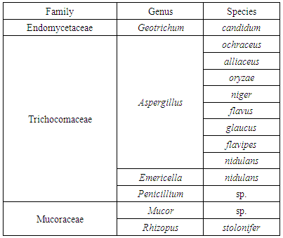
Table 2 show that, the classification position of obtained fungal isolates were classified in three families, 6 genus and 13 species as following; one species was belonging to family Endomycetaceae being Geotrichum candidum. Two species were belonging to family Mucoraceae being Mucor sp. and Rhizopus sp. The other eleven fungi were belonging to family Trichocomaceae being A. ochraceus; A. alliaceus; A. oryzae; A. niger; A. flavus; A. glaucus; A. flavipes; A. nidulans; Emericella nidulans and Penicillium sp. [21].
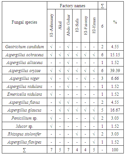
Most of fungal strains were found in El-Ashmawy and Abdo Gohar being 7 species, but Akel and El-Eman Factories were the second in fungal strains being 5 strains and the third were El-Safa and El-Faiomy being 4 strains. Obtained results is in agreement with those obtained by Florez and Mayo [25] who identified fungal isolates during manufacturing and ripening of Cabrales cheese. They found, 12 isolates of Penicillium roqueforti, 11 isolates of Penicillium, 10 isolates of G. candidum, one isolate Acremonium charticola and one unidentified isolate. G. candidum was frequently isolated from one of four cheese factories in Norway. A few isolates of other genera including Acremonium, Alternaria, Cladosporium, Mucor and Trichoderma spp. were also found on Kuflu cheese.
Table 2. The Classification of fungal isolates
     

3.4. Incident of Fungal Strains on the Ras Cheese

Table 3. shows, the presence of fungal strains of Ras cheese samples obtained from six different factories. Aspergillus oryzae was observed in all factories except Abdo Gohar and El-Eman 's factories being 39.39% while the other strains were attributed according to their percentages. Aspergillus glaucus was in the second order being16.67% that presented also in all factories except El-Ashmawy's factory. Aspergillus ochraceus was in the third order being 15.15% that observed in all factories. Aspergillus niger which the fourth order being 6.66% that presented in El-Ashmawy, Abdo Gohar and El-Faiomy factories.
Table 3. The distribution of fungal species on the Egyptian Ras cheese factories
     
The distribution of the following fungi were only in two factories, the percentage of Geotrichum candidum was 4.55% that presented in El-Ashmawy and Abdo Gohar factories. The percentage of Aspergillus flavus was 4.55% that presented in Abdo Gohar and Akel factories. The percentage of Penicillium sp. was 3.03% and was presented in El-Ashmawy and El-Eman factories. The percentage of Rhizopus sp. was 3.03% and was presented in El-Safa and El-Eman factories. Obtained results are in agreement with those obtained by Florez and Mayo [25] who identified fungal isolates during manufacture and ripening of Cabrales cheese. They found that, 12 isolates of Penicillium roqueforti, 11 isolates of Penicillium, 10 isolates of Geotrichum candidum, one isolate Acremonium charticola and one unidentified isolate. Finally, Aspergillus alliaceus and Aspergillus nidulans presented in only one factory (Abdo Gohar's factory), in a percentage of 1.52%, for each. Also, Emericella nidulans and Mucor sp. were 1.52% for each one found in El-Ashmawy factory. While, Aspergillus flavipes was found in El-Eman Factory only.

4. Conclusions

It can be concluded that, most of fungal strains were found in El-Ashmawy and Abdo Gohar being 7 strains, but Akel and El-Eman factories were the second in fungal species being 5 species and the third were El-Safa and El-Faiomy being 4 strains. Moreover, Uncontrolled fungi growth on the cheese surface must be considered as a problem and provides no guarantee of product safety for human consumption. For the ripening of Ras cheese, non-toxigenic strains of fungi should be selected

ACKNOWLEDGEMENTS

Authors like to thank Dr. Khalid Ghonim the researcher in plant pathology department, faculty of agriculture, Mansoura university for his helping.

References

[1]  Dabiza, N., and El-Deib, K., 2007, Biochemical evaluation and microbial quality of Ras cheese supplemented with probiotic strains., Polish J. food and nutrition sciences, 57(3), 255-300.
[2]  Hattem, H. E, Taleb, A. T., Manal, A. N., and Hanaa, S. S., 2012, Effect of pasteurization and season on milk composition and ripening of Ras cheese., J. Brewing and Distilling, 3(2), 15-22.
[3]  Hayaloglu, A. A., and S., Kirbag, 2007, Microbial quality and presence of moulds in Kuflu cheese., Inter. J. Food Microbiol., 115, 376–380.
[4]  Gandomi, H., Misaghi, A., Basti, A. A., Bokaei, S., Khosravi, A., Abbasifar, A., and Javan, A. J., 2009, Effect of Zataria multiflora Boiss. essential oil on growth and aflatoxin formationby Aspergillus flavus in culture media and cheese., Food and chemical toxicology, 47, 2397–2400.
[5]  Speare, R, Thomas, A. D., Shea, P. O. and Shipton, W. A., 1994, Mucor amphibiorum in the toad, Bufo marinus, in Australia., Journal of wildlife diseases, 30(3), 399-407.
[6]  Fouassier, M, Joly, D., Cambon, M., Lafeuille, H. P. and Condat, P., 1998, Geotrichum capitatum infection in a neutropenic patient. Case report and literature review., Rev. Med. Interne, 19,431-433.
[7]  Mori, T, Matsumura, M., Yamada, K., Irie, S., Oshimi, K., Suda, K., Oguri, T., and Ichinoe, M., 1998, Systemic aspergillosis caused by an aflatoxin-producing strain of Aspergillus flavus., Med. Mycol., 36,107-112.
[8]  Anstead, G. M., Sutton, D. A., Thompson, E. H., Weitzman, I., Otto, R. A., and Ahuja, S. K., 1999, Disseminated zygomycosis due to Rhizopus schipperae after heatstroke., J Clin Microbiol., 37, 2656-2662.
[9]  Kontoyiannis, D. P, Lewis, R. E., May, G. S., Osherov, N., and Rinaldi, M. G. 2002, Aspergillus nidulans is frequently resistant to amphotericin B., Mycoses, 45,406-407.
[10]  Steinbach, W. J., and Stevens, D. A., 2003, Review of newer antifungal and immunomodulatory strategies for invasive aspergillosis., Clin. Infect. Dis, 37,157-187.
[11]  Balajee, S. A., Lindsley, M. D., Iqbal, N., Ito, J., Pappas, P. G. and Brandt, M. E., 2007, Nonsporulating clinical isolate identified as Petromyces alliaceus (Anamorph Aspergillus alliaceus) by Morphological and Sequence-Based Methods., J. Clin. Microbiol., 45(8), 2701–2703.
[12]  Ghibaudo, G., and Peano, A., 2010, Chronic monolateral otomycosis in a dog caused by Aspergillus ochraceus., Veterinary dermatology, 21(5), 522–526.
[13]  Oxoid, 2006, The Oxoid Manual. 9th ed., by OXOID Limited, Wade Road, Basingstoke, Hampshire RG24 8PW, England, UK.
[14]  Ronald, M. A., 2006, Hand Book of Microbilogical Media for the Examination of Food. CRC Taylor and Francis Group Boca Raton London New York.
[15]  APHA, (American Public Health Association), 1998, Standard Methods for the Examination of Water and Wastewater 20th ed., APHA, Inc., New York.
[16]  Kure, C. F., and Skaar, I., 2000, Mould growth on the Norwegian semi-hard cheeses Norvegia and Jarlsberg., Inter. J. Food Microbiol., 62, 133–137.
[17]  Raper, K. E., and Fennel, D. I., 1965, The genus Aspergillus. Williams and Wilkins, Baltimore.
[18]  Harrigan, W. F., and McCance, M. E., 1976, Laboratory Methods in Food and Dairy Microbiology (2nd ed.), Academic Press, New York, NY, USA.
[19]  Buchta, V., and M., Otcenasek, 1988, Geotrichum candidum an opportunistic agent of mycotic diseases., Mycoses, 31,363-70.
[20]  Yoshida, K., Ando, M., Ito, K., Sakata, T., Arima, K., Araki, S., and Uchida, K., 1990, Hypersensitivity pneumonitis of a mushroom worker due to Aspergillus glaucus., Arch Environ Health, 45,245-7.
[21]  Chung, K. K. J., and J. E., Bennett, 1992, Medical mycology, (2nd ed.), Lea and Febeiger, USA.
[22]  Pitt, J. I., and A. D., Hocking, 1997, Fungi and Food Spoilage., Blackie Academic and Professional, London, UK.
[23]  Frater, J. L., Hall, G. S., and Procop, G. W., 2001, Histologic features of zygomycosis - Emphasis on perineural invasion and fungal morphology., Arch. Pathol. Lab. Med., 125,375-378.
[24]  Buommin, N. R, Filippis, E. D., Lopez-Gresa, A., Manzo, M., Carella, E., Petrazzuolo, A., and Tufano, M. M. A., 2009, Bioprospecting for antagonistic Penicillium strains as a resource of new antitumor compounds., World J. Microbiol., 24(2), 185–95.
[25]  Florez, A. B., and B., Mayo, 2006, Microbial diversity and succession during the manufacture and ripening of traditional, Spanish, blue-veined Cabrales cheese, as determined by PCR-DGGE., International Journal of Food Microbiology, 110, 165–171.
[26]  Santi, M. D., M., Sisti;, E., Barbieri, G., Piccoli, G., Brandi, and V., Stocchi, 2010, A combined morphologic and molecular approach for characterizing fungal microflora from a traditional Italian cheese (Fossa cheese)., Inter. Dairy J., 465-471.
[27]  Cathrine, F. K., and I., Skaar, 2000, Mould growth on the Norwegian semi-hard cheeses Norvegia and Jarlsberg., Inter. J. Food Microbiol., 62, 133–137.
[28]  Kure, C. F., Y., Wasteson, J., Brendehaug, and I., Skaar, 2001, Mould contaminants on Jarlsberg and Norvegia cheese blocks from four factories., Inter. J. Food Microbiol., 70, 21–27.
[29]  Florez, A. B., P. A., Martin, T. M. L., Diaz, and B., Mayo, 2007, Morphotypic and molecular identification of filamentous fungi from Spanish blue-veined Cabrales cheese and typing of Penicillium roqueforti and Geotrichum candidum isolates., International Dairy Journal, 17, 350–357.
[30]  Pattono, D., A., Grosso, P. P., Stocco, M., Pazzi, and G., Zeppa, 2013, Survey of the presence of patulin and ochratoxin A in traditional semihard cheeses., Food Control, 33,54-57.
[31]  Diaz, L. T. M., R. M. T., Blanco, G. M. C., Arias, G. M. L., Fernandez, and G., Lopez, 1996, Mycotoxins in two Spanish cheese varieties., Inter. J. Food Microbiol., 30, 391-395.
[32]  Erdogan, A., M., Gurses, and S., Sert, 2003, Isolation of moulds capable of producing mycotoxins from blue mouldy Tulum cheeses produced in Turkey., Inter. J. Food Microbiol., 85, 83-85.
[33]  Asta, D. C., D. J., Linder, G., Galaverna, N. E. A., Dossena, and R., Marchelli, 2008, The occurrence of ochratoxin A in blue cheese., Food Chemistry, 106, 729-734.
[34]  Abdel-All, S. M, M. A., Abd-El-Ghany, and M. M., Motawee, 2008, Inhibition of Aspergillus growth and aflatoxins production in some dairy products. The 3ed annual conference of quality education development in Egypt and the Arab region to achievement the requirements of job markets in the globalization age (strategic vision). Faculty of Specific Education Mansoura University, 1108-1120.
[35]  Berlkten, D., and M., Kivanc, 2012, Fungal contamination of some food, their mycotoxin and effects of antifungal agents on these fungi., Microbes in Applied Research, Current advanced and challenges, World Scientific Publishing Co.
[36]  Nasser, L. A., 2001, Fungal contamination of white cheese at the stage of consumption in Saudi Arabia., Pakistan J. Biological Sciences, 4(6), 733-735.
[37]  Ando, H., K., Hatanaka, I., Ohata, Y. Y., Kitaguchi, A., Kurata, and N., Kishimoto, 2012, Antifungal activities of volatile substances generated by yeast isolated from Iranian commercial cheese., Food Control, 26,472-478.
[38]  Sharma, J., A., Singh, R., Kumar, and A., Mittal, 2013, Partial purification of an alkaline protease from a new strain of Aspergillus oryzae AWT 20 and its enhanced stabilization in entrapped Ca-alginate beads., Internet J. Microbiol., 2(2), 1-14.
[39]  Hassan, H. N., and S. A., El-Deeb, 1988, The inhibitory effect of water and acetone extraction of certain plants as anti-fungal agents on growth and aflatoxins production by molds and yeast isolated from cheese ripening rooms., J. Agric. Res., Tanta Univ., 14, 162-171.
[40]  El-Deeb, S. A., E. E., Kheadr, N., Zaki, and Y. M., Shoukry, 1992, Formation and the penetration of aflatoxin in experimental cheese by Aspergillus parasiticus., Egyptian J. Food Sci., 15-20.
[41]  Anwar, A., and Y., Sabah, 2010, The components effect of cinnamon oil extracted in inhibition of isolated molds from Kashkawan cheese., Damascus J. of agricultural Science, 26(2), 287-300.
[42]  Barrios, M. J., L. M., Medina;, M. G., Cordoba, and R., Jordano, 1997, Aflatoxin producing strains of Aspergillus flavus isolated from cheese., J. Food Protection, 192-194.
[43]  Taniwaki, M. H., A. D., Hocking, J. I., Pitt, and G. H., Fleet, 2001, Growth of fungi and mycotoxin production on cheese undermodified atmospheres., Inter. J. Food Microbiol., 68, 125–133.
[44]  Cheong, E. Y. L., A., Sandhu, J., Jayabalan, T. T. K., Le, N. T., Nhiep, H. T. M., Ho, J., Zwielehner, N., Bansal, and M. S., Turner, 2014, Isolation of lactic acid bacteria with antifungal activity against the common cheese spoilage mould Penicillium commune and their potential as biopreservatives in cheese., Food Control, 46, 91–97.
[45]  Lynch, K. M., A. M., Pawlowska, B., Brosnan, A., Coffey, E., Zannini, A., Furey, and P. L. H., McSweeney, D. M., Waters, and E. K., Arendt, 2014, Application of Lactobacillus amylovorus as an antifungal adjunctto extend the shelf-life of Cheddar cheese., Inter. Dairy J., 34,167-173.