-
Paper Information
- Previous Paper
- Paper Submission
-
Journal Information
- About This Journal
- Editorial Board
- Current Issue
- Archive
- Author Guidelines
- Contact Us
Journal of Microbiology Research
p-ISSN: 2166-5885 e-ISSN: 2166-5931
2014; 4(3): 136-140
doi:10.5923/j.microbiology.20140403.02
Isolation of Actinomycetes from Soil
 Abstract
 Reference
 Full-Text PDF
 Full-text HTML
Stephen Kugbere Agadagba
Department of Biochemistry, Faculty of Life Sciences, University of Benin, Benin City, Nigeria
Correspondence to: Stephen Kugbere Agadagba, Department of Biochemistry, Faculty of Life Sciences, University of Benin, Benin City, Nigeria.
| Email: |  |
Copyright © 2014 Scientific & Academic Publishing. All Rights Reserved.
The main focus of this study was to isolate some antibiotic producing actinomycetes strains from soil. The soil sample used was dark brown and sandy, with no vegetation covering (bare). Isolation of soil actinomycetes was done by culture-dependent methods and isolates were tested for antibiotic production on selected indicator bacteria plates. The results indicated that a total of 3.56 × 10⁵ actinomycetes colonies were isolated per gram of dry soil. Furthermore, microscopic examination of the isolates indicated 6 major colony types (CTs) in the soil. Only 3 CTs were found to be active against one or more indicator bacteria, with inhibition zones that ranged from 7 mm to 12.5 mm in diameter. From the results, it was suggested that the low yield of antibiotic producing actinomycetes isolates obtained in this study, could be improved by employing a combination of several molecular analysis methods.
Keywords: Actinomycetes, Isolation, Soil sample, Culture method, Antibiotics
Cite this paper: Stephen Kugbere Agadagba, Isolation of Actinomycetes from Soil, Journal of Microbiology Research, Vol. 4 No. 3, 2014, pp. 136-140. doi: 10.5923/j.microbiology.20140403.02.
Article Outline
1. Introduction
- Actinomycetes are classified as a group of gram-positive bacteria that are unique for their spore forming abilities and formation of mycelia structures. These bacteria have been noted to serve as rich reservoirs of medicinal antibiotics and are therefore extremely relevant to scientists, pharmaceutical industries and agricultural industries [1]. A huge number of currently used antibiotics including erythromycin, streptomycin, rifamycin and gentamycin, are all products isolated from soil actinomycetes [2]. The two major groups of soil actinomycetes that serve as important sources of antibiotics are Streptomyces and Micromonospora. It has been stated that Streptomyces account for about 80% of the total antibiotic products; while Micromonospora closely follows with less than one tenth as much as Streptomyces [3]. Furthermore, previous experimental analysis, have also proven that secondary metabolites isolated from soil actinomycetes, are potent inhibitors of numerous plant pathogens [2]. For example, it has been highlighted that actinomycetes from farming soils, have the capacity to inhibit Erwina amylovora; a bacterium that causes fireblight to apples and Agrobacterium tumefaciens; the causative pathogen of crown gall disease in plants [4].The richness and diversity of actinomycetes present in any specific soil, is greatly influenced by the soil type, geographical location, cultivation and organic matter [3] amongst other factors. Numerous studies have been done by scientists to isolate actinomycetes, as sources of antibiotics. However, because actinomycetes occur widely in nature, only a small percentage of the globe and a small proportion of actinomycetes species have been screened [1]. Therefore, the present study was aimed at isolating some antibiotic producing actinomycetes strains from sandy soil without vegetation or leave covering (bare soil), by culture-dependent methods.
2. Materials and Methods
2.1. Collection and Preparation of Soil Sample
- The soil used was dark brown and sandy, with no vegetation covering (bare; soil 3). Soil sample collection was from a location between the North and East wings of Haworth building, University of Birmingham, United Kingdom, in an area that was relatively steep and not quite in the shade.
2.2. Actinomycetes Enrichment
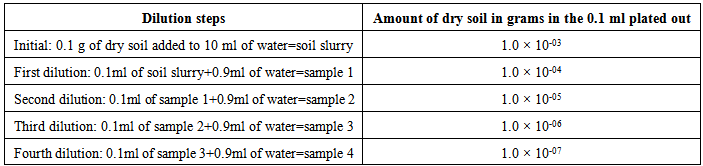
- This process was done aseptically. First, the soil slurry was made by suspending 0.1 g of the collected dry soil in 10 ml distilled water. The slurry was mixed by vortexing for 2 min and four 1 in 10 fold serial dilutions were made from the slurry. These dilutions were done in duplicates (A and B). Next, 3 ml volumes of the provided top agar were poured onto the bottom agar plates. The plates were allowed to set, after which 0.1 ml portions of each dilution, were then plated by spreading on the set chitin agar plates. All spread plates where labelled and incubated at 25 ̊C for a period of 14 days.
2.3. Examination of Plates
- On day 14, the number of colonies formed in each dilution of both groups of plates (A and B), were counted and recorded. All plates were carefully observed under the microscope to detect diversity in colonies formed. The richness, evenness and diversity index, where also calculated and recorded. Eight different colony types were then picked out with sterile forceps and streaked out on glucose yeast extract agar plates, to obtain pure single colonies. The streaked plates were then incubated.
2.4. Test for Antibiotic Production
- Single colonies of the grown actinomycetes cultures were examined with the microscope, to detect diversity in appearance. Four indicator microorganisms (Escherichia coli, Pseudomonas aeruginosa, Bacillus subtilis and Staphylococcus epidermis), were spread plated as broth cultures on 8 separate plates. A total of 8 actinomycetes were then deposited for each indicator organism, as described in the lab manual. All processes were performed aseptically, to avoid contamination.
2.5. Examination of Inhibition of Indicator Organisms
- Inhibition of indicator bacteria was accessed by measuring the diameter of clear zones formed around actinomycetes cultures, in each plate. All observations and data were recorded.
3. Results
3.1. Colony Number and Colony Diversity
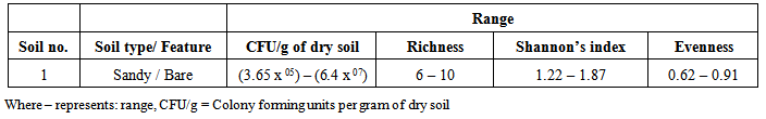
- After about 14 days of incubation, the chitin agar (top agar) was able to support the growth of only the actinomycetes and prevented growth of other soil bacteria and fungi. Cycloheximide used in the bottom agar, further prevented fungi interference by inhibiting mRNA translation, hence causing cell growth arrest and cell death of opportunistic fungi [5]. Six major colony types (richness) were observed. On microscopic examination,Colony type 1 (CT 1): Appeared as large bright white filaments, with net-like mycelia. No clear zones were observed.Colony type 2 (CT 2): Appeared as pale white branching filaments and had powdery appearance. No clear zones were observed.Colony type 3 (CT 3): Had a dark brown uniformity and crumb-like appearance.Colony type 4 (CT 4): Had a light brown appearance with cilia-like mycelia on its boundaries.Colony type 5 (CT 5): Showed a dark brown appearance, embedded with concentric circular patterns. Each pattern was separated by regions of clear zones.Colony type 6 (CT 6): Appeared as yellow colonies with beautiful mycelia and transparent boundaries.The number of actinomycetes colonies in each dilution plate did not necessarily follow the 10-fold serial dilution pattern as expected, but varied across each dilution and colony type (table 2). A total of 79 colonies were counted across all plates (table 2). Colony type 1 (CT 1) was found to have the highest colony forming units of 1.08 × 10⁵ per gram of soil (CFU/g). The lowest CFU/g obtained was 1.35 × 10⁴ and was recorded in CT 6. The total CFU/g for all colony types was calculated as 3.56 × 10⁵. The diversity index (Shannon’s index) was calculated as 1.62 and evenness as 0.91. In comparison with a parallel experiment (class sample) performed on the same soil (bare sandy soil), the highest CFU/g recorded was (6.4 × 10⁷) and the richness in the group ranged from 6 to 10 (table 3). Therefore this implies that the actinomycetes CFU/g ranged from hundredths of thousands (105) to tenths of millions (107) (table 3).
|
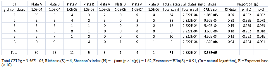 | Table 2. Counts for individual colony types (CT) and total colony types in bare soil sample |
|
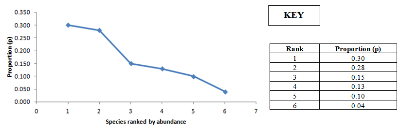 | Figure 1. Taxa abundance distribution |
3.2. Inhibition of Indicator Bacteria
- From the above (table 4 and figure 2), only 3 actinomycetes isolates (CT 4, CT 5 and CT 6) inhibited one or more indicator bacteria. Some of the zones of inhibition were present but not very obvious (figure 2). The widest inhibition zone (diameter = 12.5 mm) was recorded for CT 4 which inhibited only the growth of P. aeruginosa. The narrowest inhibition zone (diameter = 7mm) was observed in CT 6, which also inhibited two other indicator organisms (B. subtilis and S. epidermis) (table 2). Meanwhile CT 1, CT 2 and CT 3 were not seen to inhibit any of the indicator organisms.
|
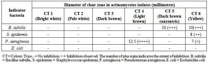
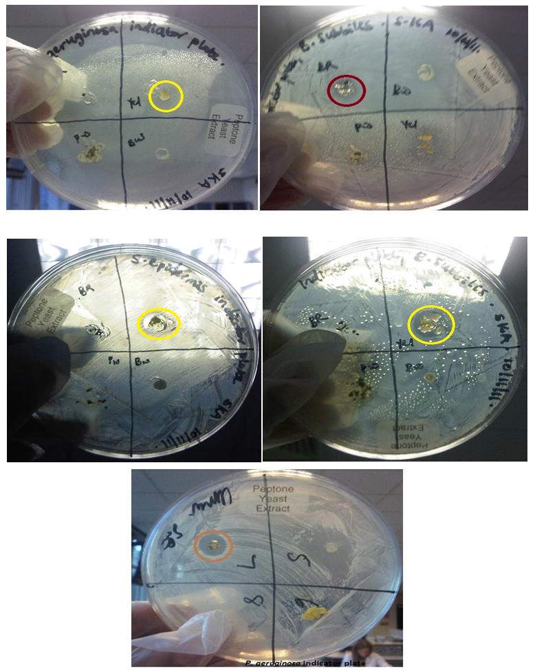 | Figure 2. Positive antibacterial activity against tested indicator bacteria by actinomycetes isolates. Dark brown circle = zone of inhibition by CT 5 actinomycetes, Yellow circles = zone of inhibition by CT 6 actinomycetes and light brown circle = zone of inhibition by CT 4 actinomycetes |
4. Discussion
- Soil actinomycetes isolated from bare sandy soil (soil type 3) in this study, were of six colony types. On initial microscopic examination, the clear zones observed in some grown actinomycetes colonies (CT 5 and CT 6), indicate that the organisms broke down the chitin for growth and development. Colony types (CT 1, CT 2 and CT 3) that did not develop clear zones, may have utilized other nutrients in the medium such as nitrates, phosphates and sulphates, in order to grow.It has been reported that 1g of soil when plated, harbours up to 10 billion microorganisms, of which about 4.2 × 10⁶ CFU/g (dry weight) are accounted for by bacteria species[6]. The number of actinomycetes (3.5 × 10⁵ CFU/g dry soil) obtained in this study was however lower than expectation. This may be partly attributed to the geographical location of the soil. As indicated in the methodology, the soil was collected from an area that was relatively steep and not quite in the shade. The steepness of the terrain, exposure to the sun rays and lack of vegetation covering of the soil (bare) may have caused leaching of actinomycetes and therefore have partly contributed to the observed low actinomycetes CFU/g of dry soil.Absence of clear zones found in three (CT 1, CT 2 and CT 3) actinomycetes isolates during final examination of chitin agar plates (table 4), shows that no antibiotics were produced in such colonies. Although, the other three isolates (CT 4, CT 5 and CT 6) were able to inhibit the growth of indicator bacteria, however the inhibition was not observed in all tested indicator bacteria. This therefore means that there was a low secretion of antibiotics by such actinomycetes isolates. A probable explanation for these results could be that the culture method used in this study, did not provide ideal conditions that should have enabled the growth of large numbers of actinomycetes and secretion of high amounts of antibiotics. Furthermore, experiments with this technique (culture-dependent methods) are quite limited in detecting the broad population of uncultured microorganisms in the soil ecosystem [6].It is therefore suggested that a combination of several molecular analysis methods such as DNA re-association and PCR-based fingerprinting techniques may extremely help to provide broader information about the total genetic diversity of soil actinomycetes community. Perhaps, such methods may lead to the improvement in isolating antibiotic producing strains of soil actinomycetes obtained in this study.
ACKNOWLEDGEMENTS
- The author wishes to thank his teachers; Prof. Charles W. Penn and Dr. Jan-Ulrich Kreft in Biosciences, University of Birmingham, UK, for their wonderful supervision during the soil practicals. I am also very grateful to my mum, my siblings, and my beautiful fiancée; Precious Onuwaje for their all-round support during the write-up of this paper.
Conflict of Interest
- The author declares no conflict of interest.