Journal of Microbiology Research

p-ISSN: 2166-5885    e-ISSN: 2166-5931

2014;  4(2): 98-103

doi:10.5923/j.microbiology.20140402.09

Hygienic Quality of Camel Milk and Fermented Camel Milk (Chal) in Golestan Province, Iran

Barat Ali Zarei Yam , Morteza Khomeiri , Alireza Sadeghi Mahounak , Seid Mahdi Jafari

Department of Food Science and Technology, Gorgan University of Agricultural Sciences and Natural Resources, Gorgan, 49138-15739, Iran

Correspondence to: Barat Ali Zarei Yam , Department of Food Science and Technology, Gorgan University of Agricultural Sciences and Natural Resources, Gorgan, 49138-15739, Iran.

Email:

Copyright © 2014 Scientific & Academic Publishing. All Rights Reserved.

Abstract

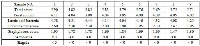
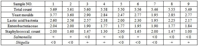
A total of 18 samples of fresh camel milk and sour camel milk (chal) were randomly collected from different retail markets with various levels of sanitation in Golestan province. Samples were subjected to determinations of pH, total count, total yeast and mould counts, Enterobacteriaceae count, lactic acid bacteria (LAB) count, total Staphylococcal count and for presence of Salmonella and Shigella. Results revealed that pH levels ranged from3.8 to 4.5 and 6.4 to 6.7 for chal and milk samples respectively. The highest and lowest total counts were determined in the ranges of 5.90 to 5.69, and 5.69 to 5.46; total yeast and mold counts were determined in the ranges of 4.11 to 3.90 and 3.77 to 1.95, and Enterobacteriaceae counts were 2.60 to 1.90 and 2.04 to 1.77, Lactic acid bacteria counts were 4.58 to 4.04 and 2.60 to 1.95, Staphylococcal counts were 1.95 to 1.30 and 2.00 to 1.00 log cfu/ml for chal and camel milk samples respectively. Salmonella and Shigella were not found in any of the chal samples, but there were traces in 8 of the raw camel milk samples. But it should be considered that chal is prepared under spontaneous fermentation, and in comparisson with pasteurized products it has low sanitation quality.

Keywords: Chal, pH, Salmonella, Sanitation Quality, Enterobacteriaceae

Cite this paper: Barat Ali Zarei Yam , Morteza Khomeiri , Alireza Sadeghi Mahounak , Seid Mahdi Jafari , Hygienic Quality of Camel Milk and Fermented Camel Milk (Chal) in Golestan Province, Iran, Journal of Microbiology Research, Vol. 4 No. 2, 2014, pp. 98-103. doi: 10.5923/j.microbiology.20140402.09.

1. Introduction

Camel milk and chal (soured camel milk) are consumed by some people in Iran and in other countries in Asia and Africa. Chal is prepared by adding water to raw camel milk and spontaneous fermentation takes place in a skin bag or a bottle at ambient temperature [23]. Camel milk and fermented camel milk have nutritional and medical properties that make them valuable foods. Chal contains some useful lactic acid bacteria [26-28]. In many regions, camel milk and chal are used to treat some diseases and to combat health problems such as dropsy, jaundice, tuberculosis, asthma, anaemia and piles [35]. Tests showed that patients with chronic hepatitis improved after consumption of camel milk [41]. Foods that promote good health and prevent disease are valued highly by today’s consumers. It should be considered that chal is made from raw milk by spontaneous fermentation, so it may contain some harmful organisms.
Dairy products generally constitute a favourable environment for the growth of yeasts, lactic acid bacteria and other acid tolerant microorganisms because they have an acidic pH and contain nutritious compounds [51-37]. Yeasts usually coexist with lactic acid bacteria in dairy products. One of the groupswill gain dominance over the other, or both groups grow together and specific interactions take place.
The aim of the present work was to assay microbial quality of camel milk and chal in Golestan province, Iran

2. Materials and Method

Samples of camel milk and chal were randomly collected from different households and retail markets with various levels of sanitation in Golestan province. The areas under investigation were the cities of Gonbad, Aghghala and Bandar Torkman. Samples were transported to the labratoary of the department of food science and technology, University of Gorgan and subjected to determination of chemical and microbial characteristics.

2.1. Compositional Analysis

Composition properties of samples were analyzed in duplicate for contents of protein, fat, ash, pH, TS, NaCl and acidity. The Kjeldahl method was used to determine total protein content of camel milk and chal, TS using a drying oven and Titrable acidity was determined by titration with 0.1 N NaOH using phenolphthalein as a color indicator until the color changed to light pink and persisted for 30 seconds, volume of NaOH was recorded and results were expressed in degrees (Dornic) [7]. Fat content was measured by the Gerber method [36] and ash by heating samples in a muffle furnace at 100°C for 1 hour, 200°C for 2 hours and 550°C overnight [30]. Levels of pH were determined using a pH meter (766knick, Germany) and NaCl evaluations were measured according tothe method cited in Bradley et al. [10].

2.2. Microbiological Assay

Microbiological characteristics of camel milk and chal such as total count, total yeast and mould count, Entrobacteriaceae, staphylococcal count and Shigella were tested according to Standard Methods for the Examination of Dairy Products [17]. Salmonella count was determined according to the method cited in Andrew and Jacobson [6], and lactic acid bacteria count was determined according to the method described in the Compendium of Methods for the Microbiological Examination of food [43]. For Salmonella count, each 25g sample was aseptically weighed and macerated and 225 mls of sterile distilled water was added. Sterile dilution was carried out using sterile distilled water as the dilutant from each dilution, 1 ml was plated using the pour plate method cited in Andrew and Jacobson [6].
For total count, total yeast and mould, Entrobacteriaceae, staphylococcal count, Shigella and lactic acid bacteria count, samples were diluted in sterile distilled water (to get readily countable numbers of microorganisms) and plated in Plate Count Agar (Mirmedia, Mp-2602, Iran) for total counts, in Baird Parker agar (Micro media, Mm0213, Hungary) for Staphylococcus aureus count, in VRBA (Micro media, Mm0114, Hungary) for Entrobacteriaceae counts, in MRS agar (Liofilchem, 610024, Italy) for lactic acid bacteria and in YGC agar (Mirmedia, Mfd, Iran) for yeast and mould counts. Plates were incubated at 37°C for 48 h for bacteria and at 30°C for 5 d for yeast and mould counts.

3. Results and Discussion

3.1. Composition Analyses

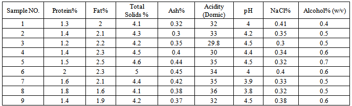
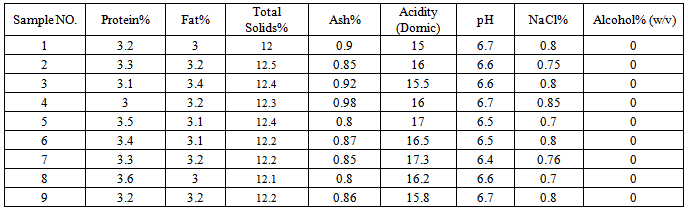
Chemical properties of camel milk and chal were determined and results are shown in Tables 1 and 2.
Results (Tables 1 and 2) revealed that pH ranged from 3.8 to 4.5 and 6.4 to 6.7 and acidity levels were 29.8 to 36 and 15 to 17.3 for chal and milk respectively. However higher values for pH and acidity were also recorded [31-29]; but the average pH recorded for camel milk was between 6.2-6.8 [22]. It can increase to 7.2 in cases of clinical mastitis [45]. Lower pH is considered inhibitory to vegetative cell growth of pathogenic microorganisms, but helpful to yeast growth, molds and lactic acid bacteria.However similar results were found in the related literature [3] but fermented foods are normally considered as safe against food-born diseases because of their low pH [21].
Table 1. Chemical Analysis of Chal Samples
     
Table 2. Chemical Analysis of Camel Milk Samples
     

3.2. Microbiological Quality

Microbial count of Chal and camel milk are shown in Tables 3 and 4. Results for chal samples showed significance in terms of yeast and mold counts (Table 3). Yeast and mould counts ranged from 4.11 to 3.9 and 3.77 to 1.95 log cfu/ml for chal and camel milk samples, respectively.
Table 3. Microbial Count of Chal Samples (log cfu/ml)
     
Table 4. Microbial Count of Camel Milk Samples (log cfu/ml)
     
Results revealed that yeasts, moulds and lactic acid bacteria counts in chal were higher in comparisson to those of camel milk. This can be attributed to the fermentation process and acidic pH of the chal samples. Benkerroum et al. [9] reported high counts (4.6 log cfu/ml) of yeasts and moulds in camel milk tests in Morroco that were higher than those of our results, also Abdel Gadir et al. [2] found a high count of yeasts in cow milk. In this study yeasts and moulds existed in all examined samples. El-Ziney and Al-Turki [17] identified yeasts and moulds in 57% of the camel milk samples with mean and maximum values of 1.9 and 5.65 log cfu/ml, respectively.
Frazier and Westhoff [20] and Almaw and Molla [34] reported that high counts of yeasts and moulds in milk are uncommon because of the natural pH level of milk that causes bacteria to predominate. El-Jakee [16] found Candida albicans in 4% of camels with clinical mastitis. Presence of yeasts might be atributed to contamination by air, water or a lack of proper hygenic practice during the production process, while the presence of moulds was usually attributed to contamination of the product by air or persons who were engaged in its preparation or transportation.
Total count recorded for chal was higher than that of camel milk and this may be attributed to growth and high yeast content of moulds and LAB (lactic acid bacteria) in Chal from the fermentation process.
In this study, the mean total counts recorded 5.56 and 5.78 log cfu/ml for camel milk and Chal respectively. The results of Wernery et al. [50] proved the total count values of camel milk (hand milking) were of 10 2 - 10 4 Cfu/ml, but they were lower than values given by Younan [52] in which total bacteria count was reported as10 3 - 10 5 Cfu/ml.
The mean of the total aerobic mesophilic bacteria count in the Qasim region reported 5 log cfu/ml with a maximum 7.15 log cfu/ml. [18] and for Saudi Arabia 5.4 log cfu/ml in average by Al Mohizea [5] and 5.6 log cfu/ml for a study on camel milk in Ethiopia by Semereab and Molla [39] and about 6 logcfu/ ml by Elgadi et al. [15].
The total bacteria count (TBC) of camel milk was reported to vary between 10² and 108 cfu/ml [39-50-38-52]. These differences show that TBC depends on several parameters such as the milk itself, contamination of the camel’s udder, milking personnel and other considerations such as transportation and containers. The results for total count in this study were in aggreement with reports from Saudi Arabia, Ethiopia and Kenya fortests on camel milk samples by Al Mohizea [5], Semereab and Molla [39] and Younan [52] respectively.
Under pastoral production conditions, environmental contamination plays a bigger role in hygiene levels of raw camel milk and fermented camel milk products than initial bacterial contamination of the camel’s milk itself [52]. If total bacterial count is determined as low, it was observed that raw milk did not to turn sour for up to 4 days when it was kept in a clean container and refrigerated [52].
Staphylococci were present in all examined samples. Highest and lowest counts recorded for chal and camel milk samples were 1.95, 1.30 and 2.00, 1.00 log cfu/ml, respectively. Also Abdel Gadir et al. [1], Barbour et al. [8], Chaffer et al. [12], Semereab and Molla [39], Tuteja et al. [45] and Elgadi et al. [15] reported the presence of Staphylicoccus spp. in different camel milk samples. El- Ziney and Al- Turki [18] reported the mean count of S. aureus in camel milk samples as 2.74, while the highest level of contamination reached to 6.72 log cfu/ml, however S. aureus was detected in all tested samples in camel milk in Morrocco with an average of 5.1 log cfu/ml [9]. Obied et al. [32], Almaw and Molla [4], Sena [40] and Abdel Gadir et al. [1] reported that coagulase positive and negative staphylococci was frequently isolated from camels and can be considered as the main reason for mastitis in dromedaries.
A high count of Staphylococcus aureus presents a potential health hazard particularly for the presence of enterotoxigenic strains. The presence of enterotoxigenic strains in food does not always mean that the toxin will be produced, but it demonstrates the need for hygene thropught the stages of production.
The Enterobacteriaceae family includes E.coli, Enterobacter, Citrobacter, Klebsiella, Salmonella, Shigella, Serratia, Yersinia, Morganella and Proteus, which are isolated from animal intestines [13-24]. The existence of Enterobacteriaceae may not necessarily indicate direct fecal contamination of a camel milk or chal sample, but it shows evidence of poor sanitary practice during milking, handling and chal preparation. In the current study, mean Enterobacteriaceae counts were 2.19 and 1.88 with the minimum of 1.9 and 1.77 and the maximum of 2.6 and 2.04 for chal and camel milk samples respectively. El- Ziney and Al-Turki [18] reported mean count values of Enterobacteriaceae in camel milk as 2.7 with a maximum 6.82 log cfu/ml, and maximum total coliform value was 4.2 log cfu/ml. The total Enterobacteriaceae count in this study is in agreement with that reported in Semereab and Molla [39] and Benkerroum et al. [9] for camel milk tests in Ethiopia and Morroco.
Salmonella and Shigella organisms were not detected in any tested samples of chal, but existed in 8 of the camel milk samples. Presence of Salmonella in camel milk is in agreement with determinations in reports by El-Ziney and Al-Turki [17]. Salmonella infection is common in camels and other animals in countries worldwide. Fazil and Hofman [19], Wernery [49], Semereab and Molla [39] and Burgemeister [11] proved that the presence of Salmonella Typhimurium and S. Enteritidis in 5.8 % of the examined camels, and El-Ziney and Al-Turki [17] isolated salmonella in 24% of tested camel milk samples in Egypt.
Minimum and maximum total bacteria counts for camel milk (hand milking) reported by Valerie [45] 0.2 × 10 1 – 4.2 × 10 3 , Staphylococci, 3.9 × 10 2, Coliform, 2.8 × 10 2 cfu/ml, and no salmonella and Bacillus Cereus was found.
El-Ziney and Al-Turki [17] found presence of salmonella spp. in 8 out of 33 samples, whereas Jayarao and Henning [25] reported that the isolation rate for this organism in raw cow milk within the range of 3-9%.
As yet there have been no reports of cases of transmission of salmonella from camels to humans from milk or milk products [52]. The most common reason for the presence of Salmonella spp. in milk is through fecal contamination after heat treatment because Salmonella are inactivated during pasteurization [27]. It is necessary to consider that chal prepared from raw milk and unpasteurized camel milk may contain presence the Salmonella and Shigella and this should not be considered as unusual. Intestinal Salmonella infections are occurring in livestock in Saudi Arabia and Kuwait, but no outbreaks of salmonellosis have been reported in animals in the UAE since 1996 [33], whereas Salmonellae have been isolated from healthy camels in the UAE [48].
Salmonella and Shigella were not found in any of the chal samples and this might be due to that fact that Salmonella and shigella are already in raw milk but are not in sour chal. AlsoVlaemynck [47] stated that contamination of raw milk with Salmonella and Shigella is usually from external sources. Presence of Salmonella and Shigella in camel milk in this study wasin agreement with reports in other studies by Deutz et al. [14], Sharmanova et al. [42] and Stephan and Buther [44] for cow milk.
As yet there are no regulations for microbiological standards for camel milk and its products, therefore, values for the microbiological limits for cow milk were used to asses the quality of camel milk.

4. Conclusions

The nature of fermented dairy produce is variable from one region to another. The present study concludes that the hygienic quality determined for camel milk was low so it recommended that milking be done under hygienic conditions and then the milk should be cooled immediately; it is also recommended that chal be produced under hygienic conditions in order to prevent contamination with undesirable microorganisms especially pathogenic microorganisms. In this study there were some undesirable or pathogenic microorganisms in chal samples, but further study is needed to detect toxins that are produced by S.aureus, E.coli, spore forming bacteria and other harmful microorganisms in chal and camel milk.
These results suggest that microbial contamination was affected by the different climate conditions in the countries involved in the various reports that were considered in related studies and that these influenced determinations of microorganisms.
This study recommends that improving hygiene practice in chal production would be an effective way to decrease yeast contamination, accordingly hand washing and udder cleaning before milking seems to be an effective method to decrease microbial contamination in milk. Also, the water used in cleaning operations and added to chal (at the preparation stage) should be good quality because the microbial quality of water has an affect on the hygienein camel milk and chal.

ACKNOWLEDGEMENTS

The authors would like to acknowledge the financial support of the Iran National Science Foundation for their interest in and support of this research.

References

[1]  Abdel Gadir A E, Hildebrandt G, Kleer J N, Molla B, Kyule M, and Baumann M. Prevalence and risk factors of camel (Camelus dromedarius) mastitis based on bacteriological examinations in selected regions of Ethiopia. J. Camel Pract. Res. 12, 33 – 36, 2005.
[2]  Abdel Gadir W S, Hamad S H, Moller P L, Jakobsen M. Characterisation of the dominant microbial of Sudanese fermented milk Rob. International Dairy Journal, 11: 63-70, 2001.
[3]  Abdel- Rahman I, Dirar H and Osman M. Microbiological and biochemical changes and sensory evaluation of camel milk fermented by selective bacterial starter cultures. African J. Food Sci, 3:398-405, 2009.
[4]  Almaw G, and Molla B. Prevalence and etiology of mastitis in camels (Camelus dromedarius) in eastern Ethiopia. J. Camel Pract, Res. 7, 97 – 100, 2000.
[5]  Al Mohizea I S. Microbial quality of camel milk in Riyadh markets. Egyptian J. Dairy Sci, 14: 469-487, 1994.
[6]  Andrew W H, Jacobson A. Salmonella. In: Bacteriolodical analytical manual online. www.cfsan.fda.gov, 2001.
[7]  AOAC. Official Methods of Analysis, 15 th ed. AOAC, Arlington. VA, 1990.
[8]  Barbour E K, Nabbut N H, Frerichs W M, Al-Nakhli H M, and Almukayel A A. Mastitis in Camelus dromedarius camels in Saudi Arabia. Trop. Anim. Hlth 17, 173 – 179, . 1985.
[9]  Benkerroum N, Boughdadi A, Bennani N, Hidane K. Microbiological quality assessment of Moroccan camel milk and identification of predominationg lactic acid bacteria. World J. Microbiol. Biotechnol, 14: 645- 648, 2003.
[10]  Bradley R L J, Amold Jr, Barbano D M, Semerad R G , Smith D E and Viries B K. Chemical and Physical Methods. Inc: Standard Methods for the Examination of Dairy Products. Marshall R T (Ed), 1992.
[11]  Burgemeister R. Problem der Dromedarhaltung und Zucht in Süd-Tunesien. Diss. med. vet, Gießen, Germany, 1974.
[12]  Chaffer M, Leitner G, Glickmann A, Van creveld C, Winkler M, Saran A, and Yagil R. Determination of udder health in camels. J. Camel Pract. Res. 7, 171 – 174, 2000.
[13]  Collins C H, Lyne P M, Grange J. Collins and Lyne`s Microbiological methods. Butterworth-Heinemann, London, 1995.
[14]  Deutz A, Pless P and Kofer J. Examination of raw cow and ewe milk for human pathogens. Emahnung, 23: 359-362, 1999.
[15]  Elgadi Z A M, Albel Gadir W S and Dirar H A. Isolation and identification of lactic acid bacteria and yeast from raw milk in Khartoum state (sudan). Research Journal of Microbiology 3(3): 163- 168, 2008.
[16]  El-Jakee J. Microbiological studies on mammary glands of one humped she-camels in Egypt. J. Camel Pract. Res. 5, 243 – 246, 1998.
[17]  El-Ziney M G and Al-Turki A I. Microbiological Quality and Safety Assassment of Camel Milk (Camelus dromedarius) in Quassim Region (Saudi Arabia). Proc. International Scientific Conference on Camels, Riyadh, Saudi Arabia, 10-12 Nay 2006, pp. 2146-2159, 2006.
[18]  El- Ziney. M.G. and Al- Turki. A.I. Microbiological quality assessment of camel milk (Camelus Dromedaries) in Saudi Arabia (Qasim REGION). Applied ecology and environment research, 5 (2): 115- 122, 2007.
[19]  Fazil M A, and Hofman R R. Haltung und Krankheiten des Kamels. Tierärztl. Prax. 9, 389 – 402, 1981.
[20]  Frazier W C, Westhoff A D. Food Microbiology. McGraw-Hill book Company, New York, 1988.
[21]  Gadaga T H, Nyanga L K and Mutukumira A N. The occurrence, Growth and Control of pathogen in African fermented foods. African J. Food, Agriculture, Nutrition and Development, 4:5358-5374, 2004.
[22]  Gnan S O and Sheriha A M. Composition of Libyan camel milk. Aust.J.Dairy Technol. 41, 33-35, 1986.
[23]  Grigoryants N N. Composition of camel milk and Chal (Ru). Vop. Pit, 13: 41–45, 1954.
[24]  Hays M C, Raylea, R.C., Murphy, S.C., Carey, N.R., Scarlett, J.M., Boor, K.J. Identification and characterization of elevated microbial counts in bulk tank raw milk. J. Dairy Sci, 84:292-298, 2001.
[25]  Jayarao B M, Henning D R. Prevalence of foodborne pathogens in bulk tank milk. J. Dairy Sci, 84: 2157-2162, 2001.
[26]  Kieselev N. Bacteriological examination of Chal (Ru). Mol. Prom, 17: 31–34, 1956.
[27]  Kleer J. Lebensmittelinfektionen und -intoxikationen - Salmonella. In: SINELL, H. J. (ed): Einführung in die Lebensmittelhygiene. Parey Verlag, Stuttgart, Germany, pp. 13 – 33, 2004.
[28]  Kuliev K. The utilization of camels' milk. Mol. Promyslenn, 20: 28, 1959.
[29]  Kurt A, Cakmaker S, Caglar A. Standarsd methods for analysis of milk and milkproducts. Ataturk University Publication Center. Publication number 252/d, 1996.
[30]  Marth E H. Standarsd Methods for the Examination of Dairy Products. 14 th Ed. American Public Health Association, Washington, D.C, 1978.
[31]  Mirghani A A. Microbiological and biochemical properties of fermented camel milk (gariss). University of Khartum Sudan. African Journal of Microbiology Research Vol. 3(8) pp. 451-457, 1994.
[32]  Obied A I, Bagadi H O, and Mukhtar M M. Mastitis in Camelus dromedarius and the somatic cell content of camels’ milk. Res. Vet. Sci. 61, 55 – 58, 1996.
[33]  OIE (Office International Des Épizooties). Multiannual animal disease status. http://www.oie.int/hs2, 2004.
[34]  Pitt J I, Hocking A D. Fungi and food spoilage. Blackie Academic and Professional, London, 1997.
[35]  Rao M B, Gupta R C and Dastur N N. Camels' milk and milk products. In. J. Dairy Sc, 23: 71–78, 1970.
[36]  Rashid M and Miyamoto. Quality Evaluation of Traditional Milk "Sahi" in Bangladesh. J. Milk Sci, 54(1): 29-36, 2005.
[37]  Sela S, Pinto R, Merin U, and Rosen B. Thermal inactivation of Escherichia coli in camel milk. J. Food Prot. 66, 1708 – 1711, 2003.
[38]  Semereab T, Molla B. Bacteriological quality of raw milk of camel (camelus dromedaries) in AFAR region (Ethiopia), J. Camel Res, 8:51-54, 2001.
[39]  Sena D S, Gorakhmal, Kumar R, and Sahani M S. Detection of subclinical mastitis in camels. J. Camel Pract. Res. 7, 203 – 204, 2000.
[40]  SharmanovT Sh, Kadyrova R Kh, Shlygina O E and Zhaksylykova R D. Change in the indicators of racioactive isotope studies of the liver of patients with chronic hepatitis during treatment with whol camels and mares milk. Voprosy Pitaniya, 1: 9 -13, 1978.
[41]  Sharmanova G P, Baryshnikova E P, Nikonova N K and Androsova N L. The microbial flora of milk used in the manufacture of products for infants and children.. Pishchevaya Promyshelnnost, 7: 24- 25, 1998.
[42]  Speck M L. Compendium Methodds for the Microbiological Examination of Foods. American Public Health Association, Washington, D.C, 1976.
[43]  Stephan R and Buther. Prevalence of Coppylobacter spp, Salmonella spp. And Listeria monocytogenes in bulk tank milk samples from norh- east Swizerland. Archiv fur Lebnsmittel Hyg, 53:62-65, 2002.
[44]  Tuteja F C, Dixit S K, Ghorui S K, Deen A, and Sahani M .S. Prevalence, characterisation and antibiotic sensitivity of intramammary infections in camel. J. Camel Pract. Res. 10, 69 – 77, 2003.
[45]  Valerie E. Hygienic status of camel milk in Dubai under two different milking management systems. Phd thesis, Munchen, 2007
[46]  Vlaemynck G. Salmonella. In: The significance of the pathogenic microorganisms in raw milk. Intl. Dairy Federation, Document No. 292, Belgium, 1994.
[47]  Wernery U. The prevalence of salmonella infections in camels (Camelus dromedarius) in the United Arab Emirates. Brit. Vet. J. 148(5), 445 – 450, 1992.
[48]  Wernery U. Infectious diseases of dromedary camels. In: GAHLOT, T. K (ed.): Selected topics on camelids. The Camelid Publishers, Bikaner, India, pp. 185 – 253, 2000.
[49]  Wernery U, Johnson B, Becker H, and Martlbauer E. Microbiological status of raw dromedary milk. J. Camel Pract. Res. 9, 1 – 4, 2002.
[50]  Younan M. Milk hygiene and udder health. In: Farah, Z. and FISCHER, A. (eds). Milk and meat from the camel - Handbook on products and processing. vdf Hochschulverlag, Zürich, Switzerland, pp. 67 – 76, 2004.