-
Paper Information
- Next Paper
- Paper Submission
-
Journal Information
- About This Journal
- Editorial Board
- Current Issue
- Archive
- Author Guidelines
- Contact Us
Journal of Microbiology Research
p-ISSN: 2166-5885 e-ISSN: 2166-5931
2013; 3(6): 208-212
doi:10.5923/j.microbiology.20130306.03
Validation of a Sonication-based Method for Bacterial Dislodgement from Flocs
 Abstract
 Reference
 Full-Text PDF
 Full-text HTML
Maite Orruño, Idoia Garaizabal, Inés Arana, Isabel Barcina
Department of Immunology, Microbiology and Parasitology, Faculty of Science and Technology, Ciencia y Tecnología, Universidad del País Vasco/Euskal Herriko Unibertsitatea (UPV/EHU), Barrio Sarriena s/n, 48940 Leioa, Spain
Correspondence to: Idoia Garaizabal, Department of Immunology, Microbiology and Parasitology, Faculty of Science and Technology, Ciencia y Tecnología, Universidad del País Vasco/Euskal Herriko Unibertsitatea (UPV/EHU), Barrio Sarriena s/n, 48940 Leioa, Spain.
| Email: |  |
Copyright © 2012 Scientific & Academic Publishing. All Rights Reserved.
Microbial aggregates or flocs are the essential component of activated sludge that determines the quality of effluents and the efficiency of wastewater treatment plants. These highly aggregated structures hinder the quantification and characterization of bacterial populations embedded there. Although different treatments have been proposed to release the attached bacteria from flocs, it is difficult to evaluate how complete, and efficient the dislodgement has been, and if during the process, any cell has been damaged. Consequently, the selection of the best method becomes a difficult task. The aim of this work is to verify the suitability of sonication for the dislodgement of indicator bacteria from flocs. To achieve this goal, we have used a modified sonication-based protocol and its efficiency has been tested by using an Escherichia coli strain expressing the GFP protein. After adhesion of GFP-expressing cells to flocs and the subsequent treatment of samples, a suspension of free cells, which could be enumerated by microscopy, was obtained after the first cycle of sonication. Moreover, the number of recovered cells did not differ from that used for inoculation. Moreover, the reporter bacteria that were still present in residual aggregates (smaller in size and diffuse) after sonication were easily enumerated by epifluorescence microscopy. Thus, our data indicate that the sonication-based procedure, which allows a high recovery of bacteria with good accuracy and reproducibility, also show the minimal damage of the enumerated cells. Additionally, the use of GFP-expressing strains have been proved to be a good approach to study the efficiency of treatment methods applied to complex samples as activated sludge.
Keywords: Activated Sludge, Total Bacterial Count, Bacterial Dislodgement, Sonication
Cite this paper: Maite Orruño, Idoia Garaizabal, Inés Arana, Isabel Barcina, Validation of a Sonication-based Method for Bacterial Dislodgement from Flocs, Journal of Microbiology Research, Vol. 3 No. 6, 2013, pp. 208-212. doi: 10.5923/j.microbiology.20130306.03.
Article Outline
1. Introduction
- The activated sludge process is one of the most commonly used methods for microbiological degradation of the contaminants present in wastewater. Microbial aggregates or flocs are undoubtedly the essential component of this wastewater treatment system, which determine the quality of effluents and the efficiency of wastewater treatment plants[1, 2]. However, biological wastewater treatment processes do not completely remove or inactivate pathogenic and parasitic organisms. Floc formation permits the packaging of a large and diverse population of bacteria. Many of these organisms become bound to solid after wastewater treatment and are merely transferred to wastewater sludge. In these wastewater treatment plants, analysis of specific bacterial abundance as well as their viability and activity is the basic requirement essential to understand the overall treatment process and to determine the possible risks of exposing the ecosystems to effluents and sludges containing associated microbial contaminants. Quantification of bacterial populations by traditional culture based methods[3], as well as by the new molecular methods proposed to replace them[4], is not fully appropriate. The main problem is the presence of the highly aggregated structures (flocs) in activated sludges in which bacteria are embedded. Consequently, the difficulty of disaggregation of activated sludge flocs leads to the risk of disrupting the cells and losing their viability[5] thus causing a risk of underestimation of the real number of pathogenic bacteria. With regard to the recovery of bacteria from complex matrices, such as sludge, in order to count them, maintaining cell viability, different treatments (vortex, mechanical blenders or sonication) have been proposed in several studies[6, 7]. Among them, those based on sonication have been proposed to be more effective than the mechanical ones[8]. However, none of the proposed treatments allow to know the exact number of cells present in the untreated activated sludge, making it difficult to decide which method is better. Therefore, authors[8] chose preferably the method which releases the highest number of free single cells in the bulk liquid maintaining bacterial viability, but without knowing the efficiency of the extraction methods.Currently, the gfp gene is a useful marker used for tracking and visualizing bacteria in environmental samples [9, 10]. Introduction of the gfp gene into a bacteria results in fluorescence that can easily be detected by epifluorescence microscopy or flow cytometry. Moreover, several authors [10, 11] have demonstrated the extreme stability of the GFP protein along time and under adverse conditions. In this work, with the aid of a GFP-expressing bacterium, we have revised the method, based on sonication, described by Falcioni et al.[6], for the dislodgment of bacteria from sludge samples.
2. Materials and Methods
2.1. Wastewater and Sludge Samples
- Grab samples of activated sludge and effluent were collected from the Crispijana wastewater treatment plant. This plant, situated close to Vitoria-Gasteiz (Spain) urban area (appr. 500,000 equivalent population), was designed to treat a flow of industrial and municipal wastewater of 120,000 m3 per day[12]. Samples were collected and processed in the lab within 1-2 h after collection.
2.2. Escherichia Coli Strain
- Escherichia coli strains were isolated from Crispijana wastewater samples. Strains were identified using the API 20E test kit for the identification of enteric bacteria (bioMérieux, Marcy l'Etoile, France), and susceptibility to antimicrobials was tested using the minimum inhibitory concentration (MIC) twofold broth dilution method[13]. To obtain gfp-expressing E. coli strains, gene transfer of the mobilizable plasmid pCAC5-OmpCGFP[kindly provided by V. de Lorenzo] from E. coli cc118pir to wastewater isolated E. coli strains was mediated by the helper plasmid pRK600 in E. coli HB101. Tri-trans-conjugants show green fluorescence emission under illumination at 360 nm (Philips TLK 40W/10R). The expression of the gfp gene was confirmed by fluorescence microscopy (see below). Among the gfp-tagged strains obtained, we selected E. coli ABCGFP. The growth rate of the derivative strain was the same as that of the non-tagged strain (data not shown).For short-term storage, strain was maintained at 4ºC on LB agar supplemented with gentamicin (10 μg/ml) and chloramphenicol (30 μg/ml). Long-term maintenance of strain was achieved using MicrobankTM bacterial preservation system (Pro-Lab Diagnostics, Neston, Wirral, UK), stored at -80ºC. For inocula preparation, E. coli ABCGFP strain was cultured aerobically in LB broth supplemented with gentamicin and chloramphenicol with shaking (120 rpm) at 37ºC for 24 h. Cells were harvested by centrifugation (3,000 g for 15 min) and washed three times with sterile Phosphate Buffer Solution (PBS, Oxoid). Finally, the pellet was suspended in sterile PBS to obtain 1010 cells/ml just before being added to sludge samples.
2.3. Sample Treatment
- Prior treatment, activated sludge samples were manually mixed and subsamples were used as untreated controls. Flasks of 250 ml were filled with activated sludge diluted 1:1 with effluent water (100 ml final volume). Two series of flasks were prepared: non-inoculated and inoculated with different concentrations of E. coli ABCGFP (from 105 to 108 cells/ml). Both flasks series were incubated at 20ºC with shaking (120 rpm) for 1 hour. Previously, in order to minimize bacterial predation by protozoa and to obtain the adhesion of E. coli cells to the flocs, different exposure times were tested and finally one hour was the time selected for further studies (data not shown). After 1 h, the samples were left for 15 minutes without shaking for sludge sedimentation. Different dilutions of the samples were made (1/2, 1/10 and 1/50) in sterile PBS and treated according to the sonication protocol described by Falcioni et al.[6] with some modifications. Subsamples of 5 ml were sonicated in an ice bath for 45 s (1 to 3 cycles) with a Sonics Vibra Cell (Sonics & Materials, Inc., Meyrin/Satigny, Switzerland) at 100 W. Between each sonication cycle samples were cooled for 15 s. All samples were finally diluted 1:500 (v/v) with sterile PBS, vigorously vortexed and filtrated throughout 8 µm-pore-size filters (Millipore, Millipore Iberica, Madrid, Spain). The supernatants with detached bacteria were collected and total bacteria enumerated.Effect of sonication upon free E. coli cells was also tested. A suspension of approximately 107 E. coli ABCGFP/ml was prepared in sterile PBS and sonicated according to the protocol previously described. Total and culturable bacteria were determined before and after treatment.
2.4. Bacterial Counts
- In non-inoculated samples, treated or untreated, total bacteria were enumerated as described by Porter and Feig[14]. Subsamples, fixed with formaldehyde (2% final volume) and stained with 2 mg/ml final concentration of 4', 6-amidino-2-phenylindole (DAPI; Sigma-Aldrich Co.) were filtered onto 0.22-m pore-size black polycarbonate filters (Millipore). These filters were examined under a Nikon epifluorescence microscope equipped with UV-2B filter block (EX330-380 excitation filter, DM400 dichroic mirror, and BA435 barrier filter). In non-inoculated samples, green fluorescent E. coli cells were enumerated in non-stained subsamples using a B-2A filter block (EX450-490 excitation filter, DM510 dichroic mirror, and BA520 barrier filter). In both cases, at least 30 fields were examined.Colony-forming units of E. coli were enumerated by the spread plate method on LB agar. Plates were incubated for 24 h at 37ºC and green fluorescent colonies were enumerated under illumination at 360 nm (Philips TLK 40W/ 10R).
2.5. Data Analysis
- All the results from experiments presented below are means of at least three experiments, and the coefficient of variation between replicates was less than 12%. Differences between means were assessed by ANOVA, a probability lower or equal to 0.05 was considered as significant (p≤0.05).
3. Results and Discussion
3.1. Bacterial Disaggregation from Flocs
- The direct estimation of bacterial cells number in flocs is always problematic and unsuccessful as bacteria are embedded into highly aggregated structures, and fluorescent dyes stain both cells and other constituents of biological flocs (Fig. 1A) such as non-biotic particles, which constitute most of the total solid content[1].
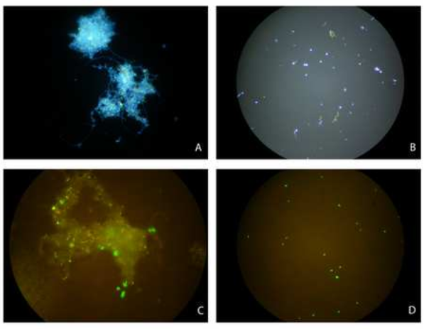 | Figure 1. Images of untreated activated sludge flocs non-inoculated (A) and inoculated with E. coli ABCGFP (C), and images of sonicated samples of non-inoculated (B) and inoculated (D) |
3.2. gfp-tagged E. Coli Cells Disaggregation from Flocs
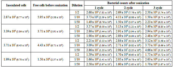
- To establish the efficiency of ultrasonication we have used an E. coli strain expressing GFP protein, E. coli ABCGFP. In a previous work[11], we demonstrated that GFP-expressing strains can be clearly distinguished from the natural community and that they do not lose fluorescence along the permanence in aquatic systems. Moreover, Olofsson et al.[19] did not detect differences between gfp-tagged and wild-type E. coli in cell surface hydrophobicity and surface charge as well as in their ability to attach to the flocs.Falcioni et al.[6] emphasized that, the fundamental difficulty in achieving the high efficiency of the dissociation of bacteria from flocculated clumps, lies in the need to keep the balance between using procedures that are hard enough to achieve a nearly complete detachment from flocs and the concomitant risk of cell disruption. Firstly, we have studied the suitability of sonication treatment for the dislodgement of flocs by using E. coli cells as a reference. The results obtained when E. coli suspensions in PBS were subjected to the designed treatment are shown in Table 1.
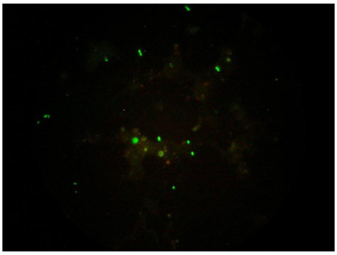 | Figure 2. Images of small aggregates detected after sonication |
4. Conclusions
- Fluorescently tagged bacteria are an excellent tool to validate the extraction method proposed for the dislodgement of bacterial populations in complex samples as activated sludge. In the case of the revised method, our data suggest that the procedure based on sonication of activated sludge samples is adequate for enumerating bacteria as it allows a high recovery of bacteria with good accuracy and reproducibility, and minimizes the damage of the cells in the suspension obtained.
ACKNOWLEDGEMENTS
- This work was supported by the research projects CTM2006-09532/TECNO from the Science and Technology Ministry of Spain, EHU08/56 from the Basque Country University and Basque Government Predoctoral Grant BF109.103 to I. Garaizabal.We also thank Prof. V. de Lorenzo and A. de las Heras for generously providing us with donor and helper strains and for information about tri-transconjugation procedure and to Prof. V. Kaberdin for assistance with the translation to English.
|
References
| [1] | E. Liwarska-Bizujkojc, “Application of image analysis techniques in activated sludge wastewater treatment processes,” Biotechnol. Lett., vol. 27, pp.1427-1433,Oct. 2005. |
| [2] | T. R. Thomsen, J. L. Nielsen, N. B. Ramsing, P. H. Nielsen, "Micromanipulation and further identification of FISH-labelled microcolonies of a dominant denitrifying bacterium in activated sludge," Environ. Microbiol., vol. 6, pp.470-479, Mar. 2004 |
| [3] | R. Girones, M. A. Ferrús, J. L. Alonso, J. Rodriguez- Manzano, B. Calgua, A. de Abreu Corrêa, A. Hundesa, A. Carratala, S. Bofill-Mas, "Molecular detection of pathogens in water – The pros and cons of molecular techniques," Water Res., vol. 44, pp.4325–4339, Aug. 2010. |
| [4] | I. Wojnowska-Baryła, A. Cydzik-Kwiatkowska, M. Zielińska, "The application of molecular techniques to the study of wastewater treatment systems," Methods Mol. Biol., vol.599, pp. 157-183, 2010. |
| [5] | B. Örmeci, K. G. Linden, "Comparison of physical and chemical methods for extraction of coliform from wastewater particles and flocs," Environ. Eng. Sci., vol. 22, pp. 459-471, Jul. 2005. |
| [6] | T. Falcioni, A. Manti, P. Boi, B. Canonico, M. Balsamo, S. Papa, "Comparison of disruption procedure for enumeration of activated sludge floc bacteria by flow cytometry," Cytom. Part B-Clin. Cytom., vol. 70B, pp. 149-153, May/Jun. 2006. |
| [7] | P. Foladori, L. Bruni, G. Andreottola, G. Ziglio, "Effects of sonication on bacteria viability in wastewater treatment plants evaluated by flow cytometry—Fecal indicators, wastewater and activated sludge," Water Res., vol. 41, pp. 235-243, Jan. 2007. |
| [8] | P. Foladori, L. Bruni, S. Tamburini, G. Ziglio, "Direct quantification of bacterial biomass in influent, effluent and activated sludge of wastewater treatment plants by using flow cytometry," Water Res., vol. 44, pp. 3807-3818, Apr. 2010. |
| [9] | Y.B. Ahn, L. A, Beaudette, H. Lee, J. T. Trevors. “Survival of a GFP-labeled polychlorinated biphenyl degrading psychrotolerant Pseudomonas spp. in 4 and 22 degrees C soil microcosms,” Microb. Ecol., vol. 42, pp.61-623, Dec- 2001. |
| [10] | A. Unge, R. Tombolini, L. Molbak, J. K. Jansson.” Simultaneous monitoring of cell number and metabolic activity of specific bacterial populations with a dual gfp-luxAB marker system,” Appl. Environ. Microbiol., vol. 65, pp. 813-821, Feb. 1999. |
| [11] | I. Arana, A. Irizar, C. Seco, A. Muela, A. Fernández-Astorga, I. Barcina, "gfp-Tagged cells as a useful tool to study the survival of Escherichia coli in the presence of the river microbial community," Microb. Ecol., vol. 45, pp. 29-38, Nov. 2003. |
| [12] | A. Muela, M. Orruño, M. L. Alonso, M. Pazos, I. Arana, R. M. Alonso, R. M. Jiménez, I. Garaizabal,M. I. Maguregui, I. Barcina, "Microbiological parameters as an additional tool to improve wastewater treatment plant monitoring," Ecol. Indic., v ol. 11, pp. 431-437, Mar. 2011. |
| [13] | Clinical and Laboratory Standards Institute, Performance Standards for Antimicrobial Susceptibility Testing. Supplement M100–S16, Wayne PA, 2006. |
| [14] | K. G. Porter, Y. S. Feig YS, "The use of DAPI for identifying and counting aquatic microflora," Limnol. Oceanogr, vol. 25, pp. 943–948, Sept./Oct. 1980. |
| [15] | S. Forster, J. R. Snape, H. M. Lappin-Scott, J. Porter, "Simultaneous fluorescent Gram staining and activity assessment of activated sludge bacteria," Appl. Environ. Microbiol., vol. 68, pp. 4772-4779, Oct. 2002. |
| [16] | A. Prorot, C. Eskicioglu, R. Droste, C. Dagot, P. Leprat, "Assessment of physiological state of microorganisms in activated sludge with flow cytometry: application for monitoring sludge production minimization," J. Ind. Microbio.l Biotechnol., vol. 35, pp.1261-1268, Nov. 2008. |
| [17] | G. Ziglio, G. Andreottola, S. Barbesti, G. Boschetti, L. Bruni, P. Foladori, R. Villa, "Assessment of activated sludge viability with flow cytometry," Water Res., vol. 36, pp. 460-468, Jan. 2002. |
| [18] | K. Y. Show, T. Mao, D. J. Lee, "Optimisation of sludge disruption by sonication,". Water Res., vol. 41, pp. 4741-4747, Dec. 2007. |
| [19] | A.C. Olofsson, A. Zita, M. Hermansson, "Floc stability and adhesion of green-fluorescent-protein-marked bacteria to flocs in activated sludge," Microbiology, vol. 144, pp. 519-528, Feb. 1998. |
| [20] | M. Furuta, M. Yamaguchi, T. Tsukamoto, B. Yim, C. E. Stavarache, K. Hasiba, Y. Maeda, "Inactivation of Escherichia coli by ultrasonic irradiation," Ultrason. Sonochem., vol. 11, pp. 57-60, Apr. 2004. |
| [21] | I. Hua, J. E. Thompson, "Inactivation of Escherichia coli by sonication at discrete ultrasonic frequencies," Water Res., vol. 34, pp. 3888-3893, Oct. 2000. |