Journal of Microbiology Research

p-ISSN: 2166-5885    e-ISSN: 2166-5931

2013;  3(1): 43-52

doi:10.5923/j.microbiology.20130301.07

Lipolytic Activity of some Strains of Klebsiella, Pseudomonas and Staphylococcus Spp. from Restaurant Wastewater and Receiving Stream

Odeyemi A. T. 1, Aderiye B. I. 1, Bamidele O. S. 2

1Microbiology Department, Ekiti State University, Ado-Ekiti, Ekiti State, Nigeria

2Biochemistry Department, Federal University of Technology Akure, Ondo State, Nigeria

Correspondence to: Odeyemi A. T. , Microbiology Department, Ekiti State University, Ado-Ekiti, Ekiti State, Nigeria.

Email:

Copyright © 2012 Scientific & Academic Publishing. All Rights Reserved.

Abstract

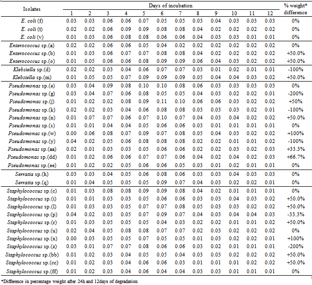
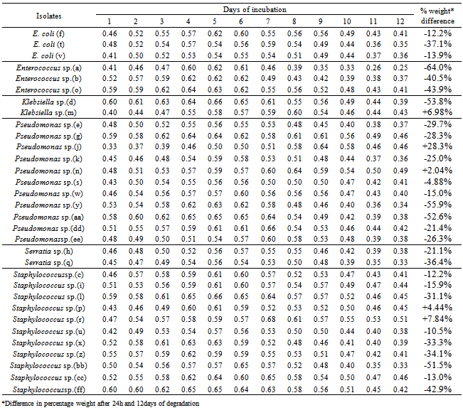
The tendency of some lipolytic bacteria isolated from restaurant wastewater and the receiving stream to biodegrade/utilizes fresh palm oil was investigated. Thirty two (32) lipolytic bacteria isolates were identified and grouped into six genera namely; Enterococcus, Escherichia, Klebsiella, Pseudomonas, Serratia and Staphylococcus. The weight of these isolates after 24h of inoculation ranged between 0.33 and 0.60mg, whereas the final weight on the 12th day of incubation was between 0.25mg and 0.51mg. Generally, the optimum growth in all the thirty two (32) lipolytic isolates in the fresh oil was observed between the fifth and seventh days. The growth rate per day were 0.02mg in Pseudomonas sp. (n); 0.03mg in Klebsiella sp. (m); 0.04mg in Pseudomonas sp. (j); 0.04mg in Staphylococcus sp. (r); and 0.05mg in Staphylococcus sp. (p).The appreciable enzymatic activity of these microbes ranged; Lipase (0.036 - 0.073 mM/min/ml) with Pseudomonas sp. (n) shown a highest lipase activity (0.073 mM/min/ml) after 12h, protease (50 - 117mM/min/ml) and amylase (7.7 - 117mM/min/ml). It appears that bacteria associated with dietary oil-rich wastewater are the novel source of environmental enzymes for possible commercial applications and may play an important role in enzyme-catalyzed organic matter cycling in domestic environments.

Keywords: Lipolytic Bacteria, Restaurant Wastewater, Palm Oil, Receiving Stream

Cite this paper: Odeyemi A. T. , Aderiye B. I. , Bamidele O. S. , Lipolytic Activity of some Strains of Klebsiella, Pseudomonas and Staphylococcus Spp. from Restaurant Wastewater and Receiving Stream, Journal of Microbiology Research, Vol. 3 No. 1, 2013, pp. 43-52. doi: 10.5923/j.microbiology.20130301.07.

1. Introduction

Lipids are one of the most important components of vegetable oil, many synthetic compounds, and emulsions are found in many pharmaceutical and cosmetic industrial effluents and municipal wastewater[1; 2]. Palm oil, obtained from the fruit of the oil palm tree, is the most widely produced edible vegetable oil in the world and its nutritional and health attributes have been well documented[3]; surpassing soybean oil as the most widely produced vegetable oil in the world[4]. It is consumed worldwide as cooking oil, in making of margarine and shortening, apart from being used as an ingredient in fat blends and avast array of food products[5].
The amount of lipid in municipal wastewater is approximately 30-40% of the total chemical oxygen demand [2].Wastewaters containing fat and oils were traditionally treated physically, which is currently considered insufficient if the fat is in its dispersed form. Biological treatment has been found to be the most efficient method for removing fat, oil and grease by degrading them into miscible molecules[6]. Therefore, manipulation of microorganisms for treatment and bioremediation purposes afford a very efficient tool for purifying contaminated effluents and natural water[7]. The use of lipase enzymes that are produced by all organisms may solve the problem, where they catalyze the synthesis or hydrolysis of fat[8].[9] evaluated a mixed culture composed of P. aeruginosa LP602, Acinetobacter calcoaceticus LP009 (both lipase – producing bacteria) and Bacillus sp. B304 (an amylase and protease producing bacterium) to lower the biochemical oxygen demand (BOD) value and lipid content of lipid-rich wastewater. Lipolytic enzymes are currently attracting enormous attention because of their biotechnological potential[10]. Among bacterial lipases, attention has usually been focused on particular classes of enzymes such as the lipases from the genus Pseudomonas, which are especially interesting for biotechnology[11]. Hence, the present work aims at investigating the biodegradability potential of some lipolytic bacteria cells in decomposing/utilizing the dietary oil as a substrate.

2. Materials and Methods

2.1. Collection of Wastewater Samples

Wastewater and water samples were obtained from different sampling points between Falegan restaurant and the receiving stream as follow:
D1 - Wastewater sample obtained at the exit of drain pipe from the wash hand basin
D2 - Wastewater sample from the drainage at a distance of 5 meters from the exit of the drain pipe
D3 - Wastewater sample from the stream at the point of entry of effluent into the stream from the drainage (at a distance of 10 meters to D2)
U1 - Water sample from the stream at 5 meters upstream from the point of entry of the effluent into the stream from the drainage
U2 - Water sample from the stream at 10 meters upstream from the point of entry of the effluent into the stream from the drainage
S1 - Wastewater sample from the stream at 5 meters downstream from the point of entry of the effluent into the stream from the drainage
S2 - Wastewater sample from stream at 10 meters downstream from the point of entry of effluent into the stream from the drainage
These samples were collected using 250ml sterile sampling bottles and immediately transported in ice buckets to the Microbiology Laboratory of Ekiti State University, Ado-Ekiti for further work.

2.2. Bacteriological Analysis

One millilitre of wastewater sample was inoculated and incubated on plate count and MacConkey agar media at 25℃ and 37℃ respectively for 24h. Pure colonies of bacteria isolates were later inoculated on sterile Tributyrin agar and incubated at 37℃ for 24h. Lipolytic activity of bacteria cells were confirmed by zone (s) of clearance around their growth.

2.3. Characterization and Identification of Isolates

The isolates were classified on the basis of their biochemical, physiological and morphological characteristics and matched against standard microbial cultures[12; 13].

2.4. Bio-degradative Activity of Bacteria Isolates

Fresh palm oil was elucidated with diethyl ether (1:1; v/v) and filtered through non-absorbent cotton wool. A known quantity (1.8ml) of the oil was inoculated with 0.2ml of standardized inoculum of lipolytic bacterial isolates and incubated at ambient temperature over the period of 12 days. The rate of degradation of the dietary oil was monitored by determining the total dry weight of the microbial cell[14].

2.5. Preparation of Wastewater and Fresh Palm Oil for Enzyme Activity

Wastewater treatment was done by distributing wastewater into five portions of 100 mL each contained in 250 mL Erlenmeyer flasks followed by inoculating 1 mL of each bacterial culture (OD6002:0) into wastewater sample contained in each flask. The flasks were kept in shaking incubator at 30℃ with 150 r.p.m. Samples were drawn from each of the flasks at intervals of 6 h for a period of 48 h and later centrifuged at 5000 x g for 30 minutes at 4℃. Cell free supernatant corresponding to growth phase was used as the crude enzyme for assay and further analysis. Also, Palm oil-containing medium were prepared with 0.2% w/v palm oil, 1.5% K2HSO4, 0.05% MgSO4, 1.0% (NH4)2SO4, 0.2% CaCl2, 0.2% FeSO4 and 0.5% yeast extract, distributed, inoculated and incubated as in the wastewater culture.

2.6. Amylase Assay

Amylase activity was assayed[15]. Briefly, 0.5 mL of properly diluted enzyme was added into a tube containing 1.5 mL of 2 % (w/v) of potato starch solution and 1 mL of 0.05 M acetate buffer, pH 5.0. The reaction mixture was incubated at 40℃ for 15 min. Then, 1 mL of the mixture was transferred to a new tube containing 1 mL of 3, 5-dinitrosalicylic acid and kept in boiled water for 10 min. The color density was determined spectrophotometrically at 520 nm. One unit was defined as 1 µmol of glucose released per minute by 1 mL of enzyme.

2.7. Lipase Assay

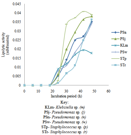
The crude enzyme preparation was the culture broth after separation of cells and particles. The enzyme was normally stored at 4℃ until used. Lipolytic activity was determined by colorimetric method based on the activity in cleavage of p-nitrophenylpalmitate (p-NPP) at pH 8.0[16]. The reaction mixture contained 180 µL of solution A (0:062 g of p-NPP in 10 mL of 2-propanol, sonicated for 2 min before use), 1620 µL of solution B (0.4% triton X-100 and 0.1 % gum Arabic in 50 mM Tris-HCl, pH 8.0) and 200 µL of properly diluted enzyme sample. The product was detected at 410 nm wavelength after incubation for 15 min at 37℃. Under this condition, the molar extinction coefficient (410 nm) of p-nitrophenol (p-NP) released from p-NPP was 15000 M1. One unit of lipase activity was defined as 1 µmol of p-nitrophenol (p-NP) released per minute by 1 mL of enzyme.

2.8. Protease Assay

Protease activity was determined[17]. 1 mL of 1.5 % casein solution, pH 7.0 was placed at 37℃ and, then, 1 mL of properly diluted enzyme sample was added. The reaction was incubated for 10 min prior to the addition of 2 mL of 0.4 M trichloroacetic acid. The solution with precipitates was altered and to 0.5 mL of the clear filtrate 2.5 mL of 0.4 M Na2CO3 and 0.5 mL of Folin reagent were added. After further 10 min of incubation, the color density developed was determined at 660 nm. One unit was defined as 1 µmol of tyrosine released per minute by 1 mL of enzyme.

2.9. Protein Determination

Protein concentration was determined using the Lowry method[18]. Reagent A: 2% NaCO3 in 0.1 N NaOH; Reagent B: 0.5% CuSO4.5H2O in 1% Na or K tartarate; Reagent C: 100ml of Reagent A + 2ml of reagent B and Reagent E: 1:2 dilution of John’s reagent water. Graded concentrations of Bovine Serum Albumin (BSA) in tubes were prepared. Then 0.3 ml of each concentration was measured into test tubes. 3 ml of reagent C was added, mixed and left for 10 min. Then 0.3ml of reagent E was added, mixed and left for 30 min. The optical density was read at 600nm. The graph of OD versus concentration of BSA was obtained and standard curve of BSA. The same was done for unknown substance and the protein concentrations from the standard curve are read off and obtained by multiplying with dilution factor. All readings were obtained in triplicates.

3. Results and Discussion

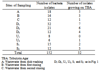
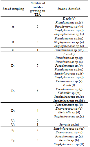
One hundred and fifty two bacteria isolates were obtained from the wastewater and the receiving stream. Only thirty two of these microbes were lipolytic in nature (Table 1). There was a reduction of about 67% of the initial number of microbial isolates after the second rinsing of the dishes (C), which may be as a result cleansing ability of combined action of water and detergent. This support the findings of[19] who reported that, washing and sanitizing treatment can play an important role in reducing microbial populations on fresh fruits and vegetables. However, this number increased about three folds at the exit of the restaurant. This may be as a result of drained wastewater having contact with the soil already contaminated with decomposed waste foods dumped nearby which may be source of nutrient for the organisms along the drainage. Microorganisms abound in the soil and are critical to decomposing organic residues and recycling soil nutrients. Since most bacteria live under starvation conditions or water stress in the soil, they have adapted to quickly reproduce when water, food, and the environmental conditions are abundant. Bacteria populations can easily double in 30 minutes[20].
Table 1. Number of bacteria isolates from wastewater and receiving stream
     
The percentage distribution of the lipolytic isolates revealed that 15.6% of these cells were obtained from dish washing, 9.38% and 3.13% of the isolates from wastewater after the first and second rinsing of the dishes respectively. Over 40.6% were isolates from the drainage, 12.5% from the point of exit of the wastewater into the receiving stream and 18.8% from the receiving stream (Table 2).
Table 2. Identification of ‘probable’ lipolytic bacteria
     
Some of the lipolytic bacteria isolates obtained from the different sampling sites were identified to the genera level as follows: A (Escherichia coli (v), Pseudomonas spp. (s & w), and Staphylococcus spp. (l & z), B (Pseudomonas sp. (aa) and Staphylococcus spp. (u & c), C (Pseudomonas sp. (dd)), D1(E. coli(f), Pseudomonas spp.(g, e, y & ee), Staphylococcus spp. (r & z)), D2 (Enterococcus sp. (a), E.coli (t), Pseudomonas sp. (j), Klebsiella sp. (m) and Staphylococcus (p & bb), D3 (Pseudomonas sp. (k), Enterococcus sp. (b), Klebsiella sp. (d) and Staphylococcus sp. (x), U2 (Serratia sp. (q), S1(Staphylococcus sp. (cc) and Enterococcus sp.(o)) and S2 (Pseudomonas sp. (n), Serratia sp. (h) and Staphylococcus sp. (ff), while the sampling point U1 had no lipolytic organisms isolated (Table 2). It is interesting to note that many isolates of the genera Pseudomonas were continuously obtained after dish washing until the wastewater drained into the receiving stream. This is because Pseudomonas is gram negative bacillus found in warm, moist environments, and can be frequently isolated from soil, water and occasionally from normal human skin[21]. Similarly, some strains of Staphylococcus spp. were detected throughout all the sampling points except after the second rinsing of the dishes. As the normal flora of the natural environment the cleaning effectiveness of the detergent led to the reduction of their population but the organic matter present shield and promotes the survival of the microbial population.
Most of the isolates (16/32, 50%) recorded maximum growth after five (5) days of biodegradation of wastewater. However, strains of Staphylococcus sp. (p) and Pseudomonas sp. (j) grew very well (weight > 0.10 mg) after 6 days. On day 12, 13/32 of the isolates still had twice the cell mass each isolate recorded after 24h incubation with Pseudomonas sp. (dd) recording over 66.7% weight increase (Table 3). This is in agreement with the report that, microbes able to degrade the contaminant increase in numbers when the contaminant is present; when the contaminant is degraded, the biodegradative population declines[22]. Only three strains of the isolates (Klebsiella sp. (m), Pseudomonas spp. (j) and (w)) showed appreciable growth rate (between 0.02 and 0.0202mg/day) within 6 days of incubation (Figure 1).
The weight of the isolates after the 1st day of incubation ranged between 0.33mg and 0.60mg, and the final weight on the 12th day ranged between 0.25mg and 0.51mg. Generally, the optimum growth in all the thirty two (32) lipolytic microbial isolates in the fresh oil was between the fifth and seventh days. The growth rate per day ranged between Pseudomonas sp. (n) 0.02, Klebsiella sp. (m) 0.03, Pseudomonas sp. (j) 0.04 and Staphylococcus sp. (r) 0.04, Staphyloccocus sp. (p) 0.05 (Table 4). This was as a result of the ability of the isolates to produce lipase as an enzyme to degrade/utilize palm oil as substrate. This is in line with the report that, many bacterial lipases, particularly those from members of the genera Pseudomonas, Bacillus, Staphylococcus, and Achromobacter have been cloned and characterized[23].
Table 3. Growth (mg) of lipolytic isolates in wastewater
     
Figure 1. Growth of selected most effective lipolytic bacteria isolates in oil-rich wastewater
Table 4. Weight (mg) of bacterial isolates in biodegraded Palm oil
     
In selecting bacteria isolates for further work, the weight of each isolate selected above was considered on the twelfth day and compared to that after the first day of incubation. The percentage weight difference was between -64% and +28.3%, showing significant difference at p ≤ 0.05. The isolation of some strains of Pseudomonas and Staphylococcus spp. with lipolytic activity in this study correlates with those of Prasad and Manjunath[24] who reported the ability of these organisms to produce highly-efficient lipases which degraded lipid-rich wastewater both individually and when used as a consortium.
The bacteria cells namely; Pseudomonas spp. (j) & (n), Klebsiella sp. (m) and Staphylococcus spp. (p & r), grown effectively in fresh oil substrate with the percentage weight difference of +28.3%, +2.04%,+6.98%, +4.44% and+7.84% respectively (Figure 2). The decomposition of dietary oil was primarily dependent on the varied lipolytic ability of the various bacteria cells as was observed by varied cell weight differences. Each organism produced specific and different amount of lipase which reflected on how the bacterial cells degraded/utilized the oil samples. The report of[25] was in support of the findings of this research work which stated that the experiments indicated the very different degradation efficiency might be due to the different reaction system of lipase from each culture. Lipase present not only catalyzed hydrolysis reaction but also catalyzed inter-esterification reaction, depending on the source of lipase and reaction condition[26; 27]. There was variation in the activities of the organisms based on the substrates in which they subjected/grown. There were more appreciable microbial growth in the palm oil as a substrate than in wastewater as related to Tables 3 and 4. The presence of some deleterious substances of detergent composition in the wastewater created adverse condition for the microbes, meanwhile the availability of enough nutrient in palm oil make it a suitable medium for microbial proliferation[27].
Microbial growth and activity are readily affected by pH, temperature and moisture[27]. Although, microorganisms have been isolated in extreme conditions, most of them grow optimally over a narrow range, so it is important to achieve optimal conditions. Meanwhile, the biological characteristics of a microorganism proposed for use in wastewater treatment should be considered carefully[28]. Indeed, it is desirable to adopt species that are Generally Regarded As Safe (GRAS). Furthermore, it is preferable that the species have limited nutritional requirements and have the ability to antagonize the growth of pathogenic species as well as the capacity to adapt to stringent environmental conditions (i.e. low temperatures and water activity, high concentrations of toxic substances).
Figures 3-7 give an illustration of the enzyme activities of Klebsiella sp. (m), Pseudomonas sp. (j), Pseudomonas sp. (n), Pseudomonas sp. (w), Staphylococcus sp. (p) and Staphylococcus sp. (r). This shows the ability of some fat-splitting enzymes lipases produced by most common bacteria. Microorganisms like Pseudomonas fragi, Staphylococcus aureus, Geotrichum candidum, Candida lipolytica, Penicillium roqueforti, and Penicillium sp. are the organisms reported to produce lipases which are active even at very low temperatures[29]. The species of Staphylococcus are slow carbohydrate fermenters producing the first trace of amylase after 6h. However, amylase production got to its peak after 18h (16.5mM/min/ml), meanwhile the highest amylase production was observed in Staphylococcus sp. (r) after 6h (17.1mM/min/ml) (Figure 3).
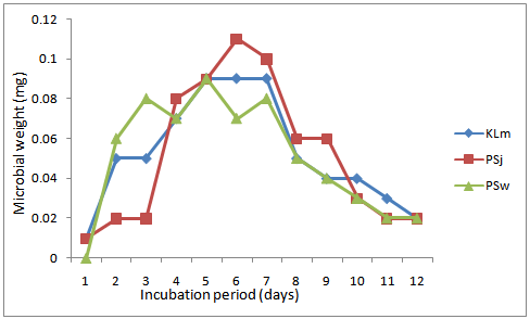
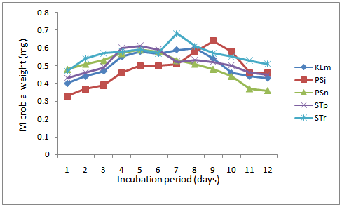
Figure 2. Growth of selected most effective lipolytic bacteria isolates in fresh palm oil
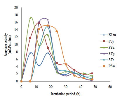
Figure 3. Time course of extracellular microbial amylase production and activity in wastewater from restaurant
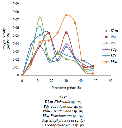
Figure 4. Time course of extracellular microbial lipase production and activity in wastewater from restaurant
Figure 4 revealed Pseudomonas sp.(w) to have shown a highest lipase activity (0.076 mM/min/ml) after 30h. Pseudomonas sp. (n) showed a lipase activity of 0.073 mM/min/ml after 12h while Staphylococcus sp. (r) exhibited 0.062 mM/min/ml at the same period. Furthermore, Klebsiella sp. (m) and Pseudomonas sp. (j) showed their highest lipase activity after 18h (0.054 and 0.053 mM/min/ml respectively). It is interesting to note that after 30h Pseudomonas sp. (j), Staphylococcus sp. (p) and Klebsiella sp. (m) still exhibited some appreciable lipase activity (0.054, 0.039 and 0.036 mM/min/ml respectively). Among bacterial lipases, attention has usually been focused on particular classes of enzymes such as the lipases from the genus Pseudomonas, which are especially interesting for biotechnology[11].
Klebsiella sp. (m) exhibited the highest protease activity (117mM/min/ml) after 12h of growth, while Staphylococcus sp. (p) showed its highest protease activity (72mM/min/ml) after 6h. Similarly, at 6h the species of Pseudomonas sp. (n), Pseudomonas sp. (w) and Klebsiella sp. (m) showed some prominent activity. Meanwhile, species of Pseudomonas (j) also exhibited high protease activity after 18h (Figure 5).
Extracellular microbial protein production during the growth phases of the test organisms was similar in all cases. Maximum extracellular protein was attained at incubation period of 15-18 h followed by a sharp decline in the amount of protein produced by microbes after 20th hour incubation period (Figure 6). This result probably indicated extracellular production of both enzymes (which are proteins) to carter for the initial breakdown of substrates (in medium) and inactive protein metabolites generated by proteolytic enzymes. These metabolites were subsequently required by the microbial cells for the initial synthesis of cellular macromolecules and growth of microbes. However, decline in the amount of protein was probably due to the loss of enzyme activity when all the available substrates were used up by the growing cells.
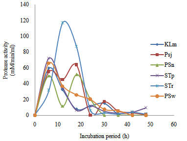
Figure 5. Time course of extracellular microbial protease production and activity in wastewater from restaurant
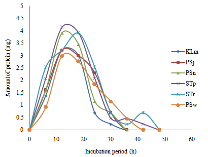
Figure 6. Time course of extracellular microbial protein production in wastewater from restaurant
Figure 7. Time course of extracellular microbial lipase production and activity in palm oil-containing medium
Figure 7 indicated lipolytic activity in palm oil-containing medium. There was delay in production of lipase in all the test organisms. This justify the report that, gradual reduction in the weight of the extracted oil over 12 days under aerobic and anaerobic conditions were due to ‘slow’ rate of utilization of the oil as source of substrate by the organisms[27]. Staphylococcus (p) sp. showed the highest lipolytic activity (0.04mM/min/ml) after 42h. Pseudomonas (J) sp. have a lipolytic activity of 0.038mM/min/ml after 48h, Klebsiella (m) sp. (0.0196mM/min/ml) after 42h , while the appreciable lipolytic activities for each of Pseudomonas (n), (w) and Staphylococcus (r) spp. was 0.036mM/min/ml after 48h respectively.
In conclusion, the ability of some lipolytic bacteria such as Pseudomonas spp. (j), (n) & (w), Klebsiella sp. (m) and Staphylococcus spp. (p) & (r) to degrade/utilize dietary oil were known to some extent. These organisms are versatile in utilizing the limited nutrient and have the ability to adapt to the toxic condition of detergent contained wastewater. This revealed that, these bacteria may be a versatile tool to treat wastewater containing palm oil generated from kitchen, restaurants and other domestic activities. These bacteria cells should be subjected to further experimental screening to proof their lipolytic nature beyond reasonable doubt. In order to obtain more detailed enzymatic information and make potential use of these enzymes in industrial and domestic processes, isolation, purification and characterization of these enzymes will be necessary.

References

[1]  Lateef, A. (2004). The Microbiology of pharmaceutical effluent and its public health implications. World J. Microbiol. Biotechnol., 20:167-171.
[2]  Chipasa, K.B., Mędrzycka, K. (2006).Behavior of lipids in biological wastewater treatment processes. Journal of Industrial Microbiology and Biotechnology, Vol. 33, No. 8, p. 635-645.
[3]  Ong, A.S.H and Goh, S.H. (2002). Palm oil: A healthful and cost––effective dietary component. Food and Nutrition Bulletin, Vol. 23, No. 1, United Nations University Press. 11-22.
[4]  Malaysian Palm Oil Board. Malaysian Oil Palm Statistics 2007. 27th ed. Bangi. 2008, pp 4-83.
[5]  Berger, K. (2005). “Review of Literature on Frying in Palm Oil.” In The Use of Palm Oil in Frying. Malaysian Palm Oil Council. Kelana Jaya. p72.
[6]  Odeyemi, A.T., Dada, A.C., Ogunbanjo, O.R. and Ojo, M.A. (2010). Bacteriological, physicochemical and mineral studies on Awedele spring water and soil samples in Ado Ekiti, Nigeria. African Journal of Environmental Science and Technology. 4(6): 319-327.
[7]  Glazer, A.N and Nikaido, H. (1995). Microbial Biotechnology: Fundamentals of Applied Microbiology. New York: W.H. Freeman and Company.
[8]  Shabtai, Y. (1991). Isolation and characterization of a lipolytic bacterium capable of growing in a low-water-content oil-water emulsion. Appl. Environ. Microbiol., 57: 1740-1745.
[9]  Mongkolthanaruk, W. and Dharmsthiti, S. (2002). Biodegradation of lipid-rich waste water by a mixed bacterial consortium. Int. Biodeterioration Biodegrad, 50: 101-105.
[10]  Arpigny, J.L. and Jaeger, K.E. (1999). Bacterial lipolytic enzymes: classification and properties. Biochem. J., 343: 177-183.
[11]  Gilbert, E.J. (1993).Enzyme. Microb. Technol., 15: 634-645.
[12]  Olutiola, P.O., Famurewa, O. and Sonntag, H.G. (1991). An Introduction to Microbiology. A Practical Approach. Printed in the federal Republic of Germany. 1991.
[13]  Cheesebrough, M. (2006). District Laboratory Practice in Tropical Countries (Part II). Cambridge University. 2nd edn., 6, pp. 50-150.
[14]  Bakken, L.R and Olsen, R.A. (1983). Buoyant densities and dry-matter contents of microorganisms: conversion of a measured, 45: 1188-1195.
[15]  Berfeld, P. Amylase. In: Colowick, S.P., Kaplan, N.O. (1955). Methods in Enzymology. Academic Press, New York, pp. 149–158
[16]  Lotrakul, P and Dharmsthiti, S. (1997). Lipase production by Aeromonas sobria LP004 in a medium containing whey and soybean meal. World Journal of Microbiology and Biotechnology, 13, 163–166.
[17]  Anson, M.L. (1938). The estimation of pepsin, trypsin, papain and cathepsin with hemoglobin. Journal of General Physiology. 22: 79–89.
[18]  Lowry, O.H., Rosebrough, N.J., Farr, A.L. and Randall, R.J. (1951). Protein determination, J. Biol. Chem., 193: 265-275.
[19]  Sapers, G.M. (2001) Efficacy of Washing and Sanitizing Methods, Food Technol. Biotechnol., 39 (4): 305–311.
[20]  Nester, E.W., Anderson, D.G., Robert, E.C.E Jr and Nester, M.T. (2007). Microbiology: A Human Perspective. Edited by Nicole Young. McGraw-Hill Publishing. NY
[21]  Botzenhart, K and Ruden, H. (1987). Hospital infections caused by Pseudomonas aeruginosa. IN:Doring, G., et al, eds. Basic Research and Clinical Aspects of Pseudomonas aeruginosa. Antibiot. Chemother., 39:1-15. Basel: Karger.
[22]  MohdKhairulNizam Bin MohdZuhan. (2008). Bioremediation of oil from domestic wastewater using mixed culture: Effect of inoculum concentration and agitation speed. Thesis submitted to the Faculty of Chemical and Natural Resources Engineering, Universiti Malaysia Pahang.
[23]  Pallavi, P., Suresh, A., Srinivas, P and Ram Reddy, S. (2010). Optimization of lipase production by Staphylococcus sp. Lp12. African Journal of Biotechnology, 9(6): 882-886.
[24]  Prasad, M.P. and Manjunath, K. (2011). Comparative study on biodegradation of lipid-rich wastewater using lipase producing bacterial species. Indian Journal of Biotechnology. 10: 121-124.
[25]  Dunhaupt, A., Lang, S and Wagner, F. (1992). Pseudomonas cepacia lipase: Studies on aggregation purification and on the cleavage of olive oil. Biotechnol. Lett., 14 (10): 953-958.
[26]  Macrae, A.R. (1983). Lipase catalyzed interesterification of oil and fat. J. Amer. Oil. Chem. Soc., 60(2): 243A-246A.
[27]  Odeyemi, A.T., Aderiye, J.B. and Adeyeye, E.I. (2011). Changes in the microflora and chemical components of domestic oil-rich wastewater. Journal of Microbiology, Biotechnology and Food Sciences, 1(1): 126-147.
[28]  Lanciotti, R., Gianotti, A., Baldi, D., Angrisani, R., Suzzi, G., Mastrocola, D. and Guerzoni, M.E. (2005). Use of Yarrowia lipolytica strains for the treatment of olive mill wastewater. Bioresource Technology, 96(3): 317–322.
[29]  Alford, J.A., Pierce, D.A. and Frank, G. (1964). Activity of microbial lipase on nature fats and synthetic triglycerides. Journal of Lipid Research, 5: 390-394.