-
Paper Information
- Paper Submission
-
Journal Information
- About This Journal
- Editorial Board
- Current Issue
- Archive
- Author Guidelines
- Contact Us
Basic Sciences of Medicine
p-ISSN: 2167-7344 e-ISSN: 2167-7352
2021; 10(2): 19-22
doi:10.5923/j.medicine.20211002.01
Received: Oct. 26, 2021; Accepted: Nov. 12, 2021; Published: Nov. 26, 2021

Third Trochanter and Hypotrochanteric Fossa in Femur: Morphology and Associated Significance
 Abstract
 Reference
 Full-Text PDF
 Full-text HTML
Rajani Singh
Department of Anatomy, UP University of Medical Sciences Saifai, Etawah 206130 UP India
Correspondence to: Rajani Singh, Department of Anatomy, UP University of Medical Sciences Saifai, Etawah 206130 UP India.
| Email: |  |
Copyright © 2021 The Author(s). Published by Scientific & Academic Publishing.
This work is licensed under the Creative Commons Attribution International License (CC BY).
http://creativecommons.org/licenses/by/4.0/

Third trochanter is defined as elongated, circular or conical tubercle at the superior part of gluteal tuberosity. It may influence the fracture break lines in pertrochanteric fractures. And may provide increased skeletal mass for the attachment of gluteus muscle, thereby improving its function. Hypotrochanteric fossa is varied expression of gluteus muscle attachment. There is scanty literature describing third trochanter and hypotrochanteric fossa. So, the study was conducted. The aim od the study is elaborate the incidence of these entities and to bring out their significance. Forty five unpaired (24 right, 21 left) femora were observed for the presence of third trochanter and hypotrochanteric fossa obtained from the osteology lab of Department of Anatomy, UP university of Medical Science. Incidences and association of hypotrochanteric fossa with gluteal ridges and third trochanter was computed. Incidences of third trochanter and hypotrochanteric fossa are 51% and 37.8% respectively. Out of total 17 femora, hypotrochanteric fossa was associated with third trochanter in 9 femora and that with gluteal ridges in 8 femora. Information about presence of third trochanter and hypotrochanteric fossa is useful for the diagnosis and management of pertrochanteric fractures and also in the study of microevolutionary trends in the anthropometric, epigenetics and comparative studies.
Keywords: Third trochanter, Hypotrochanteric fossa, Pertrochanteric fractures, Distal femur
Cite this paper: Rajani Singh, Third Trochanter and Hypotrochanteric Fossa in Femur: Morphology and Associated Significance, Basic Sciences of Medicine , Vol. 10 No. 2, 2021, pp. 19-22. doi: 10.5923/j.medicine.20211002.01.
Article Outline
1. Introduction
- The femur is the longest and strongest bone in the human body. The morphology of femur may change depending on functional requirement in day-to-day activities. One such structure is third trochanter of femur resulting from alteration in the morphology of gluteal tuberosity. Third trochanter of femur is circular, oblong or conical projection found on the superior end of gluteal tuberosity [1]. It is given the name third trochanter to delineate it from two elevations namely greater and lesser trochanters present constantly on proximal femur.Gluteus maximus inserts on gluteal tuberosity and responsible for extension of thigh and plays an important role in rising from sitting position. When gluteus muscle is overused, it exerts mechanical force on gluteal tuberosity altering its morphology resulting in formation of third trochanter [1]. In anthropometric studies, third trochanter forms an important non-metric variation in the post-cranial skeleton [2]. The third trochanter is frequently used in anthropometric, comparative and functional studies.The association, between muscle insertion and region of pertrochanteric fractures of proximal femur, has been studied thoroughly. Bone having periosteum without muscle and ligamentous attachments are more vulnerable for fractures. Hence, third trochanter can be one factor decreasing the incidence of fractures in proximal femur. Thus, variant morphology of pertrochanteric fractures is related to soft tissue attachments in concerned area of femur [3]. In addition, third trochanter may reinforce the proximal femoral diaphysis against increased ground reaction force by furnishing increased skeletal mass [3]. Moreover, third trochanter provide augmented surface area for muscle attachment resulting in increased efficacy of muscle contraction.Third trochanter in some laboratory mammals serves an important landmark for biomechanical study and accessing the medullary cavity [4].Third trochanter and hypotrochantric fossa differ in ethnic groups [3]. Scanty literature describes third trochanter and its association with hypotrochantric fossa in Indian population. Hence the study was conducted. The aim of the study is to describe the morphology of third trochanter, its association with hypotrochantric fossa along with its significance in Indian Population.
2. Material and Methods
- The study was conducted using unpaired 45 adult femora (23 right and 22 left) of unknown sex in the department of Anatomy UP university of Medical Sciences Saifai, Etawah UP India. The presence of third trochanter and hypotrochantric fossa were observed in total femora and right and left. The incidence of hypotrochantric fossa in total femora, accompanied by gluteal ridges and third trochanter were computed. Literature was explored using different data bases for the prevalence of third trochanter and hypotrochantric fossa and associated significance.
3. Results
- Third trochanter was observed in 23 out of total 45 femora constituting 51% of total. Out of total 23 femora, third trochanter was found in 13 right and 10 left femora. Vertically elongated third trochanter (Fig. 1) was detected in 22 femora and circular shaped in one femur.
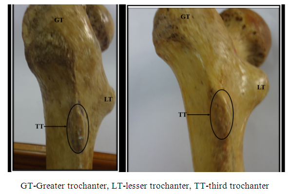 | Figure 1. Showing third trochanter |
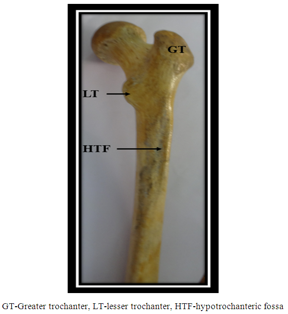 | Figure 2. Showing hypotrochanteric fossa |
4. Discussion
- Third trochanter is an important entity elucidated in anthropometric, comparative and functional studies. It acts as an essential landmark for biomechanical, densitometry studies and to access the medullary cavity. Some investigators stated that muscle insertions and ligament attachments affect the course of break lines involving trochanteric mass area depending on radiologic evaluation of subjects suffering from pertrochanteric fractures. They observed morphological correlate between muscle insertions and the course of the break line in a typical pertrochanteric fracture and concluded that bones without muscle and ligamentous attachments are more prone for fractures [5].The incidence of the third trochanter varies significantly from 17% to 72% [6,7]. The incidence of the third trochanter in present study is 51% which lies within the range described in literature but is higher than previously reported by various investigators in Indian Population (6.6%) and excavated femora from Poland (6.2%) [3,8]. In our study, third trochanter was detected more on right side (13 on right side and 10 on left side) similar to study in the whites [2]. However, left predominance of third trochanter was observed in another study in Indian Population and in Negro Population [2,3].The hypotrochanteric fossa is a depression or groove at the site of insertion of Gluteus Maximus on the femur [3]. In Gorillas, Chimpanzees and Orangutans, it is found at the muscle attachment while in Gibbons, gluteal ridge is observed [9]. However, the definite cause for the presence of gluteal ridge or hypotrochanteric fossa is unknown. Presence of gluteal ridge, third trochanter and hypotrochanteric fossa may be explained by microevolutionary theory.In present study, the incidence of hypotrochanteric fossa is 37.8% which is higher than observed by Ghosh et al in Indian population. In our current study, right sided predominance of the hypotrochanteric fossae was observed similar to study by Ghosh et al and also study in Negros but not in Whites. hypotrochanteric fossa was absent in 28 femora out of which it was absent in 15 femora with gluteal ridges and 13 femora with third trochanter. Such association of hypotrochanteric fossa with gluteal ridges and third trochanter are not described in literature for comparison.Structures akin to third trochanter are also observed in some mammals like rats, rabbits, the Eocene ancestors of whales and some primates [6,10]. There is no consensus regarding the homology of this structure in humanoids and other mammals. The third trochanter is highly evolved in Neanderthal femora but not in other anthropoids. So, interpreting third trochanter from evolutionary point of view is debatable. It is difficult to state whether the third trochanter is regressive or progressive structure [6,11].Such phenotypic developments and discontinuous skeletal traits are supposed to be regulated by genetic factors [12]. But contemporary researches reveal and emphasise on various biological and environmental factors such as age, sex, nutritional status or side dependence for non -metric traits like third trochanter and hypotrochanteric fossa in non- human and human populations [13,14].Local mechanical factors provide useful information regarding epigenesis affecting the frequency and expression of discontinuous skeletal variations [1,15]. So, third trochanter develops into various morphological forms like elongated, circular or conical. Third trochanter is an example of enthesophyte on femur which result from skeletal responses to stress. When the muscle is overused/strained there may be microtrauma in the tendon which may ultimately ossify giving rise to bony elevations [16] like third trochanter in this case. Gluteus maximus may exert a mechanical force on the gluteal tuberosity thereby changing its surface morphology forming third trochanter [1]. Enthesophytes can develop secondary to the inflammation of the enthesis occurring in seronegative spondarthropathies. Thus, third trochanter may form due to inflammation of enthesis of gluteal tuberosity in persons with seronegative spondarthropathies. Multiple idiopathic enthesophytes are features of DISH [17]. Thus, third trochanter may be manifestation of DISH. The person might be suffering from calcium metabolic disorder which predisposes for calcium deposition in tendon leading to its ossification [16] causing formation of third trochanter. Thus, if third trochanter is detected in live patient, the person should be screened for seronegative spondarthropathies, DISH and calcium metabolism.Local mechanical factors effecting incidence of discontinuous variants like third trochanter may be birthplace of epigenetic details [1,15]. Advantage of third trochanter is that it augments the insertion area for the gluteas maximus muscle thereby improving the efficiency of contraction of muscle [3].
5. Conclusions
- Incidences of third trochanter and hypotrochanteric fossa in Indian Population is 51% and 37.8% respectively which is higher than previous studies. As Third trochanter is found to be associated with seronegative spondarthropathies, DISH and calcium metabolism, so the persons with this entity should be screened for aforementioned conditions. Third trochanter at the proximal part of the femur may change the break lines in the pertrochanteric fracture patients. The knowledge of the presence of the third trochanter will be of paramount importance for the diagnosis and management of pertrochanteric fractures and also in the study of microevolutionary trends in the anthropometric and comparative studies of humans.
ACKNOWLEDGEMENTS
- No conflict of interest. No Funding has been received.