Basic Sciences of Medicine

p-ISSN: 2167-7344    e-ISSN: 2167-7352

2021;  10(1): 10-18

doi:10.5923/j.medicine.20211001.03

Received: Jun. 21, 2021; Accepted: Jul. 3, 2021; Published: Jul. 15, 2021


Will Development of Human Anatomy Revolutionize Medical Education?

Rajani Singh1, DG Jones2, Raj Kumar3, Naresh Chandra4

1Department of Anatomy Uttar Pradesh University of Medical Science Saifai Etawah, UP, India

2Department of Anatomy Otago University, Otago, New Zealand

3Department of Neurosurgery, Uttar Pradesh University of Medical Science Saifai, Etawah, UP, India

4Department of Anatomy Hind Institute of Medical Sciences, Lucknow, India

Correspondence to: Rajani Singh, Department of Anatomy Uttar Pradesh University of Medical Science Saifai Etawah, UP, India.

Email:

Copyright © 2021 The Author(s). Published by Scientific & Academic Publishing.

This work is licensed under the Creative Commons Attribution International License (CC BY).
http://creativecommons.org/licenses/by/4.0/

Abstract

Vanishing of cadaveric dissection, pruning of the curriculum and teaching schedule, the deployment of unqualified tutors/demonstrators and removal of experienced faculties with closing of Anatomy departments has eroded medical education. So tomorrows’ doctors accumulate deficient Anatomical knowledge impeding successful clinical practice. The objective of this study is to evolve a new model of Anatomy as a subject to deliver adequate Anatomy to improve clinical practice. The literature was reviewed regarding falling standards of the knowledge of Anatomy and growing failure cases. This resulted in a model for the development of Anatomy as a subject by standardizing the curriculum to be taught by medically qualified and experienced staff with the ability for standalone and collaborative research. The model will allow interaction of anatomists with clinicians/trainees by providing instant anatomical solutions during medical education. The well guided cadaveric dissection by experienced faculty will also improve the clinical practice. The model will also generate ready-made records containing variations corresponding to relevant clinical complications helping clinical trainees during clinical practice. This model will provide sufficient knowledge of Anatomy, reservoir of competent anatomical faculty and also revolutionize clinical skills among medical trainees. The aim is to ensure that the quality of medical education is enhanced.

Keywords: Inadequate anatomical knowledge, Medical education, Development of anatomy, Failure cases, Collaborative research

Cite this paper: Rajani Singh, DG Jones, Raj Kumar, Naresh Chandra, Will Development of Human Anatomy Revolutionize Medical Education?, Basic Sciences of Medicine , Vol. 10 No. 1, 2021, pp. 10-18. doi: 10.5923/j.medicine.20211001.03.

1. Introduction

The diseases are caused by changes in environmental conditions, pathogens, toxins, trauma and misuse of limbs. The results include anatomical distortions in shape, size, location and orientation of structures as well as developmental deficits that impair activities and/or the functioning of macro/microstructures, organs, limbs and bodily systems. The generation of signs and symptoms of discomforts are governed by the brain.
The preliminary diagnosis starts from physical examination through observation, palpation, percussion and auscultation involving surface Anatomy (Figure-1) [1] to assess preliminary signs and symptoms of disease.
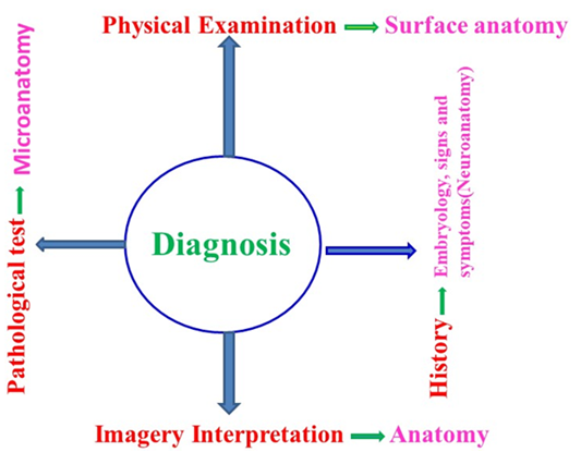
Figure 1. Showing importance of anatomy in diagnosis of diseases
The confirmation of disease is done through interpretation of images of anatomical distortions or injury to macro/micro neural structures by X-ray, CT, MRI, and endoscopy or histopathological tests. The interpretation of imagery and histopathological slides requires sound knowledge of shape, size, location, orientation, pathways, configuration and developmental deficits of structures, organs, limbs and body systems [2]. Thus anatomical investigations and diseases are interrelated and good knowledge of anatomy is an important prerequisite for an understanding of disease processes.
The perception of medical students’ understanding by Bergman and co-workers (2013) is enlightening, “One time on a scan you saw something down in the lung and then it turned out to be the stomach coming through the diaphragm. And I would never have thought of that, because to me thorax and abdomen are just two totally unrelated things (student 1f, year 2)” This clearly demonstrates the importance of Anatomy for interpretation of imagery [3]. In this study Bergman and collaborators (2013) recorded the responses of the senior medical students in these terms:- “You need it for diagnosis; you need it for physical examination, for hand-over to colleagues, for record keeping, for writing letters, in fact for understanding how certain processes work, why patients are ill and what should be done about it.” In another comment “It was the same for me [insight] that in the clerkships I suddenly thought, hey, at this point it would have been really useful if I had studied a bit more Anatomy”. Further “The neurology clerkship was mentioned by many students: “[…] I am doing neurology now and there you discover that every diagnosis, everything comes down to anatomy in the end and how things run and work, that is really awfully important.” These comments underscore the notion that medical trainees have inadequate knowledge of anatomy for clinical diagnosis.
After diagnosis, the anatomical distortions are either manipulated by surgical or medicinal treatments to get rid of discomforts of disease. The imagery guided surgical access and intervention both require detailed knowledge of the shape, size, location, orientation, configuration, pathways and developmental deficits of macro/micro structures, organs, limbs and systems [2]. Surgical restoration is also confirmed by imagery to assess the degree to which the remedy has been successful [4]. Additionally, medicinal treatment can also be precisely prescribed by using radiological interpretation to comprehend the relationship between signs and symptoms, disease, pathology and anatomically observable causative factors. It is clear from these observations that a sufficient knowledge of human Anatomy is indispensable for good quality professional medical education/profession in clinical practice [5].
Apart from this, the knowledge of normal and variant Anatomy at both macro and micro levels is essential for an understanding of newly discovered diseases and newly evolved micro-organism like Corona, Ebola and Zeka etc. Until the molecular and cellular structures of these antigens together with the microstructure of antibodies are discovered, safe and successful medicinal and surgical treatments cannot be formulated. In order to accomplish this, standalone and collaborative research in Anatomy alongside clinical, Para clinical and allied subjects need to be intensified by medically qualified anatomists and other subject experts. The role of anatomical investigations in cross disciplinary ventures of this type are .generally overlooked, when anatomical studies are relegated to a subsidiary place in biomedical investigations or are neglected in their entirety.
This neglect will not be reversed until medical students and trainees are provided with the opportunity to acquire adequate knowledge of human Anatomy [6,7,8,9,10,11,12,13]. This will not be rectified until impediments to comprehend clinical complications of health care in general and pathology, surgery, imagery and anestheology are removed, as pointed out by many specialty clinicians concerned for ways of ensuring that clinical practice is made successful for health care [4,14,15].
Inadequate anatomical training
Inadequate Anatomy has been responsible for fall in the standard of medical education for the profession [16]. Besides anatomical ignorance, there is a growing spate of litigations due to anatomical errors or ignorance leading to misdiagnosis and mistreatment [11,17,18,19,20]. Additionally, Prince and coworkers (2005a, b) noted that “In response to a questionnaire, the students at the starting phase of clinical training expressed that 18.5% of students possessed sufficient and 81.5% insufficient knowledge in Anatomy in which the deficit was serious. Not only this 64% of the students failed in the anatomical test set by students at the beginning of clinical training except graduates could fail only 26% while anatomists and clinicians failed 42% and 58% respectively [21,22]. Similarly, Bergman et al.’s (2013) experiment of perception of students also supported that there is inadequate knowledge of anatomy cultivated among the students. One student commented: “It was the same for me [insight] that in the clerkships I suddenly thought, hey, at this point it would have been really useful if I had studied a bit more anatomy” (student 8f, year 6) [3].
From above, it is evident that although sufficient knowledge of human Anatomy is essential, inadequate anatomy continues to be cultivated among medical students. There have been claims that owners of some medical schools have converted them into business hubs for earning hefty profits [11,18] so basic principles of medical education have been ignored.
A sufficient acquisition/delivery of anatomical knowledge depends on, a) students should have strong aptitude to learn a large Anatomy curriculum with the objective of creating the basis for successful clinical practice to serve humanity. b) medically qualified and experienced faculties of Human Anatomy with a strong interest in teaching and research should constitute the backbone of the teaching that should not be left entirely to tutors/demonstrators/ PBL guides and part-time clinicians. In turn clinicians involved in anatomy teaching should revisit their knowledge of Anatomy [23] and should have a broad knowledge of contemporary anatomy rather than mainly of the anatomy applying to their own [3] c) the extensive curriculum should be reorganized, redistributed and standardized at all stages of medical education and should be taught with reference to context during the preclinical years with horizontal and vertical integration for application in clinical practice. d) there is needs to be an awareness of how current anatomical research applies to clinical situations. Besides these, the inadequate delivery/acquisition has been further deteriorated by a) drastic reduction of time schedule of anatomical teaching [24,25,26], b) unsystematically pruning of parts of anatomic curriculum [24,27], c) removal of cadaveric dissection, the chief component of anatomy education and its replacement by prosections, models, charts and other e-learning resources hampering acquisition of anatomical knowledge, [24,27,28,29,30] and d) closing down of Anatomy departments in medical schools in USA, Canada and UK [18,27,31].
Two illustrations
Two illustrations of current anatomy departments and medical school teaching provide useful background data in looking at current anatomy teaching. The first is provided by the Brighton and Sussex Medical School’ (BSMS model) launched in 2003. The teaching of Anatomy curriculum is integrated horizontally and vertically based as it is on an integrated systems-based approach to cultivate academic knowledge and clinical experience. Anatomy is one of the core elements of the program and has a prominent place in the modular curriculum [8]. A multidisciplinary approach has been adopted using traditional and contemporary teaching methods. Unlike many other new medical schools, cadaveric dissection is the cornerstone of its teaching. The reason for this is that dissection is regarded as a successful means of demonstrating anatomical detail and also as a way of enhancing communication and teamwork skills, with emphasis placed on care and compassion. In this model, anatomy is also taught in the later years of the program. It is encouraging that this model has opted for a dissection based approach on the grounds that it provides a platform to help develop three-dimensional anatomical knowledge and an appreciation of anatomical variation [8]. Living anatomy sessions are pivotal to this approach, and are linked with ultrasound sessions. In this way considerable efforts have been put into teaching anatomy within a clinical setting, supplemented by the use of simulators in a Clinical Skills Laboratory. The role of anatomy within a clinical-based curriculum is encouraging.
The second illustration is from the University of Otago in New Zealand and is not intended to outline how anatomy fits into the medical curriculum, but to show that an anatomy department can be a strong research-based department if it sees itself as a research-intensive discipline within a modern university (Jones personal communication). In this case, the breadth of the department extends far beyond traditionally conceived gross anatomy. Within a department divided into biological anthropology, neuroscience and reproduction, genomics and development, clinical anatomy stands strong as an equal subgroup. It is as research-based as all the other groupings, and contributes seminally to medical teaching. Of its eight members, four have medical qualifications, one a dental qualification, one a physiotherapy background, and two have science backgrounds. All have postgraduate degrees and are research active. In addition, the teaching is supported by five teaching fellows, three of whom are medically trained. This demonstrates that it is possible to have a vigorous, research-informed anatomy department, in which medical experience is taken seriously and made available to undergraduate medical students.
Basic curricular needs
In developing an approach it is important to create interest among students and produce a reservoir of competent faculty well equipped with the ability to establish interrelationships among a large number of variables. These include signs and symptoms of disease, anatomical causation and radiological interpretation corresponding to images of normal/ variant/ distorted shape, size, location and orientation. This requires recruitment of well qualified and competent faculties [2]. Such medically qualified staff will be expected to provide the driving force for research in medical education as well as in clinically-related Anatomy. In this way they will be able to meet the future challenges and threats within Anatomy. This constitutes the basic principle of delivery/acquisition, to arrest not only the inadequate basic Anatomy during the preclinical stage but also horizontal/vertical integration of applied Anatomy during clinical training. It will also fit anatomy to contribute to clinical training, as well as postgraduate training at specialty and super-specialty levels. Therefore, the aim of the present study is to formulate, design and develop a strategy for the evolution of anatomical academics integral to the revolution of medical education. The hope is that this will improve the diagnostic and interventional relationship between Anatomy, surgery, imagery, pathology and anestheology.

2. Material and Methods

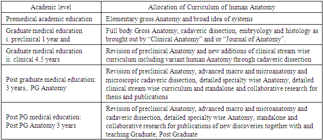
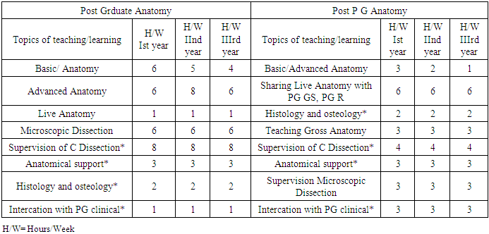
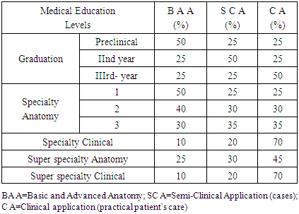
The review of literature concentrated on what the authors perceive is the inadequate knowledge of anatomy among medical graduates. Questions posed were: ‘why do trainees know too little Anatomy?’, what is the effect of the closure of anatomy departments in some countries? How does dependence upon prosections, models and charts compare with cadaveric dissection’, what parts of anatomy appear to be is irrelevant and redundant? What have been the effect of pruning syllabus of Anatomy?, ‘can clinical diagnosis and treatment be planned with inadequate anatomical knowledge?’ The search for clinical handicaps due to ignoring and neglecting the Anatomy was also done. The databases used were Researchgate, Wiley’s on line library, Google scholar, Medline, Scielo and Pubmed. Only English language articles were selected.
Scheme of Development of Human Anatomy
The development model of Anatomy will be formulated as under at Graduation, Post Graduation (PG/specialty) and Post PG (Super specialty) academic levels. The systematic development of human Anatomy will start from delivery / acquisition of balanced blend of basic and applied Anatomy sufficient to comprehend the skill and knowledge of therapeutic modalities in clinical practice. Development of Human Anatomy during graduation will be done at preclinical and at clinical training.
Preclinical Anatomy
The minimum necessary workable clinically oriented syllabus should be allocated in preclinical medical graduation brought out by the journals, Clinical Anatomy and Journal of Anatomy as detailed by Kumar and Singh, 2020 in their Model of Pedagogy with minor reorganization. Later on, the syllabus may be standardized to consist of surface Anatomy, the systems and basic concepts of structures and organs (shape size, location, configuration and landmarks). Developmental and embryological principles are to be outlined with reference to congenital defects plus organization of micro-elements in these structures. The curriculum should consist of analysis of normal histological slides/radiological images of various macro/microstructures [16] and their verification through cadaveric dissection to be applied in clinical complexities encountered during undergraduate clinical training (Table 1).
Table 1. Broad reorganization and redistribution of vast curriculum of human Anatomy
     
The curriculum of surface human Anatomy will consist of whole body surface locations’ names with internal landscape of structures, organs, limbs and systems w. r. t. landmarks with its verification on live patients and cadavers. The concepts of whole body structural disposition and systems should be clear to students [32]. Bergman et al. (2013) have commented “Neurovascular structures extending across body regions and their branching pattern are very important to know in their entirety. From students’ perspective: “Yes, you would see them run into the foot, but you had no idea where they had branched off” (student 5c & student 5b, year 4)”.
The teaching schedule of this preclinical curriculum is proposed as 1 year. After overall view of body structures and body systems as described above, the regional material will be taught in multidimensional repetitions through a blend of didactic lectures for explaining organizational and morphological concepts of developing structures.
Histology teaching and exposure to histology laboratory, demonstration classes, seminars, dissection guidelines by virtual table and pre-recorded videos and manual cadaveric dissection for practical verification of structures and retention in radiological and histological interpretation of normal/variant structures/slides for diagnosis and treatment in general and surgical access and intervention in particular should also be taught. In addition to this, the day to day cases with anatomical solutions given to 2nd year onwards students during clinical training in OPD should be collected by JRs to be discussed with the preclinical students. The new advancements in research in Anatomy by competent medically qualified faculty should be passed on to students relating them to clinical complexities.
Anatomy in clinical training at graduation
During this time, the students are slowly exposed to preliminary diagnosis starting from patients input encompassing signs and symptoms of discomforts their location, history taking and physical examination leading to anatomical causatives of disease. The preclinical syllabus covers almost entire curriculum to be applied in diagnosis but regurgitation is required. However, some new topics/variations may be additionally required for diagnosis of new diseases or new variations in structures, organs, limbs and systems through radiological images in various clinical streams to be manipulated by medicinal and surgical treatment. As a result, the curriculum of human Anatomy is to be drawn up in consultation with relevant clinicians based on clinical problems.
There is a major change in the curriculum in moving from a regional base to a system base, with interlinking structures/organs and systems. This points to the relevance of surface Anatomy in the syllabus at this level.
Development of Anatomy at postgraduate level
This pattern of systematic development of Anatomy will split PG Anatomy into Head and Neck (H N), Upper limb and Thorax (UL T), Lower limb and Abdomen, Neuroanatomy (NA) and Vascular Anatomy (VA). This could be expanded in the future, as specialties emerges. The schedule of PG Anatomy will be 3 years. In first two years the students will be learning complete human Anatomy and in the third year, they will be concentrating on their specialty and the clinical streams related to them. These specialties will provide specialized support for successful practice in the corresponding clinical specializations (Table 2) for more clinically complex situation.
Table 2. The anatomical support by Anatomy specialists in Anatomy
     
This requires very close and intimate clinical interaction to enhance the quality of clinical training.
Broad Curriculum of PG Anatomy
A brief curriculum of all the specialties of Anatomy in the specialty wise regions is presented in Table 1. A fuller anatomical curriculum should incorporate fresh additions of untaught/newly discovered variations from the research literature or explored through cadaveric dissection. These include anatomical solutions to new clinical complexities encountered during patients’ examinations, live Anatomy, advanced concepts of histological interpretation of internal microstructures, their configuration and the mapping of pathways, imagery interpretation and advanced concepts of embryology. In addition, more precise microscopic cadaveric dissection should be included for better comprehension of microstructural configuration in human organs/systems in relation to complex clinical complications. Planned surgical intervention on live patients should be demonstrated first on cadavers by surgeons/PG learners with active discussion with experienced anatomists/Post PGs of Anatomy preferably weekly. Similarly radiographs from live patients should be interpreted in association with expert radiologists, PGs, and anatomists. Also included in the curriculum should be research findings on new anatomical variations alongside understanding of the way in which pathogens invade organs and tissues though comparing normal and distorted histological slides of cells/tissues. Additionally, emphasis should be placed on phenomena such as neurovascular interrelations with compression, blockage and variations in structural pathways and configuration for surgical access and intervention.
Why super-specialty in Anatomy?
The clinical practice, disease and its diagnosis and treatment together with human Anatomy are so complexly interwoven that they cannot be separated. This is a primary requirement of successful clinical practice. “Where there is a disease, there is morbid/distorted Anatomy (structures, organs, limbs and systems) associated to it.” For example, the surgical treatment of appendicitis depends on the identification, location and configuration of variant anatomical structures detected by imagery experts and this depends on precise knowledge of Anatomy (Figure-2).
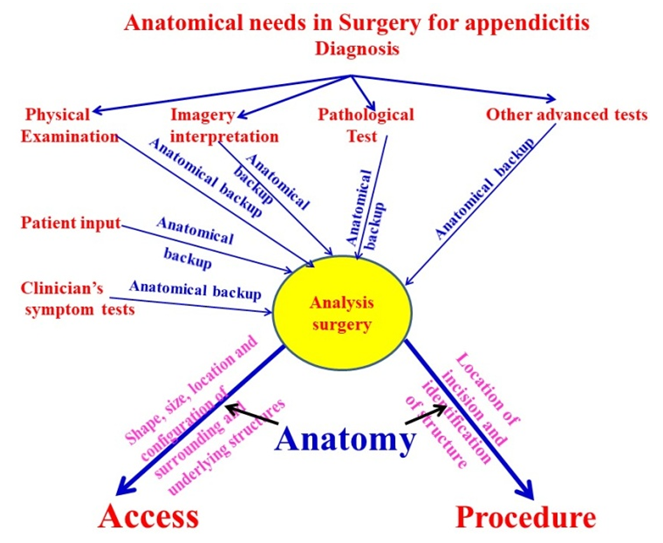
Figure 2. Showing how anatomy is indispensable for surgery e.g. in appendicitis
Post PG/super specialty professionals from all clinical streams are referred more serious patients suffering from more complicated diseases and these depend upon advanced knowledge of normal/morbid and macro/microanatomy for accurate diagnosis and treatment in clinical practice; this, in turn, requires sophisticated anatomical knowledge [2,33]. This knowledge has to be cultivated during medical education at super specialty level in all the clinical streams. In order to acquire this knowledge, whether involving new equipment to investigate internal microstructures following injury and repair, or the spread of complex vascular pathways related to atherosclerosis [34], ongoing anatomical research is required.
The emergence of new diseases requires understanding of the molecular structure of pathogens and the formulation of drugs which do not damage other structures and protective antibodies. This requires intensive, integrated and collaborative research in Anatomy with other disciplines. The need for this is becoming increasingly clear with the advent of the revolution in information technology among the public concerning the reaction of medicines in clinical practice.
In light of these developments, the systematic development of Anatomy as a subject provides firm base to revolutionize safer and secured clinical practice for advanced diagnosis and treatment. Therefore, a new academic super specialty level in Anatomy has been planned to be added like super specialties in clinical streams in general and surgery. This will supplement specialized knowledge of anatomical solutions currently unavailable and will advance excellent health care. This requires a revolution in standalone and collaborative research in Anatomy from the gross to molecular levels.
The development of Anatomy at super specialty level
Though the syllabus of super-specialty (Anatomy) should be designed by both anatomical and clinical experts a broad outline of this course corresponds to Neurosurgery, Cardiac Surgery and Gastroenterology as follows.
1. Neuroanatomy (CNS & PNS)
2. Cardiovascular Anatomy and
3. Anatomy of Alimentary tract
Broad Curriculum and Scheduling
The Broad teaching/learning curriculum of Post PG Anatomy (Table 1) will consist of the routine regurgitation of already learned Anatomy with fresh additions of anatomical variations from gross to cellular and molecular levels. This will include all the components of gross Anatomy, advanced histological interpretation like fascicular correlation/tracking and in depth advanced developmental concepts. Radiological interpretation strengthened by verification through cadaveric dissection and planning of the microstructures to fascicular level. The amalgamation of diversified interdisciplinary collaborative (with specialties and allied sciences) research of variations at macro/micro-level focused on clinical complications in the field of super specialization is indispensable in this curriculum. The analysis of Anatomy of live patients through imagery is to be done in relation to cadaveric knowledge. The curriculum should also consist of studying anatomical deformations as they correspond to signs and symptoms of diseases, impairment and malfunctions of systems / organ / limbs. The duration of super specialty in Anatomy will be 3 years similar to clinical super specialties. The schedule of learning/teaching is presented in Table 3.
Table 3. Scheduling of PG/Post PG Anatomy
     
First year should be allocated for complete Anatomy, the second year for Anatomical specialty and the third year for clinical super-specialty related Anatomy.
Evaluation system in developing Anatomy as A subject
The present system of evaluation based on passing the examination should be completely overhauled. The system of evaluation through different pattern of examinations in graduation, PG (specialty) and Post PG (super specialty) should test the practical and theoretical knowledge of Anatomy, its comprehension and ability to apply in clinical practice (Table 4).
Table 4. Evaluation Pattern of Anatomy in Medical Education
     
For this the examination pattern should be divided into testing of basic knowledge of anatomy including its application in diagnosis and treatment of disease. Thus keeping above factors in mind, theoretical examination papers must possess 50% objective, 25% short answer type and 25% explanatory questions to test definitive terminological, logical development, comprehension and applications of human Anatomy on case to case bases to clinical science for health care respectively. As regards practical examination, the students should be exposed to patients depending on academic level for diagnosis and treatment through written and oral examination exactly on the pattern of the theory examination besides cadaveric knowledge testing. These answer books should be objectively and strictly evaluated. The true and stringent evaluation will motivate junior students as per perception experiment of Bergman et al. (2013): “The stricter assessment is a frequently mentioned method as an incentive for them to study Anatomy harder [3].

3. Results and Discussion

Our analysis of this model and the available literature found that the deficiencies producing inadequate knowledge of Anatomy can be eliminated and the quality of clinical practice refined. “There is a general feeling among junior students that Anatomy is boring associated with memorizing vast amounts of factual knowledge (Vast and rote)” “Anatomy is perceived as learning the names of structures” “Well, Anatomy is just to crame. There is nothing to understand about anatomy (student 3b, year 3)” [3]. These factors creating disinterest in Anatomy among students can be eliminated by reorganizing, redistributing and standardizing the curriculum. This has been done by teaching Anatomy through repeating topics by lectures, demonstration classes, dissection and interaction and exposure to clinical cases together with providing continuous and consistent anatomical solutions to problem based learning. In this way, the material is retained, and fresh motivation can be kindled among students as long as the classes are taught in a coherent manner by medically qualified and well experienced faculty.
A standardized/reorganized curriculum at various stages of medical education will eliminate widely variable curriculum of Anatomy and this will facilitate migration of students across various countries, American and British medical schools in particular [18,35]. The variations in curricula are currently so great that the anatomical knowledge acquired by medical students, even within the same country incompatible. The variability across countries is far greater, so that the acquired knowledge and expertise in clinical practice leads to the difficulties experienced by migrating students.
The provision of competent faculty in this model will not only provide easily comprehensible anatomical knowledge through amalgamation of best and thoughtful teaching methodology but also facilitate the advancing knowledge of anatomy needed to combat any future threats of grey areas in diagnosis and treatment.
Interdisciplinary collaborative research by medically qualified, expert and experienced anatomical faculties will enhance the quality of medical education by explaining difficult to comprehend clinical grey areas. These include variations in macro/microstructures for safe clinical practice supported by anatomical analysis and solutions to the diagnosis of disease and treatment at all levels of medical education. This morphological research will be used to help formulate drugs to efficiently control the antigens and protect against from complications due to drug reactions. The amalgamation of research on variant structures, their fascicular constituents, pathways, distribution of nerve fibers and their configuration and identification for neuro-microsurgery at fascicular level will add another dimension to medical practice [2,33]. Knowledge like this may prove useful when confronted by epidemics due to new pathogens or antigens like Corona/Ebola viruses. While advances in all cognate fields are required when confronted by situations such as these, anatomically-based clinical advances may contribute substantially to the clinicians’ armamentarium, and thus decrease mortality and morbidity.
For surgeons the availability of precise and well informed anatomical background, and the ability to practice extensively on cadavers will tremendously enhance the confidence of the novice surgeon and enhance the precision of surgery with minimal damage to surrounding structures.
This will not only improve clinical practice but also produce semi-clinical anatomists to teach Anatomy teaching more in context with clinical problems. The model will facilitate meeting the future challenges of medical education and synergize the practical and theoretical interaction between clinical and non-clinical faculty and students. Thus the scheme will develop not merely anatomy academics to produce future competent faculties for Anatomy but also enhance clinical skills by supplementing anatomical solution by exploring anatomical variations and expanding morbid anatomy to overcome complications of diagnosis and treatment.

4. Conclusions

This aim of this model of development of Anatomy is to reorganize and standardize the Anatomy curriculum to be taught by qualified and experienced faculties of Anatomy with a strong background in standalone and collaborative research. Its goal is to decrease the rate of failure due to inadequate anatomical knowledge at all levels of medical education and to restore the falling credibility of the medical profession. The graduate, PG and post PG trainees will be exposed to one to one synergistic interaction and cadaveric dissection to help them understand imagery interpretation, disease and surgical access and procedure. In addition, it is intended to assist in helping understand malfunctioning of structures and systems and hence to comprehend molecular interactions of pathogens and antibodies for formulating and administering the drugs. In this way the development of Anatomy will contribute to improve clinical health care of human beings.

References

[1]  Standring, S. (2012) Evidence-based surface anatomy, Clin Anat. 25, pp. 813–5.
[2]  Kumar, R. and Singh, R. (2020) Model pedagogy of Human Anatomy in Medical Education, Surg Radiol Anat. 42, pp. 355-365 https://doi.org/10.1007/s00276-019-02331-7.
[3]  Bergman, M. Esther., Anique, B.H. de Bruin., Andreas, Herrler., Inge, W.H. Verheijen., et al. (2013). Students’ perceptions of anatomy across the undergraduate problem-based learning medical curriculum: a phenomenographical study, BMC Medical Education. 13, pp. 152.
[4]  Singh, R., Shane Tubbs, R., Gupta, K., Man Singh., et al. (2015). Is decline of Anatomy is hazardous to medical education/ profession? A review, Surg Radiol Anat. 37(10), pp. 1257-1265.
[5]  Beahrs, O.H. (1991) Gross anatomy in medicine, Clin Anat.4, pp. 310–2.
[6]  Bhangu, A., Boutefnouchet, T., Yong, X., Abrahams, P., et al. (2010). A three year prospective longitudinal cohort study of medical students’ attitudes toward anatomy teaching and their career aspirations. Anatomical Sciences Education. 3, pp.184–90.
[7]  Dusseauj, Knutson, D. and Way, D. (2008) Anatomy correlations: introducing clinical skills to improve performance in anatomy, Fam Med. 40(9), pp. 633-637.
[8]  Evans, D.J.R. and Watt, D.J. (2005) Provision of Anatomical Teaching in a New British Medical School: Getting the Right Mix, Anat Rec (part B: new anat.). 284, pp. 22-27.
[9]  Fitzgerald, J.E., White M.J., Tang, S.W., Maxwell-Armstrong, C.A., et al. (2008). Are we teaching sufficient anatomy at medical school? The opinions of newly qualified doctors, Clin Anat. 21, pp. 718–24.
[10]  Smith, J.A. (2005) Can anatomy teaching make a comeback? ANZ Journal of Surgery. 75, pp93.
[11]  Turney, B.W. (2007) Anatomy in a Modern Medical Curriculum, Ann R Coll Surg Engl. 89(2) pp.104-107. https://publishing.rcseng.ac.uk/doi/10.1308/003588407X168244.
[12]  Singh, Rajani. and Tubbs, R Shane. (2015) Should a Highly Skilled Surgeon be an Advanced Anatomist first? - A View Point, Basic Sciences of Medicine. 4(4), pp. 53-57.
[13]  Waterston, S.W. and Stewart, I.J. (2005) Survey of clinicians' attitudes to the anatomical teaching and knowledge of medical students, Clin Anat. 18(5), pp. 380-4.
[14]  Fasel, Jean H. D., Morel, Philippe. and Gailloud, Philippe. (2005) A survival strategy for Anatomy, Lancet. 365 (9461), pp. 754.
[15]  Raftery, A. (2006) Anatomy teaching in the UK, Surgery. 25(1), pp1–2.
[16]  Leveritt, S., Mcknight, G., Edwards, K. L., Pratten Margaret, K., et al. (2016). What anatomy is clinically useful and when should we be teaching it? Anat Sci Edu. 95, pp. 468-75.
[17]  Bergman, M., Cees, P.M., VanDerVleuten, Albert J. J. and Scherpbier, A. (2011). Why don’t they know enough about anatomy? A narrative review: Medical Teacher. 33, pp. 403–409.
[18]  Cahill, D.R., Leonard, R.J. and Marks, S.C. JR. (2000) Standards in Health Care and Medical Education, ClinAnat. 13, pp. 15.
[19]  Ellis, H. (2002) Medico-legal litigation and its links with surgical, Anatomy. Surgery. 20 pp. i-ii.
[20]  Goodwin, H. (2000) Litigation and surgical practice in UK, Br J Surg. 87(8), pp 977-979.
[21]  Prince K.J., Scherpbier, A.J., van Mameren, H., Drukker, J., et al. (2005a). Do students have sufficient knowledge of clinical anatomy? Med Educ. 39(3), PP 326–332.
[22]  Prince, K.J., Boshuizen, H.P., van der Vleuten, C.P. and Scherpbier, A.J. (2005b) Students’ opinions about their preparation for clinical practice, Med Educ. 39(7), pp. 704–712.
[23]  Cornwall, Jon. (2013) Perhaps we don’t know what we thought we knew: Why clinicians need to re-visit and re-engage with clinical anatomy, Australas Med J, 6(6), pp 339-340.
[24]  Drake, R.L., Mcbride, J.M., Lachman, N. and Pawlina, W. (2009) Medical education in the anatomical sciences: the winds of change continue to blow, Anat Sci Educ. 2, pp. 253–259 https://doi.org/10.1002/ase.117.
[25]  Lockwood, A.M. and Roberts, A.M. (2007). The anatomy demonstrator of the future: an examination of the role of the medically qualified anatomy demonstrator in the context of tomorrow’s doctors and modernizing medical careers, Clin Anat. 20, pp. 455-459.
[26]  Parker, L.M. (2002) What’s wrong with the dead body? Use of the human cadaver in medical education, Med J Aust. 176 (2), pp 74-76.
[27]  Tibrewal, Saket. (2006) The anatomy knowledge of surgical trainees: the trainer's view, Bull R Coll Surg Engl. 88, pp. 240-242.
[28]  Older, J. (2004) Anatomy: a must for teaching the next generation, Surgeon. 2(2,) pp79-90.
[29]  Turney, B.W., Gill, J. and Morris, J.F. (2001) Surgical trainees as anatomy demonstrators: revisited, Ann R Coll Surg Engl (Suppl). 83, pp. 193–5.
[30]  Jones, DG. (1997) Reassessing the importance of dissection: a critique and elaboration, Clin Anat. 10, pp. 123–127.
[31]  Craig, S., Tait, N., Boers, D. and Andrew, D. M.C. (2010) Review of anatomy education in Australian and New Zealand medical schools, ANZ J Surg. 80, pp. 212-216.
[32]  Roche, A., Hunter, L., Pocock, N. and Brown, D. (2009) Physical examination of the foot and ankle by orthopaedic and accident and emergency clinicians, Injury. 40, pp. 136–8.
[33]  Chandra, N. and Singh, R. (2019) Tracking of fascicles of Sartorius and pectineus nerves-a key to neurosurgery, J Clin Diagn Res. 13(1), pp. 1-8.
[34]  Singh, R. and Tubbs, R Shane. (2018) Histological verification of atherosclerosis due to bends and bifurcation in carotid arteries predicted by hemodynamic model, J Vasc Bras. 17(4), pp. 280-289.
[35]  General Medical Council (UK). (2009). Undergraduate medical education: Reports from schools and publications. London, UK: General Medical Council. Available at: http://www.gmc-uk.org/education/undergraduate. Accessed Aug 2, 2019.