-
Paper Information
- Paper Submission
-
Journal Information
- About This Journal
- Editorial Board
- Current Issue
- Archive
- Author Guidelines
- Contact Us
Basic Sciences of Medicine
p-ISSN: 2167-7344 e-ISSN: 2167-7352
2020; 9(3): 44-45
doi:10.5923/j.medicine.20200903.02
Received: Dec. 3, 2020; Accepted: Dec. 22, 2020; Published: Dec. 28, 2020

Twin Foramina in Posterior Third of an Adult Hard Palate and Their Significance
Rajani Singh
Department of Anatomy, UP University of Medical Sciences, Saifai Etawah, India
Correspondence to: Rajani Singh, Department of Anatomy, UP University of Medical Sciences, Saifai Etawah, India.
| Email: |  |
Copyright © 2020 The Author(s). Published by Scientific & Academic Publishing.
This work is licensed under the Creative Commons Attribution International License (CC BY).
http://creativecommons.org/licenses/by/4.0/

Hard palate is formed by union of maxillary process of palatine bone and horizontal plate of palatine bone during development of foetus in 12th week. Three types of foramina, greater palatine allowing greater palatine nerves and vessels, lesser palatine and incisive foramina allowing passage of lesser palatine and nasopalatine nerves respectively are normally present in hard palate. The purpose of study is to report two novel foramina in hard palate and to bring out associated clinical significance. The author observed two new foramina one on either side of intermaxillary sulcus at the junction of anterior 2/3rd and posterior 1/3rd of hard palate during scanning of base of skulls for any abnormality in the Department of Anatomy of my native institute. The diameters of the right sided foramen was 6 mm while that of on left sided was 5 mm. The distance of foramen from midline on the right side was 3 mm while that of on left side was 2 mm. The distance of foramen on the right side from the centre of inferior border of hard palate was 13 mm while that of left side was 10 mm. The hard palate separates nasal cavity and oral cavity and essential for speech, feeding and respiration. The anomalous foramina observed may create problems during speech, feeding and respiration. These foramina are of paramount importance to speech therapists, maxillofacial surgeons, physicians and academicians for new entity not described in literature.
Keywords: Hard Palate, Intermaxillary sulcus, Greater palatine foramen, Lesser palatine foramen
Cite this paper: Rajani Singh, Twin Foramina in Posterior Third of an Adult Hard Palate and Their Significance, Basic Sciences of Medicine , Vol. 9 No. 3, 2020, pp. 44-45. doi: 10.5923/j.medicine.20200903.02.
1. Introduction
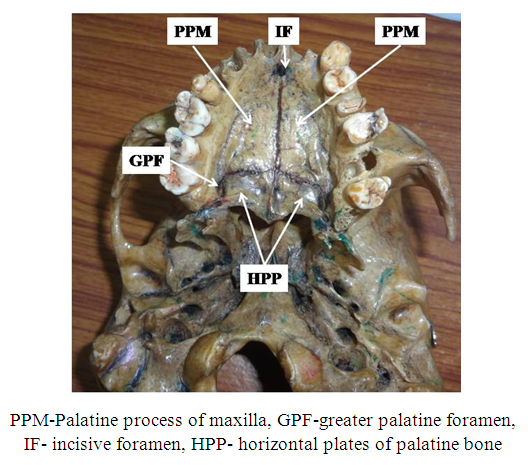
- Palate consists of hard palate and soft palate. Normally anterior 2/3rd of hard palate is formed by palatine process of maxillae and posterior 1/3rd by horizontal plates of palatine bones (Fig. 1).
 | Figure 1. Showing normal morphology of hard palate |
2. Case Report
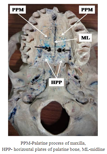
- During scanning of inferior surface of adult dry human skulls in the Department of Anatomy UPUMS Saifai Etawah, author detected twin foramina at the junction of anterior 2/3rd and posterior 1/3rd of hard palate. These foramina were present on either side of midline (Fig. 2).
 | Figure 2. Showing two foramina (Black arrows) at the junction of anterior 2/3rd and anterior 1/3rd of hard palate |
3. Discussion
- Normally, greater palatine, lesser palatine and incisive foramina are found in hard palate. Variations in number and location of these foramina have been reported in literature as cited below.Saralya and Nayak studied 132 unsexed skulls and found GPF opposite 3rd molar in 74.6% cases [2]. However it was observed in 54.87% cases out of 80 human skulls at same location in the Brazilian population [3]. Das et al. in their study found GPF medial to 3rd molar tooth in 42.7% of skulls [4]. But Author observed two new and unique foramina at the junction of anterior 2/3rd and posterior 1/3rd of hard palate in addition to normal three foramina. No literature is available describing these new foramina. Author expects that these might have developed due to incomplete fusion of palatine process of maxillae with horizontal plates of palatine bone. Genes SHH, BMP-2, FGF-8 are responsible for development of hard palate [5]. Varying degree of mutations in these genes may be the cause of these abnormal foramina. Since hard palate separates oral cavity and nasal cavities, these foramina becomes conduit between oral and nasal cavities creating helm of clinical complications to the patients.The hard palate is essential for feeding, respiration and speech. So these new foramina may cause signs and symptoms related to difficulty in feeding, respiration and speech. Thus the patients revealing these signs and symptoms caused by presence of these foramina may be treated by plugging them. The morphometry of these foramina will be highly useful in diagnosing and treating of such patients. The defective hard palate is fatal to mammals due to suckling problem and is used for mastication. Similary these foramina may create in suckling in human beings also. The hard palate is essential to produce palatal consonants “J” and “T” [5]. Therefore, these new foramina may cause difficulties during suckling, mastication and defective pronunciation of “J” and “T”. Besides this two abnormal foramina may be confused for abnormal structure causing misinterpretation of radiographs.
4. Conclusions
- The twin foramina observed by the author are unique and are of paramount importance to speech therapists, maxillofacial surgeons for treating various complications in suckling, mastication, defective pronunciation of “J” and “T”, academicians for new entity and radiologists to avoid misinterpretation of radiographs.
 Abstract
 Reference
 Full-Text PDF
 Full-text HTML