Basic Sciences of Medicine

p-ISSN: 2167-7344    e-ISSN: 2167-7352

2020;  9(1): 10-14

doi:10.5923/j.medicine.20200901.03


Surgical Anatomy of Thyroid Gland - A Comprehensive Review

Rajani Singh

Department of Anatomy, UP University of Medical Sciences, Saifai, Etawah, UP

Correspondence to: Rajani Singh, Department of Anatomy, UP University of Medical Sciences, Saifai, Etawah, UP.

Email:

Copyright © 2020 The Author(s). Published by Scientific & Academic Publishing.

This work is licensed under the Creative Commons Attribution International License (CC BY).
http://creativecommons.org/licenses/by/4.0/

Abstract

Thyroid gland is the largest endocrine gland and thyroid diseases are common health problems especially in females. Thyroid disorders like goitre, adenoma and carcinoma can be manipulated mainly through surgical interventions. A thorough and in depth knowledge of thyroid anatomy is essential for safe thyroid surgery to minimise postoperative complications. Thus the aim of present study was to elucidate surgical anatomy of the thyroid gland to update and consolidate the information associated with thyroid gland for ready use for future endocrinologists and thyroid surgeons. The literature search was carried out using data bases, Scielo, Scopmed, medline, Web of Science, Meditext, Amed, pubmed and Wiley online library and terms related to thyroid gland were used to search literature. Literature search brought out complete anatomy of thyroid gland and associated clinical significance which will be of utmost use as ready reference to thyroid surgeons and endocrinologists.

Keywords: Thyroid gland, Thyroid anatomy, Thyroid surgeons, Endocrinologist

Cite this paper: Rajani Singh, Surgical Anatomy of Thyroid Gland - A Comprehensive Review, Basic Sciences of Medicine , Vol. 9 No. 1, 2020, pp. 10-14. doi: 10.5923/j.medicine.20200901.03.

1. Introduction

The thyroid gland is enshrined on the front of the neck below adam's apple along the front of the trachea. Thyroid ailments are common health problems reported to endocrino-pathologists and 5% of the world population are found to be affected by various thyroid diseases [1] namely goitre, thyrotoxicosis, adenoma, carcinoma etc. These thyroid disorders are associated with enlargement of the gland and can be manipulated by medical and surgical therapy [2]. Hence to carry out safe and effective thyroid surgery, thorough knowledge and understanding of thyroid anatomy and pathology is imperative [3]. For performance of surgical interventions like total thyroid lobectomy and total thyroidectomy with minimal risk of invasion to neurovascular structures associated with thyroid and parathyroid glands comprehension of thyroid anatomy is very essential. Considering immense surgical implications pertaining to damage to neurovascular structures of thyroid during thyroidectomy, the study highlighting surgical anatomy of the thyroid gland has been carried out. The study will be updating and consolidating present and past research work and make the information available for ready use to future researchers, thyroid surgeons to carry out thyroid surgery with minimal damage to neurovascular structures of thyroid gland.

2. Material and Methods

Literature search was conducted in the department of Anatomy UPUMS Saifai during September 2019- December 2019 using the data bases appended below: Scielo, Scopmed, medline, Web of Science, Meditext, Amed, pubmed and Wiley online library. Papers containing original data were selected and secondary references retrieved from bibliographies. The search was restricted to English language articles and selected reference text books. Various terms used to search literature were as illustrated below: Thyroid gland, surgical anatomy of thyroid gland, recurrent laryngeal nerve (RLN), variations of recurrent laryngeal nerve, variations of external laryngeal nerve, blood supply of thyroid gland, clinical implications of recurrent laryngeal nerve and clinical implications of external laryngeal nerve. Data after surveying was consolidated and presented here.

3. Results and Discussion

Location of Thyroid Gland:
Thyroid gland is the largest endocrine gland. It is situated opposite C5- C7 and T1 vertebra (Fig 1A) embracing anterolateral part of trachea [3].
The gland is related medially to trachea and oesophagus and carotid sheath laterally. The gland is covered anterolaterally by sternocleidomastoid and three ribbon muscles, sternohyoid, sternothyroid and superior belly of omohyoid muscles. There are numerous variations in the shape and extent of thyroid gland. The alterations in shape and size of the thyroid gland along with moderation of its anatomical borders occur under the conditions, thyroiditis, malignancy, goiter, substernal goiter, hypothyroidism, prior cervical surgery and prior radioiodine ablation [2].
Thyroid gland comprised of left and right lobes which are connected to each other by isthmus, a horizontal band of thyroid tissue. Each thyroid lobe extends from mid-thyroid cartilage superiorly to the 4th or 5th tracheal ring inferiorly and common carotid arteries laterally. Though thyroid lobes are normally flat or globular yet partially cover the trachea posteriorly by a three-dimensional shape.
Variations are observed in the occurrence of lobes, isthmus, arterial supply, venous drainage and innervation pattern of thyroid gland. These are discussed one by one as under:
Pyramidal lobe:
A variation of thyroid lobes is also seen having a third lobe known as pyramidal lobe (PL) besides two normally found lateral lobes. Pyramidal lobe may project upward from the isthmus or from one of the lobes of thyroid gland (Fig 1B). Occasionally, a fibrous or fibromuscular band known as levator glandulae thyroidae may extend from hyoid bone to either isthmus or PL.
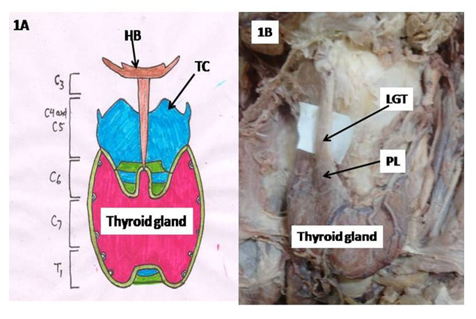
Figure 1. Showing (A) location and extent of thyroid gland, (B) pyramidal lobe arising from right lobe of thyroid gland HB-Hyoid bone, TC-thyroid cartilage, C5, C6, C7, C8- cervical vertebrae, PL-Pyramidal lobe, LGT-Levator glandulae thyroidae
The pyramidal lobe can be long, short and stubby, bifid, or absent. A bifid PL is seen as paired structures on both sides of the midline. Braun found PL in 55 percent of individuals (32/58) in a cadaveric study, and its incidence was more in males than in females. Scintigraphic imaging may not be able to investigate PL as scintigraphic imaging detects functions and not morphology. Therefore the anterior cervical region must cautiously be examined during surgery for removing remnant thyroid tissue, PL in total thyroidectomy [4] when complete removal of the thyroid is indicated. The presence of the PL may be misinterpreted during a preoperative diagnosis on ultrasonographic or scintigraphic images. According to Geraci et al (2008), the PL was identified in 50% of cases during preoperative investigations by ultrasonography or Tc-99m pertechnetate scintigraphy [5]. Hence the presence of PL preoperatively cannot deterministically be detected. Therefore, the anatomic study is imperative for safer thyroid surgery. The thyroid disease may relapse due to the remnant PL if total thyroidectomy is indicated [6] as PL consists of thyroid tissue which is inactive when normal thyroid gland is present and become active only after thyroidectomy.
For effective results of postoperative radioactive iodine treatment in patients with differentiated thyroid carcinoma, the PL should be completely excised [5] otherwise the PL will absorb most of the radioactive iodine reducing the therapeutic benefit of treatment. The primary thyroid disease are occasionally, reported to occur in the PL. A follicular carcinoma developed on the tip of the PL was removed along with PL, thyroid isthmus and prelaryngeal lymph nodes. The patient remained free of complications and diseases after postoperative 15 month follow up [7]. Two cases of malignant tumour in PL with papillary carcinoma in one and multiple papillary carcinoma in another was reported by Zivic et al [8]. In such cases, the information about the presence, position, types, relationships and size of the PL may be used to ensure safer partial thyroidectomy in order to preserve thyroid function after surgical treatment. Also as PL is the site of malignancy, it should be completely sectioned during thyroidectomy to prevent recurrence of malignancy.
The pyramidal lobe and the band develop form remnants of the epithelium and connective tissue of the thyroglossal duct [9].
Thyroid isthmus:
Thyroid isthmus overlies 2nd to 4th tracheal ring. The thyroid isthmus can be wide, long or even absent, and may have a PL.
Thyroid capsule:
Thyroid gland is enveloped by two capsules namely true and false. True capsule is formed by condensation of connective tissue of the gland and false capsule is derived from pretracheal layer of deep cervical fascia. Dense capillary plexus is located deep to true capsule. Main vessels lie between true and false capsules. It is difficult to ligate vessels in capillary plexus while large vessels laying between true and false capsules can be easily ligated. Hence during thyroidectomy, thyroid gland is removed along with true capsule.
Tubercle of Zuckerkandl:
Emil Zuckerkandl described Tubercle of Zuckerkandl (ZT) in 1902 as the processus posterior glandulae thyroideae [10]. But the structure was first observed by Otto Madelung in 1867 as the posterior horn of the thyroid [11]. Zuckerkandl's tubercle is defined as posterolateral extension of the lateral lobes composing of thyroid tissue only [12]. It should be included in the Nomina Anatomica as the “processus posterior glandulae thyroideae” described by Zuckerkandl. Recurrent laryngeal nerve, inferior thyroid artery (ITA) and inferior laryngeal nerve are intimately related with ZT. The ZT and its intimate relationship with RLN has been described in literature [13,14]. Because of close relation of above structures to the ZT, thyroid surgeons must take utmost care during thyroidectomy to identify and preserve RLN, ITA and inferior laryngeal nerve.
Ectopic thyroid tissue:
Abnormalities in development during embryogenesis may result in ectopic thyroid tissue. Lingual thyroid tissue along the path of the thyroglossal duct is the most common site of thyroid ectopy. Ectopic thyroid tissue can be benign or malignant [15]. Malignant transformation of ectopic thyroid tissue is rare. However, if thyroid tissue is found in the lateral cervical lymph nodes (lateral aberrant thyroid), a metastasis of a malignant thyroid tumor should be excluded [15].
BLOOD SUPPLY:
The thyroid gland is irrigated mainly by superior and inferior thyroid arteries but in 3% of individuals thyroidea ima artery also supply besides above arteries.
Superior thyroid artery:
Superior thyroid artery originates from external carotid artery. It descends down in intimate relationship with external laryngeal artery and on reaching just near the upper pole of the gland, the external laryngeal nerve deviates medially away from the thyroid gland. Hence during thyroid surgery, the superior thyroid artery is ligated as near the gland as possible to preserve the external laryngeal nerve. In addition to this, the superior thyroid artery also irrigate superior parathyroid glands in approximately 15 percent of cases. Besides this the superior thyroid artery is an important landmark for identification of the superior laryngeal nerve, which courses with the artery and about 1 cm from the superior thyroid pole it divides into internal and external laryngeal nerves [16]. Thus knowledge of variations in the origin and course of superior thyroid artery is very essential to neurosurgeon.
Inferior thyroid artery:
Inferior thyroid artery arises from thyrocervical trunk. But it may originate from common carotid or subclavian or arch of aorta. It ascends upward and medially to reach the base of the gland where it gives terminal branches. The terminal part is intimately related to RLN so the artery should be ligated as away from the gland as possible to preserve RLN. But the nerve may be anterior (Fig 2A) or posterior or in between the two branches (Fig 2B) of the ITA.
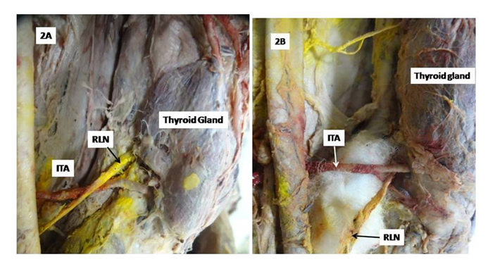
Figure 2. Showing (A) recurrent laryngeal nerve lying anterior to inferior thyroid artery, (B) recurrent laryngeal nerve lying between branches of inferior thyroid artery ITA-inferior thyroid artery, RLN- recurrent laryngeal nerve
Thus identification of RLN during thyroid surgery is very essential to avoid iatrogenic injury to this nerve. The inferior thyroid artery also supplies the inferior parathyroid glands and approximately 85 percent of superior parathyroid glands. Hence ITA gains additional importance during surgery to prevent ischemia to parathyroid glands.
Superior thyroid artery and ITA branches anastomose along the posterior border of the lateral lobe of the thyroid gland. Since parathyroid glands lie along the posterior border of lateral lobes of the thyroid glands, therefore the anastomose between superior and inferior thyroid arteries is an important guide to locate parathyroid glands during thyroid surgery as their inadvertent removal may cause hypoparathyroidism culminating into teteny.
Thyroidea ima artery:
The thyroidea ima artery may arise from the aortic arch or innominate artery and courses to the inferior portion of the isthmus or inferior thyroid poles [3]. The thyroidea ima artery can be quite enlarged in patients with thyroid disease such as goiter or hyperthyroidism. It has also been reported to be a compensatory artery when one or both of the inferior thyroid arteries are absent and in a few cases the only source of blood to the thyroid gland [11]. Thus, information regarding the occurrence of this artery is very essential. It varies in origin, size, blood supply and termination and occurs in only 3-10% of the population [17]. Thyroid ima artery is very important because of its relatively small size and infrequent presence but it can cause complications such as severe bleeding in surgery of the thorax, trachea, thyroid or parathyroid glands [18] if variations in the origin and course of this artery is not known to surgeons. Identification of this artery is very essential during tracheostomy and thyroidectomy [19] as it is smaller than the other thyroid vessels, and originates from one of the large vessels, injury to it while performing the surgery may cause complications such as severe hemorrhage and significant blood loss [18]. The artery, if damaged may draw back into the mediastinum and further complicate the condition by causing hemorrhage and clots in the thoracic cavity.
Nerve supply:
Superior laryngeal nerve bifurcates into external laryngeal and internal laryngeal nerves. External laryngeal nerve innervates cricothyroid muscle. As it is intimately related to superior thyroid artery and about 1 cm away from superior pole of thyroid gland it deviates medially. Hence superior thyroid artery should be ligated as close to gland as possible to avoid damage to external laryngeal nerve. Inadvertent damage to the external laryngeal nerve during thyroidectomies is reported to be as high as 58% [20]. In case of external laryngeal nerve palsy, cricothyroid is paralysed resulting into deep and hoarse voice, decreased range and volume, and vocal fatigue [21] and may include aspiration due to bowing and inferior displacement of the vocal fold. These conditions occur and are more pronounced in women, professional speakers and in singers. This nerve is also likely to be damaged during thyroid and parathyroid surgery, carotid endartectomy and anterior approaches to cervical spine [22]. This nerve may be inadvertently stretched, ligated or transected and presence of inflammation, tethering, malignancy, previous surgery, increases the risk of trauma to this nerve. Hence it is imperative to identify and preserve the nerve to decrease the morbidity rates due to its injury. Umpteen number of variations have been reported in literature inviting controversy in identification and retention of this nerve during surgery.
Cernea et al classified the positional interrelationship of external laryngeal nerve w. r. t. superior thyroid artery above the superior pole of thyroid gland (Fig 3) as under.
Type-1: Nerve crosses the superior thyroid artery one cm or more above the superior pole (Fig 3A). Type 2a: It crosses the superior thyroid artery at a distance of <1 cm above the superior pole (Fig 3B). Type 2b: It crosses the superior thyroid artery below the upper border of thyroid gland (Fig 3C).
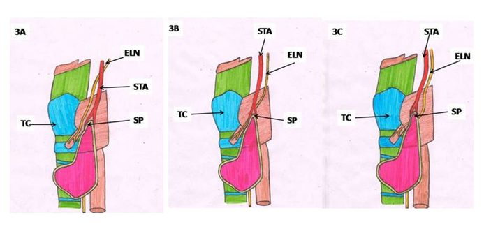
Figure 3. Schematic diagram displaying Cernea classification. (A) Type-1 (B) Type-2a (C) Type-2b ELN-external laryngeal nerve, STA-superior thyroid artery, SP- superior pole of thyroid gland, TC- thyroid cartilage
In both 2a and 2b types, the nerve is more likely to be damaged [16] as conventionally the superior thyroid artery is ligated as near the gland as possible during thyroid surgery and both 2a and 2b types external laryngeal nerve is very closely related to superior thyroid artery. Therefore, knowledge of 2a and 2b types should be kept in mind to prevent damage to external laryngeal nerve.
Internal branch:
The internal branch of the superior laryngeal nerve is sensory to the larynx. The internal branch enters the larynx through the thyrohyoid membrane superior to the external laryngeal branch.
Recurrent laryngeal nerve:
Recurrent laryngeal nerve innervates all muscles of larynx except cricothyroid. It lies very close to ITA near the gland. To protect this nerve during thyroid surgery, ITA should be ligated away from the gland. In most of the cases the RLN is related anterior to the inferior thyroid artery (Fig 2A). But it may be posterior to or in between the branches of inferior thyroid artery (Fig 2B). The variant relationship of RLN to ITA alerts the surgeon for its identification during thyroid surgery to prevent it from damage during thyroid surgery. Injury to the RLN is most frequent when a branch of the ITA is ligated, which often results in the nerve being clipped or resected with the arterial branch [23]. Transient post-operative RLN paralysis occurs in approximately 3-8% of cases and permanent paralysis in 0.3-3% of cases [24] culminating in symptoms of occult hoarseness in unilateral lesions to stridor and acute airway obstruction in bilateral damage [25]. Hence, it is very essential to identify the anatomical variants of the RLN in order to preserve the nerve and its function during surgery.
Voice symptoms are frequently found in women, professional speakers and singers as reported by Hayward. Hayward reported RLN palsies in 0.3-8% of cases, as a complication of thyroid surgery [26]. Hence, identification of the RLN is a vital step in preventing RLN injury and despite controversy, visualization of the RLN is still usually the proposed first step in thyroidectomy [27].
Although recent monitoring advances have allowed intraoperative neuro-monitoring to curb the incidence of RLN injury [28], visual identification of the RLN remains the gold standard for RLN injury prevention [29].

4. Conclusions

Knowledge of anatomical variations of thyroid gland and neurovascular configuration coupled with their variations help in proper identification of thyroid gland, associated vessels and nerves for routine surgical procedure during and after neck surgeries of thyroid in order to avoid major complications. Detailed thyroid anatomy and its associated anatomical variants are of paramount importance to endocrinologist and neck surgeons, so that these anomalies are taken into account in diagnosis and treatment.

References

[1]  Hussein Muktyaz, Yadav Birendra, Saxena Dhiraj et al. Anatomical variations of thyroid gland and its clinical significance in north Indian population. G.J B.A.H.S. 2 (2): 12-16, 2013.
[2]  Strachan MWJ, Walker BR. Endocrine disease. In: Boon NA, College NR, Walker BR, Hunter JAA. Eds. Davidson’s principles and practice of medicine. 20th Ed. New Delhi: Elsevier Churchill Livingstone; 2006; p. 744-62.
[3]  Bliss RD, Gauger PG, Delbridge LW. Surgeon's approach to the thyroid gland: surgical anatomy and the importance of technique. World J Surg. 24: 891, 2000.
[4]  Braun EM, Windisch G, Wolf G et al. The pyramidal lobe: clinical anatomy and its importance in thyroid surgery. Surg Radiol Anat. 29: 21, 2007.
[5]  Geraci G, Pisello F, Li Volsi F et al. The importance of pyramidal lobe in thyroid surgery. G Chir. 29(11–12): 479–482, 2008.
[6]  Mohebati A, Shaha AR. Anatomy of thyroid and parathyroid glands and neurovascular relations. Clin Anat. 25(1): 19–31, 2012.
[7]  Ogawa C, Kammori M, Onose H et al. Follicular carcinoma arising from the pyramidal lobe of the thyroid, J Nippon Med Sch. 76(3): 169–172, 2009.
[8]  Zivic R, Radovanovic D, Vekic B et al. Surgical anatomy of the pyramidal lobe and its significance in thyroid surgery. S Afr J Surg. 49(3):110, 112, 114 passim, 2011.
[9]  Moore KL, Dalley AF. Clinically oriented anatomy 5th ed. Lippincott Williams- Wilkins: 2006; Pages: 1083-5.
[10]  Zuckerkandl. https://en.wikipedia.org/wiki/Zuckerkandl%27s_tubercle. Accessed in Dec 2019.
[11]  Thyroidimaartery. https://en.wikipedia.org/wiki/Thyroid_ima_artery. Accessed in Dec 2019.
[12]  Costanzo M, Caruso LA, Veroux M et al. The lobe of Zuckerkandl: an important sign of recurrent laryngeal nerve. Annali Italiani di Chirurgia. 76 (4):337–341, 2005.
[13]  Serpell JW. New operative surgical concept of two fascial layers enveloping the recurrent laryngeal nerve. Annals of Surgical Oncology. 17(6):1628–1636, 2010.
[14]  Yun JS, Lee YS, Jung JJ et al. The Zuckerkandl’s tubercle: a useful anatomical landmark for detecting both the recurrent laryngeal nerve and the superior parathyroid during thyroid surgery. Endocrine Journal. 55(5): 925–930, 2008.
[15]  Kousta E, Konstantinidis K, Michalakis C et al. Ectopic thyroid tissue in the lower neck with a coexisting normally located multinodular goiter and brief literature review. Hormones (Athens). 4: 231, 2005.
[16]  Cernea CR, Ferraz AR, Nishio S et al. Surgical anatomy of the external branch of the superior laryngeal nerve. Head Neck. 14:380, 1992.
[17]  Chaurasia BD. Garg Krishna, Mittal, PS et al. eds. BD Chaurasia's Human Anatomy: Head-Neck and Brain. Vol. 3 (6th ed.). CBS Publishers. 2012; p. 143.
[18]  Sagaya Raj, Azeem Mohiyuddin, Shuaib Merchant et al. "Thyroidea Ima Artery: A Report of Two Cases". International Journal of Head and Neck Surgery. 5 (2): 89-90, 2014.
[19]  Pratt G Wyatt. "The Thyroidea Ima Artery". Journal of Anatomy. 50 (3): 239-242, 1916.
[20]  Barczynski M, Bellantone R, Brauckhuff M et al. External branch of the superior laryngeal nerve monitoring during thyroid and parathyroid surgery: International Neural Monitoring Study Group standards guideline statement: IONM During Thyroid Surgery. Laryngoscope. 123:S1–S14, 2013.
[21]  Kierner A, Ainger M, Burian M. The external branch of the superior laryngeal nerve: its topographical anatomy as related to surgery of neck. Arch. Otolaryngol. Head Neck Surg. 124: 301-303, 1998.
[22]  Furlan J, de Magalhaes R, de Aguiar ET et al. Localization of the superior laryngeal nerve during carotid endarterectomy. Surg Radiol Anat. 24: 190-193, 2002.
[23]  Sturniolo G, D’Alia C, Tonante A et al. The recurrent laryngeal nerve related to thyroid surgery. Am J Surg. 177: 485-488, 1999.
[24]  Serpell J W, Yeung M J, Grodski S. The motor fibers of the recurrent laryngeal nerve are located in the anterior extralaryngeal branch. Annals of surgery. 249: 648-652, 2009.
[25]  Jeannon J P, Orabi A A, Bruch G A et al. Diagnosis of recurrent laryngeal nerve palsy after thyroidectomy: a systematic review. International journal of clinical practice. 63, 624-629, 2009.
[26]  Hayward NJ, Grodski S, Yeung M et al. Recurrent laryngeal nerve injury in thyroid surgery: A review. Aust N Z J Surg. 83:15-21, 2013.
[27]  Sun SQ, Zhao J, Lu H et al. An anatomical study of the recurrent laryngeal nerve: Its branching patterns and relationship to the inferior thyroid artery. Surg Radiol Anat. 23: 363-369, 2002.
[28]  Randolph GW, Dralle H, Abdullah H et al. Electrophysiologic recurrent laryngeal nerve monitoring during thyroid and parathyroid surgery: International standards guideline statement. Laryngoscope. 121 Suppl: S1-16, 2011.
[29]  Page C, Monet P, Peltier J et al. Non-recurrent laryngeal nerve related to thyroid surgery: report of three cases. J Laryngol Otol. 122:757-761, 2008.