-
Paper Information
- Paper Submission
-
Journal Information
- About This Journal
- Editorial Board
- Current Issue
- Archive
- Author Guidelines
- Contact Us
Basic Sciences of Medicine
p-ISSN: 2167-7344 e-ISSN: 2167-7352
2017; 6(1): 14-17
doi:10.5923/j.medicine.20170601.03

Lactogenic Hormones and Histological Evaluation in Female Lactating Wistar Rats Treated with Fermented Soya Bean and Vitamin C Supplements
 Abstract
 Reference
 Full-Text PDF
 Full-text HTML
Malgwi I. S.1, Tanko Y.2, Eze E. D.3, Kawu M. U.4, Salami H. A.1, Emmanuel N. S.2, Mohammed A.2
1Department of Human Physiology, College of Medical Sciences, University of Maiduguri Borno, Nigeria
2Department of Human Physiology Faculty of Medicine, Ahmadu Bello University Zaria, Nigeria
3Department of Physiology, Faculty of Biomedical Sciences, Kampala International University, Kampala, Uganda
4Department of Veterinary Physiology, Faculty of Veterinary Medicine, Ahmadu Bello University Zaria, Nigeria
Correspondence to: Malgwi I. S., Department of Human Physiology, College of Medical Sciences, University of Maiduguri Borno, Nigeria.
| Email: |  |
Copyright © 2017 Scientific & Academic Publishing. All Rights Reserved.
This work is licensed under the Creative Commons Attribution International License (CC BY).
http://creativecommons.org/licenses/by/4.0/

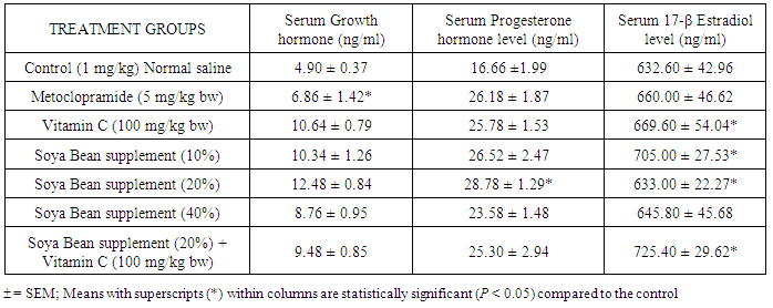
This study was designed to evaluate the effect of fermented Soya bean and Vitamin C supplement on some lactogenic hormones and mammary gland histology in female lactating Wistar rats. Thirty five (35) adult female rats were used for this study. At parturition, the animals were randomly divided into seven (7) groups of five (5) rats each (n=5) and treated as follows: Group I: (Normal control) was given normal feed and distilled water, orally (1 ml/kg bw), Group II: metoclopramide (5 mg/kg bw), Group III: 100 mg/kg bw of Vitamin C, Groups IV, V and VI were given soya bean supplement thus; 10%, 20% and 40%, respectively. Group VII was co-administered with 20% soya bean supplement and Vitamin C (100 mg/kg bw). Treatment was done for the period of ten (10) days at 06:00 pm daily. The result of serum level of Growth hormone showed significant increase (P < 0.05) in the groups treated with vitamin C (VIT C), Soya Bean (SB) 10%, SB 20% and SB 20% + VIT C. compared to the control group. Progesterone level was significantly high (P <0.05) in the groups treated with metoclopramide (26.18 ± 1.87), VIT C (25.78 ± 1.53), SB 10% (26.52± 2.47) and SB 20% (28.70 ± 1.29) when compared to the control (16.66 ± 1.99) group. A significant difference (P < 0.05) in serum 17-β Estradiol level was recorded in the groups treated with VIT C (669.60 ± 54.04), SB 10% (705.00 ± 27.53), and SB 20% + VIT C (725.40 ± 29.62) compared to the control (632.60 ± 42.96) group. All the treated groups showed an improvement in alveoli distension in the mammary gland, enhancing its capacity to accommodate increased milk volume during lactation. Therefore, supplementation with Soya bean and vitamin C should be encouraged in lactating mothers.
Keywords: Soya bean, Vitamin C, Growth hormone, 17-β Estradiol, Progesterone and Mammary gland
Cite this paper: Malgwi I. S., Tanko Y., Eze E. D., Kawu M. U., Salami H. A., Emmanuel N. S., Mohammed A., Lactogenic Hormones and Histological Evaluation in Female Lactating Wistar Rats Treated with Fermented Soya Bean and Vitamin C Supplements, Basic Sciences of Medicine , Vol. 6 No. 1, 2017, pp. 14-17. doi: 10.5923/j.medicine.20170601.03.
Article Outline
1. Introduction
- One of the major roles of the endocrine system is to coordinate mammary function with the reproductive state of the animal. This physiological synchronization is a very complex process that involves the action and interaction of multiple hormones, as well as the interplay between hormones in circulation and local regulation of the mammary response to these hormones [1]. During lactation, several key hormones are involved in the regulation of mammary cell number, secretory activity, and consequent milk production potential. Prolactin (PRL) is mainly synthesized and secreted by the lactotrophic cells of the pituitary gland [1]. Growth hormone releasing hormone, secreted from the hypothalamus, stimulates the secretion of Growth hormone from the pituitary gland. Somatostatin also secreted from the hypothalamus, inhibits the release of Growth hormone. Oxytocin is synthesized in the paraventricular nucleus (PVN) and supraoptic nucleus (SON) and axonally transported to the neural lobe where it is briefly stored until release from terminals. Axons of the oxytocin neurons passing through the median eminence already form synaptic boutons, which contact the capillary loops of the median eminence. Indeed, oxytocin release into the long portal vessels has been well-established [2]. Oxytocin release is controlled by a positive feedback mechanism; for example, oxytocin release in the PVN stimulates further release of oxytocin by neurons in the same region [3]. The mammary gland development during all stages is influenced by some factors, which certainly are the endocrine hormones that interact with the different growth factors and the epithelial and mesenchymal constituents [4]. The development and function of the mammary gland occurs through a cyclical process that changes during the physiological states of pregnancy, lactation and involution. During pregnancy, maternal hormones in circulation are responsible for the stimulation of the mammary gland development which ensures a sufficient number of mammary cells to produce milk during lactation [5]. This study was aimed at evaluating the effect of fermented Soya bean and Vitamin C supplement on some lactogenic hormones and mammary gland histology in female lactating Wistar rats.
2. Materials and Methods
2.1. Animal Grouping and Drug Administration
- Group I: (Normal control) was given normal vital feed pellets and distilled water, orally (1 ml/kg), Group II: metoclopramide (5 mg/kg bw), Group III: 100 mg/kg bw of Vitamin C., Groups IV, V and VI were given soya bean supplement thus; 10%, 20% and 40%, respectively. Group VII: was co-administered with 20% soya bean supplement and Vitamin C (100 mg/kg bw). Administration was carried out orally for a period of ten (10) days. Ethical approval was obtained from the ethical committee of Ahmadu Bello University, Zaria on animal handling, consistent with standard animal welfare guideline.
2.2. Sample Collection
- At the termination of experiment, the rats were anaesthetized by chloroform inhalation in a closed chamber and blood samples obtained via cardiac puncture into specimen bottles and allowed to clot and separated by centrifugation at 2,000 × g for 10 minutes using Centrifuge Hettich (Universal 32, Made in Germany) at an average room temperature of 23°C and the supernatant obtained was used for biochemical assays. Inguinal mammary gland tissues, two from each group were harvested using a pair of scissors and a forceps, and thereafter deposited in a labelled test tube containing a physiological solution; 10 % formal saline.
2.3. Mammary Gland Histology
- The method of H and E staining technique was used for the histological preparation. The mammary gland tissue sections obtained from the experimental rats and stored in 10% formal saline were hydrated in descending grades of alcohol from 100%, 95%, 90% and finally 70% [6]. Each of the steps lasted for three (3) minutes and the tissues were washed in a running tap water and stained afterward with haematoxylin for twenty five (25) minutes, washed with water and then differentiated in acid alcohol. The tissues were then counter stained with eosin and blued in Scott water. Afterward, the tissues were hydrated in ascending grades of alcohol and cleared in xylene for three (3) changes in five (5) minutes each. The tissues were then mounted with cover slips using a mounting media. The tissues were then viewed under a light microscope and the photomicrographs taken.
2.4. Statistical Analysis
- All data were expressed as mean ± standard error of the mean (mean ± SEM). Statistical significance was carried out using one way analysis of variance (ANOVA) followed by Tukey’s post-hoc test. All statistical analysis was evaluated using SPSS version 20.0 software and Microsoft Excel (2007). Values of P < 0.05 were considered significant.
3. Results
|
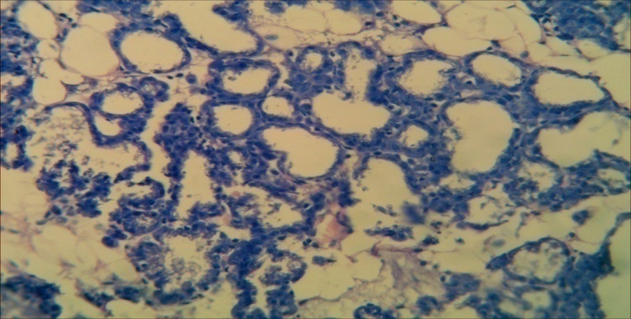
3.1. Effect of Administration of Soya Bean Supplement and Vitamin C for 10 Days on Histology of the Mammary Gland
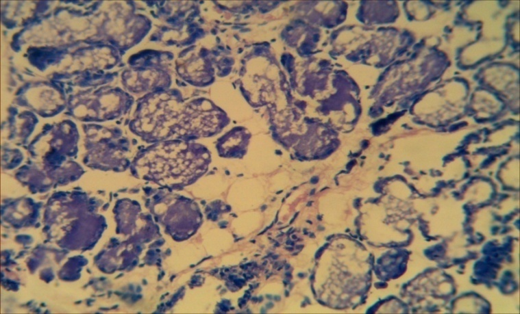
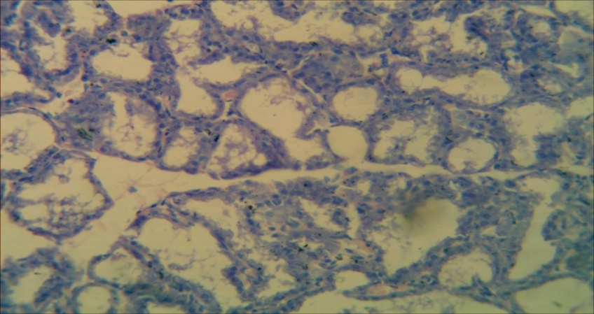
- Photomicrograph sections of the mammary gland following 10 days of oral administration are presented as follows; CNTRL = Rats treated with normal saline (1 ml/kg bw), METCL = Rats treated with Metoclopramide (5 mg/kg bw), VIT C 100 mg/kg bw= Rats treated with Vitamin C (100 mg/kg), SB 10% = Rats treated with Soya bean supplement (10%), SB 20% = Rats treated with Soya bean supplement (20%), SB 40% = Rats treated with Soya bean supplement (40%), SB 40% + VIT C 100 mg/kg bw = Rats treated with co-administration of Soya bean Supplement (40%) and Vitamin C (100 mg/kg bw).
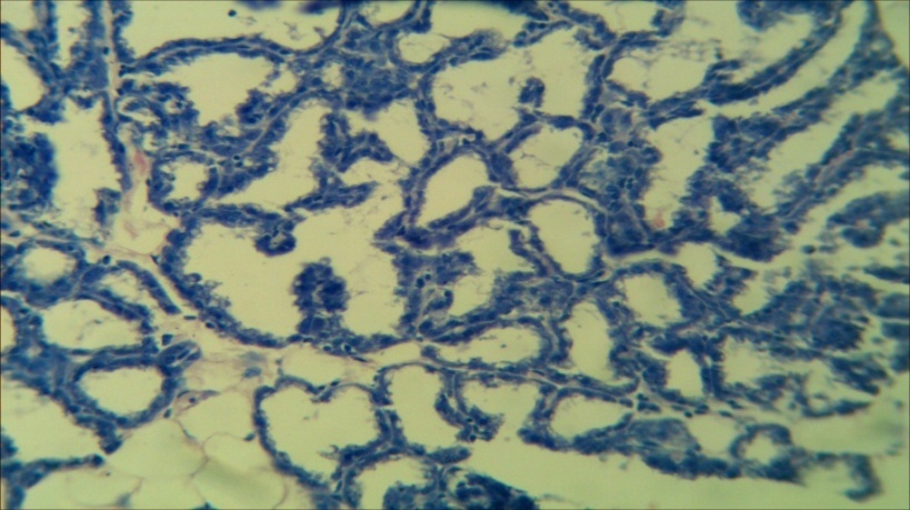 | Plate 1. Control (x250) |
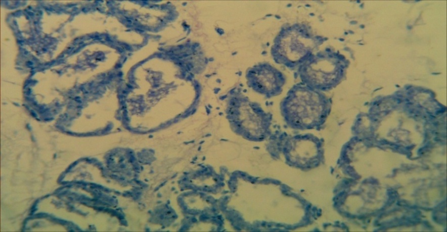
 | Plate 2. Metoclopramide (5 mg/kg bw) (x250) |
 | Plate 3. Vitamin C (100 mg/kg bw) (x250) |
 | Plate 4. SB 10% (x250) |
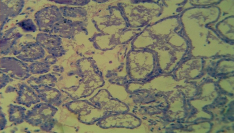
 | Plate 5. SB 20% (x250) |
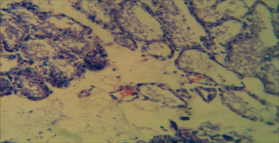
 | Plate 6. SB 40% (x250) |
 | Plate 7. SB 20% + Vitamin C (100 mg/kg bw) (x250) |
4. Discussion
- In the present study, the increase in serum level of growth hormone in the metoclopramide treated group was not significant (P> 0.05). The VIT C treated group showed a significant increase in serum growth hormone. This result could have been due to the anti-oxidant activity of VIT C, allowing it to directly scavenge of free reactive oxygen species (ROS), which could have interfered with normal hormonal release. There was a significant increase (P< 0.05) in serum growth hormone level in the SB 10%, 20% and the SB 20% + VIT C. This result could have either been due to a possible direct stimulatory activity of this supplement at the said doses on the somatotropic cells of the pituitary gland or an inhibitory activity on somatomedin secretion, both of which would result in increased level of growth hormone [7]. The action could also have been attributed to the activity of this supplement on Growth hormone releasing hormone (GHRH) from the hypothalamus. However the supplement at 40% tends to have a sort of less stimulatory action on growth hormone level when compared to the 10% and 20% respectively which could have been from down regulation of receptors of somatotropic cells of the adenohypophysis. The result of this current study showed significant increase (P< 0.05) in the serum level of progesterone hormone in the treated groups except SB 40% and SB 20% + VIT C which showed a non significant increase (P> 0.05) compared to the control. Soya bean is the richest dietary source of bioactive phytoestrogen called isoflavones. This result agrees with Duncan [8] who showed that consumption of soya bean increased progesterone hormone level in female Wistar rats. However it is in contrast to the work of Serag [9] who showed that administration of soya bean decreased serum progesterone level in female rats.A possible stimulatory activity of soya bean supplement on the Gonadotrophic releasing hormone secreting cells of the hypothalamus which stimulates the secretion of follicle stimulating hormone (FSH) and Luteinizing hormone (LH) resulting in an increased progesterone secretion from the ovaries could have caused this result. From the present study, the result showed a significant increase (P< 0.05) in the serum level of 17-β Estradiol in all treated groups except SB 40% which showed a non significant increase (P> 0.05) compared to the control. Isoflavones bind to and activate estrogen receptors (ER) resulting in ER-dependent gene transcription through both ER isoforms, generally with higher binding affinity estrogen receptors. This causes a consequent release of 17-β Estradiol as shown in the treated groups.In this present study, the mammary tissue of the control (plate 1) showed normal histological appearance of alveoli with milky secretions in the lumen. There was however no much alveoli distension as shown in some of the treated groups which is one of the indicators of milk accumulation. With slightly more alveoli distension in the metoclopramide group (plate 2) compared to the control. These pro-lactational changes observed in the mammary glands could be attributed to the activities of these lactogenic hormones which play key roles in mammary gland development involving both alveoli distensions and ductal system development. There were also pockets of lymphocytic infiltration into the mammary tissue in the metoclopramide treated group (plate 2). The 20% supplement treated group (plate 5) showed increased alveoli distension and increased accumulation of milky secretions in the alveoli when compared with the control group (plate 1). This result could be due to the activity of the supplement on serum growth hormone levels, which is known to cause tissue growth and development. The presence of numerous mitotic figures within the epithelial cells in the SB 20% treated group (plate 5) is indicative of higher cellular division and proliferation, thus resulting in increased milk synthesis and secretion, relative to plate 3, plate 4, plate 6 and plate 7 which showed no significant pro lactational changes. This result agrees with the findings of [7], who reported the distension of alveoli and mitotic presence as a pro-factor for increased secretory activity and milk synthesis in mammary epithelial cells. In conclusion, Soya bean and Vitamin C supplementation should be encouraged in lactating mothers.