-
Paper Information
- Next Paper
- Paper Submission
-
Journal Information
- About This Journal
- Editorial Board
- Current Issue
- Archive
- Author Guidelines
- Contact Us
Basic Sciences of Medicine
p-ISSN: 2167-7344 e-ISSN: 2167-7352
2012; 1(5): 23-29
doi: 10.5923/j.medicine.20120105.01
The Physiological Consequences Induced by DNA after its Release from the Cell
 Abstract
 Reference
 Full-Text PDF
 Full-Text HTML
Aziz Ferradji
Pharmacist specialized in biochemistry 37 Rue Larbi Ben M’hidi. Algiers, 16000, Algeria
Correspondence to: Aziz Ferradji , Pharmacist specialized in biochemistry 37 Rue Larbi Ben M’hidi. Algiers, 16000, Algeria.
| Email: |  |
Copyright © 2012 Scientific & Academic Publishing. All Rights Reserved.
It has been demonstrated that collagen fibres play an important role in the process of the cell assembly in order to form the human tissues, this mechanism is due to the adhesive characteristic of those fibres from which has been explained their involvement in the platelets aggregation after a vascular breach that uncovers collagen. However, it is known that DNA fibres are strongly anionic which makes them able to adhere to the surrounding cells after their liberation. Hence, we might hypothesize that nucleic acids are also involved in the cell assembly and in the platelets aggregation when they interact with the extracellular fluids. Therefore, the purpose of this work is to give a new insight into the physiological consequences induced by the release of cell products after apoptosis.
Keywords: Apoptosis, DNA Fibres, Coagulation, Cell Assembly, Tissue Pigmentation
Article Outline
1. Introduction
- The experiences stated in this paper demonstrate that DNA has several similarities with collagen including:1. The existence as long and firm fibres in an aqueous medium2. Abundance in the interstitial fluids3. Adhesive characteristic4. Platelets aggregation in the bloodstreamAlso, this article proves that DNA fibres participate in the pigmentation of tissues by adsorbing different pigments coming from the metabolism of haemoglobin, which might provide a better understanding into the pigmentation of the skin and the hair.
2. Materials and Methods
2.1. The Sedimentation of Nucleic Acids as White Fibres in the Aqueous Mediums
- The existence of DNA as white fibres can be proved by a simple experience which principle is based on the freezing-defrosting process of a blood sample taken on an anticoagulant tube (Figure 1).The first stage consists in freezing 5 millilitres (ml) of a blood sample during 20 minutes (it is preferable to discard the plasma after the centrifugation), then the sample is defrosted. This process induces the destruction of all bloodcells (red and white). The defrosted blood is transferred into a 20-ml tube to which we add about 15 ml of distilled water, and then we let the mixture incubate in an ambient temperature for 30 minutes. During this incubation, we notice the appearance of white filaments decanting into the bottom of the tube and those filaments have the same aspect as those obtained in the classical method of DNA extraction with pure alcohol(1).Also, those DNA fibres remain in the same form if they are transferred into Nacl 0.9% solution with pH = 7, which reflect the physiological conditions of the bloodstream.
2.2. Interpretation of the Experience
- It has been proved that DNA is a macromolecule that exists as long white fibres compacted in the nucleus. Thus, according to the experience described in the figure1, we deduce that the major consequence of apoptosis is the release and the accumulation of DNA fibres which size enhances because they are loosened after their liberation from the cell that compacts them. This phenomenon explains perfectly the swelling of inflammatory regions where cells are subject to necrosis. On the other hand, the enhancement of the size of DNA fibres might be explained by the liberation of histones that are responsible of the compaction of DNA fibres within the cell. It should be noted that histones are hydrosoluble (they belong to globular proteins) and are linked to DNA with hydrogen bonds, which indicates that, after the lysis of the cell, DNA will systematically get bared and loosened, because histones will be dissolved in the different fluids of the human body. In this experience, the addition of the distilled water leads to the dissolution of histones and the precipitation of DNA (Figure 2).
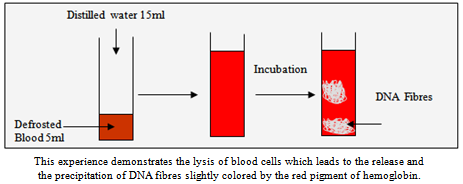 | Figure 1. The sedimentation of DNA in an aqueous medium |
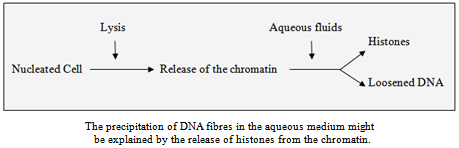 | Figure 2. The cause of DNA sedimentation in an aqueous medium |
2.3. The Involvement of DNA in the Platelets Aggregation
- The second experience provides us the evidence that DNA fibres are involved in the platelet aggregation. In fact, if we analyse the method of the leukocytes extraction(1) from a whole blood sample (10 ml) mixed with ammonium chloride 0.16M (35 ml), we would notice that leukocytes decant as small clots into the bottom of the tube that contains the mixtureThis experience indicates that the extraction of leukocytes is mediated by the action of ammonium chloride (NH4Cl) that engenders the rupture of blood cells. The lysis of the red cells induces the release of haemoglobin, but when it comes to leukocytes, the lysis induces the liberation of DNA fibres that are responsible of the platelets aggregation around the released nucleus (Figure 3), from this, we deduce that the sedimentation of the white cells by NH4Cl is due to the adhesion of platelets to the broken leukocytes which leads to the acceleration of their decanting. As a result, we might understand that the main cause of the platelets aggregation in the blood circulation is the release of DNA fibres after the lysis of leukocytes, and this can be confirmed if we process the freezing-defrosting experience on a partially coagulated blood, we would notice the appearance of small clots tied up to DNA fibres.
2.4. The Involvement of DNA Fibres in the Formation of Tissues
- This experience consists in demonstrating that DNA fibres are adhesive and abundant in the interstitial fluids, but before we process this experience we should ask the following question: everyday, millions of cells die in the human body, and they are replaced by new cells, so how DNA fibres are eliminated? , Knowing that the amount of the released DNA is too high to be digested by enzymes. So there must be a track that conducts those fibres in order to be eliminated. To this end, if we mix the saliva with a small amount of pure alcohol, we would notice the precipitation of white fibres that have the same aspect as those obtained in the classical method of DNA extraction (1). The figure 4 represents the simplest method of DNA extraction from the human body, from which we conclude that apoptosis induces the release of DNA fibres into the interstitial fluids in order to be eliminated through salivary glands which explains perfectly why saliva is viscous.On the other hand, if we repeat the same experience by mixing pure alcohol with a tiny amount of natural glue that is supposed to be rich of collagen, we would notice the appearance of DNA filaments in the mixture (the same result obtained in the Figure 4) which demonstrates that DNA is adhesive just like a glue and therefore can be involved in the cell assembly in order to form tissues and this characteristic might be explained by the anionic charge of nucleic acids.
2.5. DNA Fibres and the Pigmentation of Tissues and Hairs
- It is known that Haemoglobin exists as a red pigment in the red cells that circulate in the bloodstream, this colour is maintained by the double positive charge of iron Fe (++) that is held by the molecule of heme, in the meantime, if we mix a small amount of non coagulated blood (about 50 microlitres) with 1 ml of different anionic chemical components ( like citrate, acetate, EDTA…) that have the capacity to adhere to Fe (++), we would notice that Haemoglobin changes its colour to another one that is specific to the nature of the component, also we would notice that the intensity of the coloration is directly proportional to the amount of haemoglobin.
 | Figure 3. Platelet aggregation following the lysis of leukocytes |
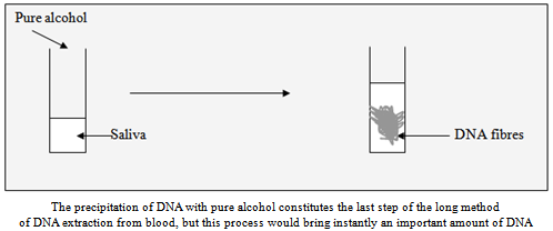 | Figure 4. The simplest and the most effective method of DNA extraction |
- Besides, it is known that the oxidation of Fe (++) to Fe (+++) changes the colour of the blood to brown, which means that if the lysis of red cells occurs in tissues containing enzymes that extract electrons from the iron of haemoglobin, then the tissue would be coloured in brown, this concept suggests that haemoglobin plays an important role in the pigmentation of tissues in the human body, and therefore it is necessary to study all the biochemical reactions involved in the metabolism of haemoglobin that happens after its release from the red cells. Also, it is important to mention that the cells within the tissue in which haemoglobin is liberated, are regularly submitted to apoptosis that induces systematically the release of DNA fibres which are highly adhesive, and thus they can bind to the oxidised haemoglobin. This hypothesis leads to the concept that the basic of the tissue pigmentation is carried out in two stages: 1. The transformation of the released haemoglobin into another metabolite with different colour that depends on the type of the reaction.2. The coloration of DNA fibres by the metabolite of haemoglobin. In this case, the colour’s intensity of the tissue depends on the amount of DNA fibres as well as the metabolite of haemoglobin. Furthermore, if we heat a piece of meat, in which myoglobin is very abundant, we would notice that the colour turns to brown which reflects the oxidation of the iron according to the reaction represented in the figure 9.This reaction gives the irrefutable evidence that the darkness of the skin depends on the charge of the iron held by the pigments that colour the DNA fibres involved in the cell assembly.As DNA fibres and haemoglobin are involved in the pigmentation, it would be logical to hypothesize that the hair is the result of packing old blood cells together, in parallel, it has been demonstrated that the composition of the hair might provide some information about the state of the bloodstream, This correlation allows us to state that the hair is produced after the lysis of red and white cells in the hair follicle according to the two stages stated above. In other words, the hair is composed of compacted DNA fibres coloured by the metabolised haemoglobin which colour depends on the nature of the reaction that takes place in the hair follicle.
3. Discussion
- The purpose of this analysis is to deliver to the scientific community the concept that apoptosis is the source of DNA fibres that play important structural roles in the human body. In fact, the anionic charge of nucleic acids provides them the adhesive characteristic which makes them able to adhere to the surrounding cells. Also, if we establish a link between DNA fibres and the blood clotting, we would come to the conclusion that the thrombosis process is carried out in two concomitant stages:1. The conversion of fibrinogen into insoluble fibrine.2. Adhesion of platelets to the DNA fibres liberated by leukocytes.
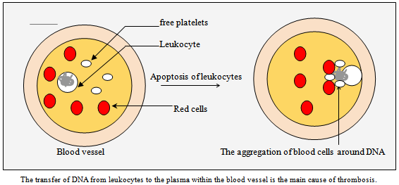 | Figure 5. thrombosis process |
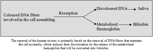
- Hence, we understand that the structure of blood clots are essentially formed by DNA fibres that attract red cells and platelets, moreover, the presence of leukocyte’s enzymes in the serum of a coagulated blood, as well as their absence in the plasma of non-coagulated blood, give us the irrefutable evidence that the white cells are submitted to necrosis during the coagulation process. Notably, the platelets aggregation in the bloodstream, that is described in the pathology of atherosclerosis, is due to the apoptosis of leukocytes induced by the action of cardiovascular risk factors, for instance, when macrophages are gorged with LDL-cholesterol, they will systematically settle on the inflammatory regions called commonly atheroma plaques located on the vessel walls, which means that macrophages would be doomed to apoptosis if they are immobilised by the atheroma plaque, and this is the main cause of the blood clotting (Figure 5).Regarding the pigmentation of tissues that is mediated by the combined effect between the metabolite of haemoglobin and the adhesion of DNA fibres, this mechanism might provide us another insight into the pathology of Albinism which is caused by the lack of pigmentation of the skin, the hair and the eyes, in fact, this disorder could be explained by the following deficiencies: - The inhibition of the conversion of haemoglobin into the appropriate pigment.- The incapacity of DNA fibres to adsorb the metabolised haemoglobin: The paleness observed in this pathology, reflects the white colour of DNA fibres.However, if we observe the red eyes of the albinos hamsters, we would logically incriminate the first deficiency in the pathology of albinism As well, Recent finding published in Pigment Cell Research (2) suggests that melanin is able to effectively ligate metal ions, which corresponds to the characteristic of haemoglobin and its metabolites, on the other hand, the presence of an ion makes melanin capable to adhere to the anionic nucleic acids after their release from the cells, Therefore, if we establish a correlation between the tissue pigmentation described in this paper and the finding of that research(2), it would become necessary to verify whether melanin comes from the metabolism of tyrosine or haemoglobin.The concept that DNA is involved in the cell assembly might provide a better understanding into the ageing process, from which we can hypothesize that the amount and the quality of DNA fibres vary from a young to an old skin, and the whiteness of the hair during the ageing process reflects the white colour of DNA fibres that are incapable to adsorb the metabolite of haemoglobin.Furthermore, According to the mechanism of the hair growth we might explain the cause of the hair loss during chemotherapy that lowers the activity of the bone morrow in which occurs the production of blood cells that are the precursors of the hair. This concept allows us to understand further the effects of chemotherapy on the apoptosis of leukocytes within the hair follicle and in the immune system. Additionally, the compacted and coloured DNA Within the hair follicle is dried under the effect of the greasy characteristic of the sebum that allows the dehydrating of DNA fibres and leads to the appearance of dried and greasy hairs. This mechanism indicates that acne is not only due to the excess of the sebum production but also due to the exaggeration of the lysis of red and white cells that is responsible of the accumulation of DNA fibres that might disrupt the hair growth and leads to the inflammatory swelling of the hair follicle.The question raised in this paper concerning the removal of DNA fibres after the cell death indicates that salivary glands belong to the track of DNA fibres elimination, and this concept might help researchers to study further this track in the human body that goes from tissues in which DNA is released, to salivary glands which allows us to extrapolate new concepts about the renewal of tissues that is based on two major activities: Mitosis and Apoptosis because it is obvious that this renewal is based on the resorption and the removal of DNA fibres involved in the cell assembly, that will be replaced by new DNA fibres coming from apoptosis. On the other hand, the white colour of DNA fibres obtained from the saliva with pure alcohol (figure4), indicates that the resorption of DNA from the human tissues induces their discoloration, which is why it becomes clear that the oxidised haemoglobin, that is released after the resorption of DNA, will be converted to bilirubin (Figure 6).
4. Medical Approaches
- The study of this article might provide new medical approaches that allow the fabrication of new drugs that have the capacity to dissolve the DNA fibres of the clots formed in the bloodstream in order to be resorbed by the interstitial fluids and eliminated through salivary glands. On the other hand, DNA fibres should be classified among medical adhesives that are able to stop haemorrhages that occur during surgical procedures, in fact, the use of DNA fibres as bioadhesives would have the advantage to be non-toxic and biocompatible with the human body, and more importantly, they would be of interest in the treatment of haemophilia because, according to this paper, the disruption of the blood clotting might be explained by the slowdown of leukocyte’s apoptosis as it provides DNA fibres that are essential for the closing up of vascular breaches.
 | Figure 6. The resorption of DNA fibres involved in the cell assembly |
 | Figure 7. The formation of a cancerous tissue |
 | Figure 8. Therapeutic concepts concerning the treatment of cancers |
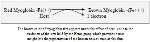 | Figure 9. Oxidation of the iron within Myoglobin |
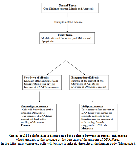
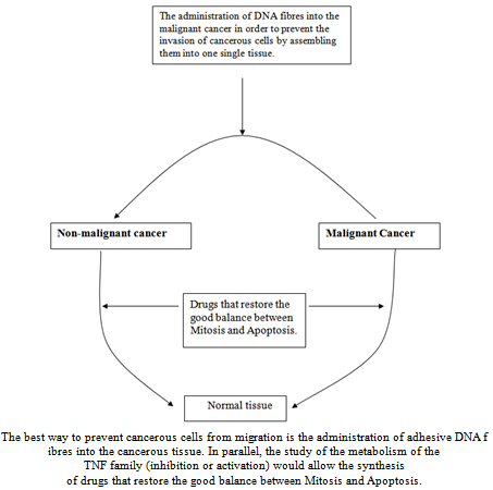
- Additionally, the structural role of DNA fibres in the human body is of a big importance in the understanding of the aspect of the tumour tissues. In fact, according to its common definition, a cancerous cell refuses to die, which leads to the lack of DNA fibres that are necessary for the cell assembly, in this case, the cancerous cells will be free to migrate and proliferate throughout the human body (metastasis). To this end, the administration of adhesive DNA fibres would be recommended in order to retain the cancerous cells into one place (the source) so that they might be submitted to apoptosis (Figure 7 and 8).As TNF family has been reported as inflammatory, pro-coagulant and anti-cancerous it would be logical to state that TNF enhances the amount of DNA fibres by activating apoptosis and therefore it is necessary to study themetabolism of TNF family and their interactions with different molecules in order to bring new medical approaches regarding the disorders related to the increase or the decrease of the extra cellular DNA fibres, for instance, the swelling of inflammatory regions is due to the accumulation of DNA fibres caused by the exaggeration of necrosis and therefore the anti-TNF drugs are beneficial in the treatments of inflammatory disorders such as polyarthritis.
5. Conclusions
- The major physiological consequence of apoptosis in the human body is the release and the accumulation of DNA in the extracellular fluids such as Plasma or Lymph which raises a controversial question: Why this data has never been described in the literature? But if we study the helical structure of Collagen as well as its similarities with DNA we would come to the conclusion that Collagen fibres are not proteins but rather nucleic acids, in addition, this article brings the hypothesis that the role of the lymphoid system is the removal and the elimination of the extra cellular DNA from different tissues of the human body. As for red blood cells, the consequence of their lysis is the Tissue pigmentation that is effected by haemoglobin, as a result, the darkness of the skin, observed in certain populations, is simply due to the oxidation of the iron held by the Heme group.