American Journal of Materials Science

p-ISSN: 2162-9382    e-ISSN: 2162-8424

2017;  7(3): 64-70

doi:10.5923/j.materials.20170703.04


Characterization of Pectin Biofilms with the Addition of Babassu Mesocarp and Whey Protein Concentrate

Lopes I. A.1, Santos Jr J.1, Da Silva D. C.1, Da Silva L. J. S.1, Barros A. K.2, Villa-Vélez H. A.1, Santana A. A.1

1Chemical Engineering Coordination, Federal University of Maranhão, São Luís, Brazil

2Department of Electrical Engineering, Federal University of Maranhão, São Luís, Brazil

Correspondence to: Villa-Vélez H. A., Chemical Engineering Coordination, Federal University of Maranhão, São Luís, Brazil.

Email:

Copyright © 2017 Scientific & Academic Publishing. All Rights Reserved.

This work is licensed under the Creative Commons Attribution International License (CC BY).
http://creativecommons.org/licenses/by/4.0/

Abstract

Biofilms are defined as flexible films prepared from biological materials with the potential for application in the pharmaceutical and food areas, their use depending on various parameters such as the mechanical properties, barrier properties and water solubility, amongst others. The present work aims to characterize pectin (P) biofilms composed of babassu mesocarp (BM) and whey protein concentrate (WPC), prepared by the casting method. Thus physical attributes such as moisture content, water soluble mass, thickness, water vapor permeability and degradation were determined. Fourier-transform infrared spectroscopy and scanning electron microscopy were used in order to determine the chemical composition and surface structure, respectively, of the biofilms. Calcium crosslinked and non-reticulated films were considered. The biofilms composed of 100%P and 50%P+50%WPC were more hygroscopic than those composed of 50%P+50%BM and 50%P+25%BM+25%WPC. The lowest water vapor permeability and solubility values were found for the films plasticized with 50%P+50%BM. The films plasticized with 50%P+50%BM and 50%P+25%BM+25%WPC showed very similar functional attributes regarding their application as food wrappings and/or production bags.

Keywords: Babassu mesocarp, Casting method, Physical characterization, Whey protein concentrate, Glycerol

Cite this paper: Lopes I. A., Santos Jr J., Da Silva D. C., Da Silva L. J. S., Barros A. K., Villa-Vélez H. A., Santana A. A., Characterization of Pectin Biofilms with the Addition of Babassu Mesocarp and Whey Protein Concentrate, American Journal of Materials Science, Vol. 7 No. 3, 2017, pp. 64-70. doi: 10.5923/j.materials.20170703.04.

1. Introduction

Biofilms are natural polymers formed from biological, animal or vegetable materials such as microorganisms, lipids, proteins and polysaccharides. These materials, obtained from the growth of microorganisms or from animal protein and plants, are currently replacing the plastic materials synthetized from petroleum due to their environmental friendliness [1, 2]. Recently, the production of biofilms combining different polysaccharides, proteins and lipids has been the focuse of research to improve their functionality (e.g., mechanical and barrier properties), showing synergistic advantages when compared with the pure components [3].
Pectin is an anionic polysaccharide widely used in the food industry due its high availability in citrus peels, with low extraction costs, high solubility, good gelling properties, high biocompatibility and simple modification by chemical and biochemical processes. In addition, pectin has great potential for use in the preparation of biofilms, as food coverings and drug coating, amongst others [4, 5]. The characteristics of biofilms composed of pectin have been reported by Giancone et al., [6], where films made from high methoxyl pectin showed barrier properties comparable to commercial biodegradable film packaging and, according to Fishman et al., [7], where films made from mixtures of pectin/starch/plasticizer resulted in a very definite loss of brittleness of the film, making them much more flexible. Thus the physical, thermal and mechanical properties of pectin-based films, depends on the aggregated substances, such as plasticizers and emulsifiers, as well as the coverings used on the matrix.
Amongst the diversity of raw material that have been researched to produce biofilms, the babassu mesocarp and whey protein concentrate are two possible compounds that can be mixed with the filmogenic matrix containing pectin. The babassu (Orbignya phalerata Mart.) mesocarp is produced during the separation of the babassu kernel, being widely marketed in the states of Maranhao, Piaui and Tocantins (Brazil) [8]. The dried babassu mesocarp flour is used as a substitute for cassava flour, and serves as food for both humans and animals [9], as well as in natural medicines for the treatment of multiple diseases [10-12]. Chemically, the babassu mesocarp flour contains 60% of starch (gelatinization temperature of the granules in the range from 63 to 73°C) [13] and a significant amylose content and its polymeric structure is highly crystalline, showing promising applications in the form of elastic matrices [14, 15]. On the other hand, the whey protein concentrate contains globular proteins such as β-lactoglobulin, α-lactoalbumin and bovine serum albumin, presenting both thermo-reversible and thermo-irreversible gels when heated above 65-70°C, with wide applications for use in the food industry as an emulsifier [16]. Moreover, the emulsifying capacity depends on the composition of the matrix considered (e.g., ionic strength and pH), the processing to which it is submitted and the storage conditions that it experiences during its lifetime, e.g., heating, cooling, mechanical agitation [17, 18].
Both compounds show promising applications in the production of biofilms, since films prepared with pure pectin are completely soluble in water, besides having poor mechanical properties. Thus the combinations could provide new characteristics which should be researched. In addition, studies concerning the use of babassu and whey protein concentrate in the confection of biofilms have not been reported in the literature. Thus the present work aims to study the influence of the mixture of pectin/babassu mesocarp/whey protein concentrate, with added calcium chloride (crosslinking agent) and glycerol (plasticizing agent), on the chemical, barrier and mechanical properties of biofilms.

2. Material and Methods

2.1. Raw Materials and Sample Preparation

Citric pectin (P) (Isofar, Rio de Janeiro, Brazil), whey protein concentrate (WPC) (Alibra, Campinas, Brazil), anhydrous calcium chloride (Merck, Darmstadt, Germany), glycerol (Synth, São Paulo, Brazil) and babassu mesocarp (BM) were used in this research.
Babassu mesocarp (BM) was obtained from in nature coconut. The coconut was first peeled and the fiber and mesocarp extracted with a steel knife. The mesocarp was then milled in a ball mill (Model 460*600, Yongsheng, China) and sieved through a No. 400 mesh screen (Tyler series, W.S. Tyler, USA) in order to obtain a powder with a diameter of 37 μm. The chemical characteristics of the BM were determined according to the Standard Analytical Methods [19], obtaining values for moisture content of 1.66±0.03 g/100g (w.b.), protein content of 4.03±0.58 g/100g (w.b.), fat content of 0.43±0.10 g/100g (w.b.), and ash content of 2.34±0.08 g/100g (w.b.). The carbohydrate content was obtained by difference, obtaining a value of 91.54±0.08 g/100g (w.b.).

2.2. Film Preparation

Biofilms were prepared according to the methodology of Santana and Kieckbusch [20] with modifications. Citric pectin (P) together with babassu mesocarp (BM) and/or whey protein concentrate (WPC) and glycerol (3 ml) were dissolved in distilled water to a total volume of 200 ml, with constant magnetic stirring. After dissolution, 30 ml of the cross-linking solution was added and the temperature increased to 70°C. The cross-linking solution (Ca2+) was added slowly to the solution containing the citric pectin with babassu mesocarp and/or whey protein concentrate, using a peristaltic pump (model 77120-70, Masterflex, USA) with a maximum flow rate of 0.6 ml/min, to avoid local gelling. The films were obtained by the casting technique, i.e., aliquots of 50 g of the hot solution were poured into round Plexiglas pans (area = 172.03 cm2) and slowly dried at 40°C in an oven with air recirculation (model 099EV, Fanem, Brazil) for 18 to 20 hours. The dried films were removed from the support and stored at 52% RH and 25°C for 48 hours. In all, four formulations were studied, 100%P, 50%P+50WPC, 50%P+50%BM and 50%P+25%BM+25%WPC.

2.3. Characterization of the Biofilms

Film thickness
The thickness of the biofilms (δ) was controlled by pouring a constant mass (50 g) into the film forming solution over the support. The thickness of the conditioned films was measured using a digital micrometer (model MDC-25S, Mitutoyo, Japan). Measurements were taken at five different positions on the film surface and the mean value reported.
Moisture content
The moisture contents (w) of the four formulations were determined in a vacuum oven (model MA030, Marconi, Brazil) at 60°C for 24 h using the gravimetric method AOAC 934.06 [19]. The analyses were carried out in triplicate and reported on a dry weight basis (d.b.).
Matter dissolved in the water
The mass dissolved in the water (S) was determined in triplicate according to the methodology proposed by Irissin-Mangata et al. [21]. The mass of a piece of film was determined and then immersed in 50 ml of distilled water under mild shaking action (175 rpm) at 25 ± 5°C for 24 h, in an orbital controlled temperature shaker (model 3545-40-EA, Termo Fisher Sci Inc, USA). The sample was then transferred to a vacuum oven (60°C for 24 h) to determine the final dry matter. The soluble matter was expressed as a function of the initial dry matter according to Eq. (1):
(1)
where w is the moisture content (d.b.), mi is the initial mass (g) and mf is the final mass (g).
Water vapor permeability
Water vapor permeability (WVP) was determined using the ASTM E95-96 method [22] at 25°C in a small Plexiglas cell (25 ml) completely filled with calcium chloride granules in order to maintain the relative humidity (RH) of the system at 0%. The film was fitted tightly to the removable cell lid, covering a central rectangular opening (38.5 cm2). The cell was placed inside another hermetic Plexiglas jar (≈500 ml) containing a saturated NaCl solution (Synth, São Paulo, Brazil) at the bottom, and submitted to intermittent agitation to keep the RH of the system at 75% and hence maintain a constant difference in water vapor pressure. After the system attained steady-state conditions (≈2 h), the cell weight was measured every 12 h for 3 days using an analytical balance (model AP210, Ohaus, Switzerland) and the water vapor permeability [(g.mm)/(m2.day.kPa)] calculated from Eq. (2):
(2)
where, δ is the thickness of the film (mm), Ae is the area of the exposed surface of the film (m2), ΔPw is the partial water pressure difference across the film (kPa), G is the permeation rate (g/day) calculated by linear regression of the mass gain vs. time, and F is a correction factor that takes the additional diffusion resistance in the stagnant air gap between the surface of the calcium chloride layer and the film into account. In the geometry used, this correction was close to 1.0 and was therefore not considered [23].
Biodegradation test
The biodegradation was determined according to the ASTM G160-98 method [24] under specified experimental conditions, breeding ground, soil pH between 6.5-7.5, relative humidity between 85-95%, temperature 30±2°C and analysis time of two weeks. Figure 1a shows a small packaging scheme of the test piece bodies. To determine the resistance to biodegradation, samples of each biofilm were weighed and placed in a container with mineral (ground), vegetable (grass) and animal (insect) matters, and exposed to the action of nature (living organisms, rain, wind and sun) (Figure 1b). After two weeks, samples were taken from the environment and weighed to determine the percent degradation (D %) according to Eq. (3):
(3)
where m is the mass after the exposure time (g) and mi is the initial mass of the sample (g).
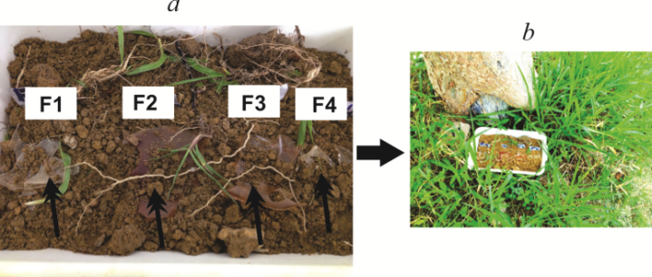
Figure 1. Biodegradation analysis: (a) Biofilm packaging composed of 100%P (F1), 50%P+50%WPC (F2), 50%P+50%BM (F3) and 50%P+25%BM+25%WPC (F4) and, (b) biofilms in the natural environment

2.4. Morphology

Scanning electron microscopy (SEM) was employed to study the surface and cross-sectional morphologies of the biofilms produced. All the biofilms were dried in a vacuum oven at 70°C for 24 h and the samples mounted onto stubs, sputter coated with gold in a vacuum chamber and photographed (magnifications of x145, 265, 360, 620, 1,450, 3,100, 4,100 and 5,000) using the scanning electron microscope (model Phenom Pro, Phenom-World, Netherland) operated at 5 kV.

2.5. Fourier-transform Infrared Spectroscopy (FTIR)

FTIR-ATR spectroscopy of the biofilms was carried out using a spectrometer (model IR-Prestige, Shimadzu, Japan) with 128 scans between 400 and 5000 cm-1 and 4 cm-1 resolution.

2.6. Statistical Analysis

All regressions were carried out using the Statistica V9 (Statsoft, Tulsa, USA) software. In addition, an analysis of variance and Tukey test were employed to determine any statistically significant differences (p < 0.05) between the averages.

3. Results and Discussion

3.1. Biofilm Characterizations

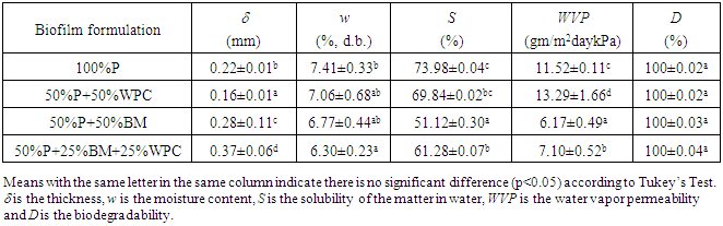
Biofilms composed of pectin with the addition of babassu mesocarp and/or whey protein concentrate were formulated and prepared by the casting technique, obtaining the following four matrices 100%P, 50%P+50WPC, 50%P+50%BM and 50%P+25%BM+25%WPC. Visually, the 100%P and 50%P+50%WPC biofilms showed a slightly yellowish, translucent and homogeneous surface, flexible texture and were easily removed from the Plexiglas mold. On the other hand, the 50%P+50%BM and 50%P+25%BM+25%WPC biofilms showed a brown homogeneous surface and low flexibility as compared to the biofilms without BM (Fig. 2). Table 1 shows the filmogenic characteristics of thickness, moisture content, the matter dissolved in water, water vapor permeability and biodegradation data for the four formulations.
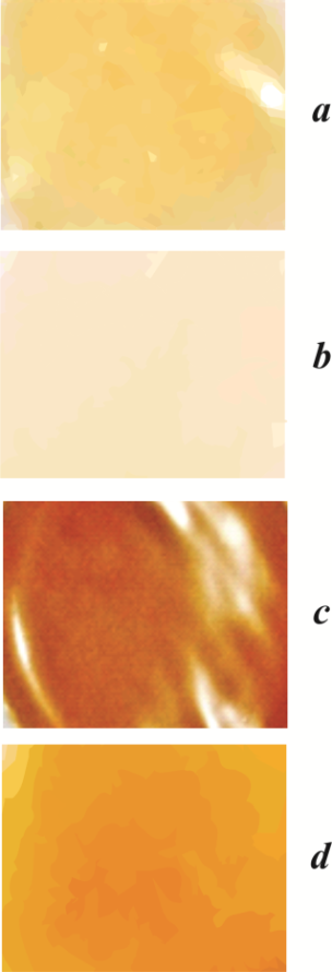
Figure 2. Visual aspects of the four pectin biofilms with added babassu mesocarp and whey protein concentrate. (a) 100%P, (b), 50%P+50%WPC, (c) 50%P+50%BM (F3) and (d) 50%P+25%BM+25%WPC
Table 1. Characterization of the biofilms made from the four formulations composed of citric pectin (P), babassu mesocarp (BM) and whey protein concentrate (WPC)
     
Table 1 shows that the biofilms composed of 100%P and 50%P+50%WPC showed thickness values (δ) that were 57% less than the biofilms composed of 50%P+50%BM and 50%P+25%BM+25%WPC. These results can be explained by the particle size of the babassu mesocarp of 37 um, this therefore being the minimum limit to forming a homogeneous biofilm. Moreover, the thickness of the films composed of the ternary mixture of 50%P+25%BM+25%WPC increased even more than the binary mixtures of P with BM and/or WPC. According to Silva et al. [5], this tendency at the macroscopic level can be attributed to an increase in molecular volume due to a more intense plasticizing effect at higher glycerol and citric pectin concentrations.
The moisture contents (w) of the biofilms ranged from 6.30 to 7.41% (d.b.), the formulations with 100% pectin showing slightly higher moisture contents than the other formulations. This behavior was also observed by Bierhalz et al. [25] who attributed it to the high hydrophobicity of the pectin. For the other three formulations, the low moisture contents can be attributed to the intumescence power of the whey protein concentrate [26] and babassu mesorcarp [13].
The water solubility (S) of the four formulations presented values between 51.12 and 73.98%. These values were higher than those reported by Silva et al. [5] of between 8.8 and 37.2%, by Seixas et al. [4] of between 32.88 and 51.98% and by Bierhalz et al. [25] of between 16.72 and 25.66, all for biofilms composed of pectin and alginate. In the preent study, one can observe that the solubility decreased as the concentration of BM increased. This was because the BM promoted crosslinking of the polymer chains, and hence as the BM concentration increased, so the intermolecular bonds became more cohesive and organized in the matrix, making the biofilms more resistant to dissolution. Although high film solubility may be desirable in some applications, the low solubility of edible films is one of the most important requirements for food and pharmaceutical applications [3].
The water vapor permeability (WVP) ranged from 6.17 to 13.29 g.mm/m2.day.kPa for the four formulations, these values being similar to those found for biofilms composed of zein-oleic acids [27], high methoxyl pectin [6] and of PLA/nanoclay composite [28]. In general terms, the biofilms composed of citric pectin with BM presented moderate barriers to water vapor permeability. Also, the values found for WVP for the four formulations also exhibited an inverse relationship with the values for solubility (Table 1). This behavior can be attributed to the low level of reticulation with Ca2+ in the formulations. Furthermore, for each type of formulation, the WVP increased with film thickness. In ideal homogeneous polymeric films, the permeability constants are independent of thickness. Water transport through hydrophilic films, however, is extremely complex due to non-linear moisture sorption isotherms and water content-dependent diffusivity [20, 29]. In the present study, the interfacial equilibrium with humid air promoted an incipient structure relaxation in the outer portion of the cross section of the film, increasing the permeation rate in this surface layer. The thicker film reduced the relative contribution of this layer to the total resistance and hence the WVP increased [20, 25].
The biodegradability test (D) showed complete biodegradation in the four biofilms. The degradation analysis was carried out for 7 days. During the first five days of monitoring, the weather was stable and the films suffered little degradation. However, on the last 2 days there was torrential rain and the films were completely biodegraded. This can be explained by the fact the natural macromolecules of the biofilms produced were formed of proteins and carbohydrates that are degradable in biological systems due to hydrolysis followed by oxidation. On the other hand, not all the biofilms were completely solubilized, since the action of the rain accelerated the biodegradation process. Furthermore, the films were also attacked by insects and microorganisms, which removed some of the biodegradable components. According to Tripathi et al. [30] and Chandra and Rustgi [1], during the biodegradation process in the soil, the amorphous fraction of the material is exposed to attack by microorganisms and the microbial degradation results in an increase in the degree of crystallinity of the biofilms.

3.2. Morphology Results

Figure 3 shows the SEM images for the formulations 100%P, 50%P+50%WPC, 50%P+50%BM and 50%P+25%BM+25%WPC, respectively.
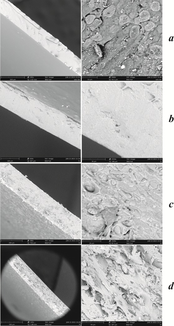
Figure 3. SEM images of the cross sections (left) and surfaces (right) of the four pectin biofilms with added babassu mesocarp and whey protein concentrate. (a) 100%P, (b), 50%P+50%WPC, (c) 50%P+50%BM (F3) and (d) 50%P+25%BM+25%WPC
Cross sectional images of the biofilms composed of 100%P and 50%P+50%WPC show a continuous and compact filmogenic matrix. Fig. 3 shows the formation of lumps on the surface of the biofilms composed of 100%P, a characteristic attributed to the gelling process of the pectin and the roughness of the surface. Moreover, as can be seen from the physical characterization, this formulation showed greater dissolution in water. A study by Cavallaro et al., [31] showed the peculiar effect on the wettability of the biomaterial generated by biofilms based on pectin/polyethylene glycol, which was ascribed to the increase in surface roughness, as confirmed by the SEM analyses, showing the surface to be very rough with many craters (size of ca. 2 μm).
Biofilms composed of 50%P+50%WPC showed micrographs with a homogeneous surface. In some areas, the surfaces presented small fragmentations and a sponge-like structure. The cross-sectional morphology was also smooth and jellylike in these films, although an incipient stratification could be identified. Similar results were reported by Fang et al. [32] for biofilms prepared with WPC+20% glycerol. These authors showed that the addition of Ca2+ caused greater protein aggregation in the biofilm, marked by the irregular but continuous surface. The cross-section revealed a sponge-like structure, in which the whey protein aggregates appeared to be bound by fine wires to form a continuous web. The existence of the net was reflected in the increase in tensile strength of this film.
The biofilms composed of 50%P+50%BM and 50%P+25%BM+25%WPC were shown to be brown and have a voluminous appearance, due to the presence of the babassu mesocarp, which has a brown color. However, both formulations showed a homogeneous, continuous and uniform aspect. The micrographs of the films with BM showed an intense formation of blocks on the surface, as well as in the cross section, due to the low solubility of the BM. This confirms the observation made from the visual aspect of the film. The blocks distributed over the surface impart the feeling of roughness and the bulky cross section, with an undulated fractured conformation which differs considerably from the well-aligned arrangement of the 50%P+50%WPC films. In general terms, the biofilms with BM in their composition showed an increase in roughness of the surface. They also became more flexible and consequently less resistant to tension, with lower permeability to water vapor since the area displayed became relatively greater. According to Arzate-Vásquez et al. [33], a strong film must have a more compact cross section.

3.3. Fourier-transform Infrared Spectroscopy (FTIR)

The FTIR-ATR spectra obtained for the different biofilms can be seen in Figure 4. The characteristic pectin peaks can be observed at around 3400, 2900, 1700, and 1050 cm-1, attributed to the O-H, COO- (asymmetric), COO- (symmetric) and C-O-C stretching of the biopolymers, respectively. The peaks in the 1700 – 500 cm-1 region corresponded to several vibrations of the carbohydrate ring [34].
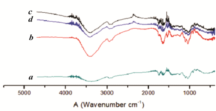
Figure 4. FTIR-ATR spectra of the four biofilm formulations. (a) 100%P, (b), 50%P+50%WPC, (c) 50%P+50%BM (F3) and (d) 50%P+25%BM+25%WPC
A decrease in the overall absorbance in the 3290 cm-1 region can be visualized for the formulation 50%P+50%BM. This peak appeared to be due to O-H stretching vibrations, associated with polar interactions between film components and unbound water present in the biofilms. The babassu mesocarp with pectin, together with calcium, promotes an increase in intermolecular hydrogen bonding on the polymer, and consequently, the decrease in intensity of this O-H peak can be related to a reduction in polymer-water interactions and moisture absorption [35]. This behavior is consistent with the moisture content, matter dissolved in water and water vapor permeability observed for the formulation 50%P+50%BM. Furthermore, the presence of WPC with BM and P also promoted shifts in the wave numbers of asymmetric and symmetric COO- vibrations. For a possibly asymmetric COO- peak, the wave numbers were 3002, 2998, 3000 and 3000 cm-1 for the biofilms 100%P, 50%P+50%WPC, 50%P+25%BM+25%WPC and 50%P+50%BM, respectively. Thus the possibly symmetric vibration occurred at 2400 cm-1 for all the formulations.
Maniglia and Tapia-Blácido [14] studied the FIRT spectra of starch and fibers from babassu mesocarp, showing a high crystallinity index of the starches and the corresponding fiber residues. Since starch was the main component in these materials, the crystallinity index was mainly related to this structure, suggesting that the functional properties of the starch from babassu mesocarp could make it suitable for application as a food ingredient and in biodegradable film production.

4. Conclusions

Biofilms prepared by blending pectin with babassu mesocarp and whey protein concentrate presented improved properties when compared to the films prepared from the pure polymers. The composite biofilms composed of 100%P and 50%P+50%WPC showed translucent and homogeneous appearances. Moreover, the biofilms prepared with 50%P+50%BM and 50%P+25%BM+25%WPC presented similar structures, confirming the strong presence of BM in the formulation, making a stronger and more compact biofilm. In the present study a filmogenic solution composed of 50%P+50%BM with the addition of glycerol (3%, v/v) and CaCl2 (1% m/v) showed the best characteristics amongst the four biofilm formulations.

ACKNOWLEDGEMENTS

The authors are grateful to the central analytical laboratory of CCET/UFMA and to Professors Arão Pereira da Costa Filho and Maria da Gloria Almeida Bandeira for offering their laboratories for the experimental procedures.

References

[1]  Chandra, R. and Rustgi, R., 1998, Biodegradable polymers. Progress in Polymer Science, 23(7), 1273-1335.
[2]  Lee, S. Y., 1996, Bacterial polyhydroxyalkanoates. Biotechnology and Bioengineering, 49(1), 1-14.
[3]  Garcı́a, M. a. A., Pinotti, A., Martino, M. N., and Zaritzky, N. E., 2004, Characterization of composite hydrocolloid films. Carbohydrate Polymers, 56(3), 339-345.
[4]  Seixas, F. L., Turbiani, F. R. B., Salomão, P. G., Souza, R. P., and Gimenes, M. L., 2013, Biofilms composed of alginate and pectin: effect of concentration of crosslinker and plasticizer agents. Chemical Engineering Transactions, 23, 1693-1698.
[5]  Silva, M. A., Bierhalz, A. C. K., and Kieckbusch, T. G., 2009, Alginate and pectin composite films crosslinked with Ca2+ ions: Effect of the plasticizer concentration. Carbohydrate Polymers, 77(4), 736-742.
[6]  Giancone, T., Torrieri, E., Di Pierro, P., Cavella, S., Giosafatto, C. V. L., and Masi, P., 2011, Effect of Surface Density on the Engineering Properties of High Methoxyl Pectin-Based Edible Films. Food and Bioprocess Technology, 4(7), 1228-1236.
[7]  Fishman, M. L., Coffin, D. R., Onwulata, C. I., and Konstance, R. P., 2004, Extrusion of pectin and glycerol with various combinations of orange albedo and starch. Carbohydrate Polymers, 57(4), 401-413.
[8]  Silva, M. V. V., Sales, J. F., Silva, F. G., Rubio-Neto, A., Alberto, P. S., and Pereira, F. D., 2012, The influence of moisture on the vitro embryogermination and morphogenesis of babassu (Orbignya Phalerata Mart.). Acta Scientiarum Agronomy, 34, 453-458.
[9]  Carneiro, A. P. M., Pascoal, L. A. F., Watanabe, P. H., Santos, I. B., Lopes, J. M., and Arruda, J. C. B., 2009, Farelo de babaçu em rações para frangos de corte na fase final: desempenho, rendimento de carcaça e avalição econômica. Ciência Animal Brasileira, 10(1), 40-47.
[10]  Pinheiro, M. M. G., Boylan, F., and Fernandes, P. D., 2012, Antinociceptive effect of the Orbignya speciosa Mart. (Babassu) leaves: Evidence for the involvement of apigenin. Life Sciences, 91(9–10), 293-300.
[11]  de Souza, P. A. V. R., Palumbo Jr, A., Alves, L. M., de Souza, V. P., Cabral, L. M., Fernandes, P. D., Takiya, C. M., Menezes, F. S., and Nasciutti, L. E., 2011, Effects of a nanocomposite containing Orbignya speciosa lipophilic extract on Benign Prostatic Hyperplasia. Journal of Ethnopharmacology, 135(1), 135-146.
[12]  Rego, T. J. A., Fitogeografia das plantas medicninais no Maranhão. 1. Ed. São Luís (MA): EDUFMA, vol. 1, 1995.
[13]  Cinelli, B. A., López, J. A., Castilho, L. R., Freire, D. M. G., and Castro, A. M., 2014, Granular starch hydrolysis of babassu agroindustrial residue: A bioprocess within the context of biorefinery. Fuel, 124, 41-48.
[14]  Maniglia, B. C. and Tapia-Blácido, D. R., 2016, Isolation and characterization of starch from babassu mesocarp. Food Hydrocolloids, 55, 47-55.
[15]  Baruque Filho, E. A., Baruque, M. d. G. A., and Sant’Anna Jr, G. L., 2000, Babassu coconut starch liquefaction: an industrial scale approach to improve conversion yield. Bioresource Technology, 75(1), 49-55.
[16]  Kinsella, J. E. and Whitehead, D. M., "Proteins in Whey: Chemical, Physical, and Functional Properties", in Book "Proteins in Whey: Chemical, Physical, and Functional Properties", E.K. John, Editor. 1989, Academic Press. p. 343-438.
[17]  Demetriades, K., Coupland, J. N., and McClements, D. J., 1997, Physical Properties of Whey Protein Stabilized Emulsions as Related to pH and NaCl. Journal of Food Science, 62(2), 342-347.
[18]  McClements, D. J. and Keogh, M. K., 1995, Physical properties of cold-setting gels formed from heat-denatured whey protein isolate. Journal of the Science of Food and Agriculture, 69(1), 7-14.
[19]  AOAC, "Official methods of analysis". 2007, AOAC International: Gaithersburg.
[20]  Santana, A. A. and Kieckbusch, T. G., 2013, Physical evaluation of biodegradable films of calcium alginate plasticized with polyols. Brazilian Journal of Chemical Engineering, 30, 835-845.
[21]  Irissin-Mangata, J., Bauduin, G., Boutevin, B., and Gontard, N., 2001, New plasticizers for wheat gluten films. European Polymer Journal, 37(8), 1533-1541.
[22]  ASTM, "Standard test method for water vapor transmission of materials". 1995, ASTM International: West Conshohocken.
[23]  McHugh, T. H., Avena-Bustillos, R., and Krochta, J. M., 1993, Hydrophilic Edible Films: Modified Procedure for Water Vapor Permeability and Explanation of Thickness Effects. Journal of Food Science, 58(4), 899-903.
[24]  ASTM, "Standard practice for evaluating microbial susceptibility of nonmetallic materials by laboratory soil burial". 1998, ASTM International: West Conshohocken.
[25]  Bierhalz, A. C. K., da Silva, M. A., and Kieckbusch, T. G., 2012, Natamycin release from alginate/pectin films for food packaging applications. Journal of Food Engineering, 110(1), 18-25.
[26]  Bernard, C., Regnault, S., Gendreau, S., Charbonneau, S., and Relkin, P., 2011, Enhancement of emulsifying properties of whey proteins by controlling spray-drying parameters. Food Hydrocolloids, 25(4), 758-763.
[27]  Pena-Serna, C. and Lopes-Filho, J. F., 2013, Influence of ethanol and glycerol concentration over functional and structural properties of zein–oleic acid films. Materials Chemistry and Physics, 142(2–3), 580-585.
[28]  Rhim, J.-W., Hong, S.-I., and Ha, C.-S., 2009, Tensile, water vapor barrier and antimicrobial properties of PLA/nanoclay composite films. LWT - Food Science and Technology, 42(2), 612-617.
[29]  Schwartzberg, H. G., "Modelling of gas and vapour transport throuugh hydrophilic films", in Book "Modelling of gas and vapour transport throuugh hydrophilic films", M. Mathlouthi, Editor. 1986, Elsevier Science Publishing Co.: New York. p. 115-135.
[30]  Tripathi, S., Mehrotra, G. K., and Dutta, P. K., 2010, Preparation and physicochemical evaluation of chitosan/poly (vinyl alcohol)/pectin ternary film for food-packaging applications. Carbohydrate Polymers, 79(3), 711-716.
[31]  Cavallaro, G., Lazzara, G., and Milioto, S., 2013, Sustainable nanocomposites based on halloysite nanotubes and pectin/polyethylene glycol blend. Polymer Degradation and Stability, 98(12), 2529-2536.
[32]  Fang, Y., Tung, M. A., Britt, I. J., Yada, S., and Dalgleish, D. G., 2002, Tensile and Barrier Properties of Edible Films Made from Whey Proteins. Journal of Food Science, 67(1), 188-193.
[33]  Arzate-Vázquez, I., Chanona-Pérez, J. J., Calderón-Domínguez, G., Terres-Rojas, E., Garibay-Febles, V., Martínez-Rivas, A., and Gutiérrez-López, G. F., 2012, Microstructural characterization of chitosan and alginate films by microscopy techniques and texture image analysis. Carbohydrate Polymers, 87(1), 289-299.
[34]  Papageorgiou, S. K., Kouvelos, E. P., Favvas, E. P., Sapalidis, A. A., Romanos, G. E., and Katsaros, F. K., 2010, Metal–carboxylate interactions in metal–alginate complexes studied with FTIR spectroscopy. Carbohydrate Research, 345(4), 469-473.
[35]  Tapia-Blácido, D. R., Sobral, P. J. A., and Menegalli, F. C., 2010, Potential of Amaranthus cruentus BRS Alegria in the production of flour, starch and protein concentrate: chemical, thermal and rheological characterization. Journal of the Science of Food and Agriculture, 90(7), 1185-1193.