-
Paper Information
- Paper Submission
-
Journal Information
- About This Journal
- Editorial Board
- Current Issue
- Archive
- Author Guidelines
- Contact Us
Journal of Nuclear and Particle Physics
p-ISSN: 2167-6895 e-ISSN: 2167-6909
2016; 6(4): 88-93
doi:10.5923/j.jnpp.20160604.03

Evaluation of Radiation doses and Radiation Risk in Teaching Sohag Hospital, Egypt
 Abstract
 Reference
 Full-Text PDF
 Full-text HTML
S. Harb
ERML Lab., Physics Department, Faculty of Science, South Valley University, Qena, Egypt
Correspondence to: S. Harb , ERML Lab., Physics Department, Faculty of Science, South Valley University, Qena, Egypt.
| Email: |  |
Copyright © 2016 Scientific & Academic Publishing. All Rights Reserved.
This work is licensed under the Creative Commons Attribution International License (CC BY).
http://creativecommons.org/licenses/by/4.0/

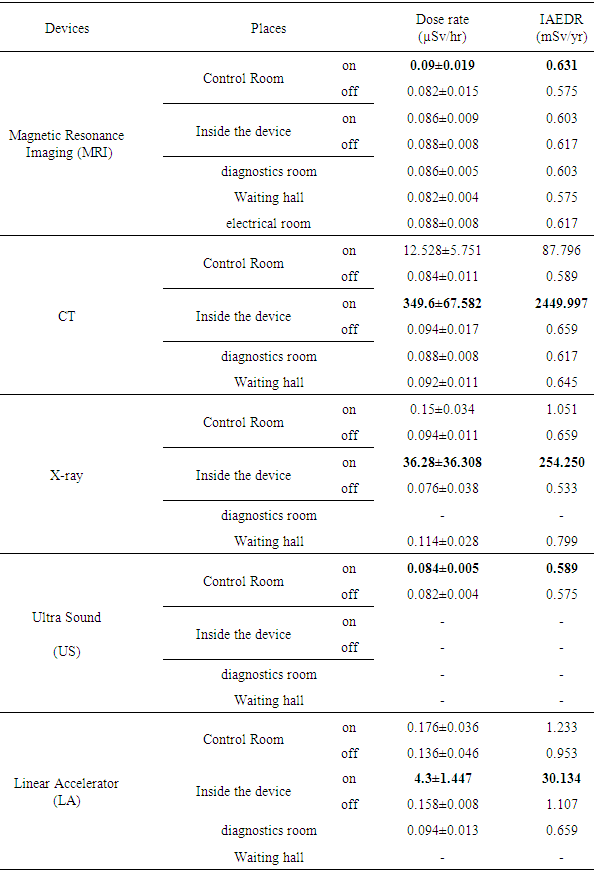
Measurement of background ionizing radiation level at Teaching Sohag Hospital, Egypt was carried out using Thermo Scientific RadEye B20 Multi-Purpose Survey Meters. Radiation doses from MRI, CT, X-ray, US and linear accelerator departments and its related risks to the patients from were analyzed. The results indicated that the ambient dose rate and indoor annual effective dose rate (IAEDR) are high when the devise is ON. Ambient dose values ranged from 0.082±0.015 to 0.09±0.019 μSv/hr, 0.084±0.011 to 349.6±67.582 μSv/hr, 0.076±0.038 to 36.28±36.308 μSv/hr and 0.094±0.013 to 4.3±1.447 μSv/hr for MRI, CT, X-ray and linear accelerator, respectively. Results obtained range from 0.07±0.014 μSv/hr to 0.186±0.024 μSv/hr with an average of 0.1±0.023 μSv/hr for indoor measurement in different departments and locations within the hospital. This present work showed that the CT, X-ray and linear accelerator systems can impart high radiation doses and increase of radiation risk to patients if optimization protocols are ignored. The radiation doses values when compared to standard of 0.274μSv/hr recommended as worldwide average natural dose of background ionizing radiation are within permissible allowed value in different departments and locations within the hospital. Therefore, different departments in Teaching Sohag Hospital are radiologically safe.
Keywords: Ambient dose equivalent, IAEDR, Radiation protection, Radiation risk, RadEye B2
Cite this paper: S. Harb , Evaluation of Radiation doses and Radiation Risk in Teaching Sohag Hospital, Egypt, Journal of Nuclear and Particle Physics, Vol. 6 No. 4, 2016, pp. 88-93. doi: 10.5923/j.jnpp.20160604.03.
1. Introduction
- Ionizing radiations are part of the human environment such as cosmic rays and naturally occurring radioactive materials. They involve electromagnetic radiations (X-rays and gamma rays) as well as corpuscular radiations (alpha, beta and neutron radiations). Ionizing radiations can activate acute effects (for instance, burns) and long-term effects (for instance, cancer and hereditary diseases), which are also known as non-stochastic and stochastic effects. Radioactive sources are used through the world for a wide variety of useful purposes in industry, medicine, research, agriculture and education. The use of radioactive sources includes risks due to radiation exposure. Exposure to ionizing radiation is extensively used by physicians and health professionals in diagnosis and in the treatment of diseases [1].Radiations in hospitals originated from three main sources, medical exposures, cosmic and terrestrial radiation and radioactivity from the background [2]. So, dose measurements are essential in every hospital to ensure compliance with acceptable reference level as well as consideration to justification and appropriate optimization. Medical exposures constitute a good percentage of indoor background ionising radiation [3].The radiographies, CT and X-rays investigations can protect life but their high level radiation doses can affect people health. International Commission on Radiological Protection (ICRP) had definite that the use of computed tomography (CT) had inclined significantly and the radiation dose from CT procedures may be too high [4]. X-ray machines and radiation emitting sources are used in hospitals for the diagnosis and treatment of diseases. X-ray examinations expose the human body to variable amounts of radiation. Depending on its location with respect to the boundaries of the irradiated body volume, a specific organ or tissue can be exposed to primary radiation completely, partly, or not at all. Moreover, the long-term outcomes after endovascular repair are not as well documented as the long-term outcomes after open repair. In particular, the exact cancer and mortality risks associated with such treatments should be evaluated [5]. Linear accelerators machines used in radiation therapy for the treatment of cancer and other diseases.Exposure of patients to radiographic examination (computerized tomography (CT), and routine exposure to x-rays), radioisotope procedures and radiation therapy have contributed to increase in background radiation and radiation levels of patients and many occupational workers [6]. As the time spent in a radiation field increases, the radiation dose received also increases. Therefore, it is best to minimize the time spent in any radiation area. As the distance from a radiation source increases, the radiation exposure decreases rapidly.The global average natural dose of background ionizing radiation to humans is about 0.274 μSv/hr [7]. Eighty percent (80%) of which results comes from nature, while the remaining 20% results from exposure to man-made radiation sources, primarily from medical imaging. Average background ionizing radiation exposure is much higher in developed countries, mostly due to various industrial and medical activities. The International Commission on Radiological Protection (ICRP) recommends that dose limits are 20mSv/year for occupational exposure (for workers engaged in radiation work) and 1mSv/year for the general public [8].The hospital has many departments and units which may contain radiation emitting devices and drugs. So, the current study was showed in order to assess the background ionization levels and its related risk to the occupational workers, patients and general publicto determine its radiation burden and to provide base-line data for future studies in Teaching Sohag Hospital in Egypt.
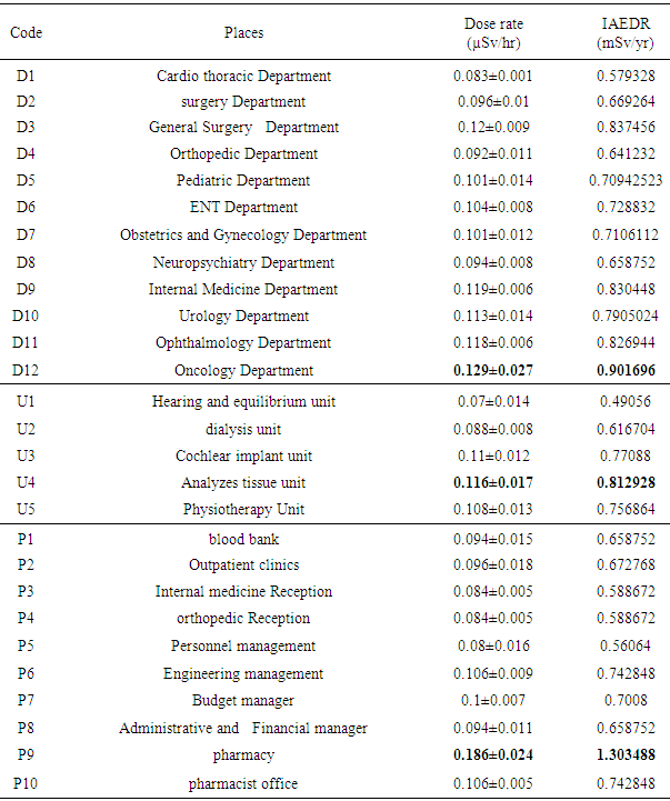
2. Material and Method
- The indoor radiation level measurement of Teaching Sohag Hospital in Egypt was obtained with a hand held dosimeter: RadEye B20 from Thermo Scientific. The selected locations for the study were: MRI, CT, X-ray, US and linear accelerator departments, Cardio thoracic, surgery, General Surgery, Orthopedic, Pediatric, ENT, Obstetrics and Gynecology, Neuropsychiatry, Internal Medicine, Urology, Ophthalmology, oncology departments and In a different place in hospital. Handheld Nuclear Radiation Monitor dosimeter develops safety in laboratories and in the hospital through rapid analysis and determination of radiation levels. The handheld monitor measures alpha, beta, gamma and x-radiation. Its safety-first calibration feature can reduce exposure for personnel. The readings were measured directly (within a minute), exposure rate was taken in μSv/hr and taken four times with each and an average taken and recorded. The indoor ambient dose rate from the survey meter was converted to the indoor annual effective dose rate in for each of the location using this relation by FaraiI [9]:
 | (1) |
3. Result and Discussion
- The MRI, CT, X-ray, US and linear accelerator are required to assess the radiation risks associated with the scanning examinations for patients and worker in Teaching Sohag Hospital, Egypt. The results are presented in Tables 1-2, while Figs. 1-4 show the obtained results compared with standard. Better knowledge about radiation due to exposure from patients is important for deciding on reasonable and appropriate precautions against unnecessary radiation exposure for employees and next-of-kin. Our results reaffirm that “undue anxiety among hospital staff with regard to exposure to radioactive patients must be placed in the proper perspective through education and training”. However, we observed that significant anxiety was still present. Perhaps particularly amongst staff not directly involved in hospital, but still in contact with the patients in other departments.
|
|
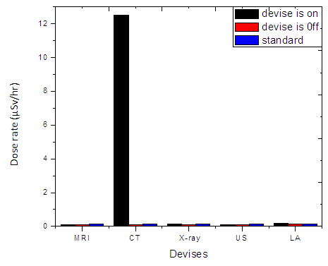 | Figure 1. Dose rate due to some devices at control rooms in Teaching Sohag Hospital |
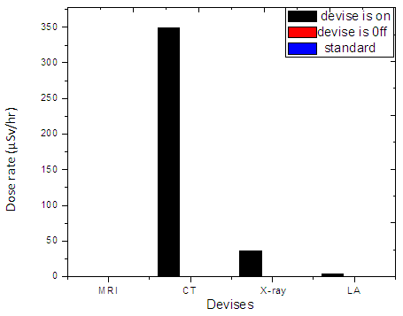 | Figure 2. Dose rate due to some devices at inside them in Teaching Sohag Hospital |
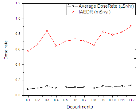 | Figure 3. Average Dose rate and IAEDR in different departments of Teaching Sohag Hospital |
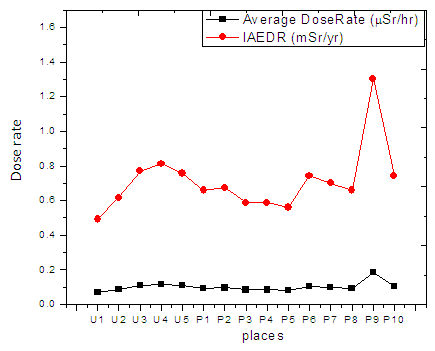 | Figure 4. Average Dose rate and IAEDR in different places in Teaching Sohag Hospital |
4. Conclusions
- Radiation dose is a very important parameter to control the quality of the MRI, CT, X-ray, US and linear accelerator services within the hospital. Dose monitoring helps to ensure the best possible protection of the occupational workers, patients and general publicand provides an immediate indication of incorrect use of technical parameters or equipment malfunction.Measurements of ambient dose rates for most of departments and location in Teaching Sohag Hospital in Egypt were performed using RadEye B20 dosimeter. The mean dose rate and indoor annual effective dose rate (IAEDR) have been obtained as 0.1±0.019 μSv/hr and 0.7±0.16 mSv/yr, respectively when the devices are OFF. While, the mean dose rate and IAEDR have been obtained as 0.22±1.2 μSv/hr and 1.47±8.2mSv/yr, respectively when the devices are operated (ON). This dose is assumed to correspond to workers and patients in the study area.The results of this study indicated that there are insignificant health hazards of 1 mSv/yr equivalent dose rate for public exposure in CT, X-ray, linear accelerator and pharmacy departments and 20mSv/yr for radiation workers in CT, X-ray and linear accelerator departments. Ionizing radiation safety monitoring and assessment have become issues of great concern environments since at high doses, ionizing radiation is carcinogenic. This study provides additional data to establish reference dose level for diagnostic radiology in Egypt. The results are also useful international and professional organizations.