
-
Paper Information
- Previous Paper
- Paper Submission
-
Journal Information
- About This Journal
- Editorial Board
- Current Issue
- Archive
- Author Guidelines
- Contact Us
International Journal of Virology and Molecular Biology
p-ISSN: 2163-2219 e-ISSN: 2163-2227
2026; 15(8): 195-198
doi:10.5923/j.ijvmb.20261508.04
Received: Sep. 12; Accepted: Oct. 8, 2026; Published: Oct. 30, 2026

Changes in Regional Lymph Node Morphology in Secondary Acute Peritonitis
 Abstract
 Reference
 Full-Text PDF
 Full-text HTML
E. А. Eshbaev1, А. Q. Xaydarov2
1Tashkent State Medical University, Tashkent, Uzbekistan
2Ferghana Medical Institute of Public Health, Fergana, Uzbekistan
Correspondence to: E. А. Eshbaev, Tashkent State Medical University, Tashkent, Uzbekistan.
| Email: |  |
Copyright © 2026 The Author(s). Published by Scientific & Academic Publishing.
This work is licensed under the Creative Commons Attribution International License (CC BY).
http://creativecommons.org/licenses/by/4.0/

Acute secondary peritonitis is the main source of morphological changes in the mesenteric lymph nodes in 18-45 year olds, which comes with relaxation of the morphofunctional areas of the lymph nodes in the first 1-3 days of peritonitis. It is determined that the development of lymph node lesions in the cortex and medulla is accompanied by the presence of lymphadenitis, various fragmented cell fragments, coarse colloidal fluid, macrophages, a small number of neutrophils, and the appearance of lymphadenitis. At the same time, the morphological features are characterized by the presence of large macrophages undergoing incomplete phagocytosis in the reticular fibers of the subcapsular and medulla layers of the lymph nodes. It is precisely these macrophages that are found in the form of adhesions to the interdigitating cells of the medulla layer of the lymph node.
Keywords: Lymph node, Peritonitis, Lymphoid follicle, Immunodeficiency
Cite this paper: E. А. Eshbaev, А. Q. Xaydarov, Changes in Regional Lymph Node Morphology in Secondary Acute Peritonitis, International Journal of Virology and Molecular Biology, Vol. 15 No. 8, 2026, pp. 195-198. doi: 10.5923/j.ijvmb.20261508.04.
Article Outline
1. Introduction
- In the world today, the lethality of purulent peritonitis is very high, reaching 20-60% [1,2,3]. When this disease occurs, it is accompanied by the development of rapidly developing endogenous intoxication and multi-organ failure. In the USA and Europe, this indicator is 10-15 per 100 thousand, in Turkey and Korea it is 12-17 per 100 thousand. In the Russian Federation and the CIS countries it is 15-20 per 100 thousand, in the countries of Central Asia, Azerbaijan, Armenia this indicator is 50-71 per 100, with a lethality of 60-85% [4,5,6,7,8]. In our country, on average, 700-1300 infants under the age of 1 die from various types of peritonitis per year. In peritonitis [9,10], the main organs actively involved in the drainage of the abdominal cavity are only the regional lymph nodes, and at 1-3 months of age, this drainage is dominated by the occurrence of physiological paralysis and slow blood circulation, while at 3-6 months, the weakening of intestinal peristalsis and drainage in the regional lymph nodes, insufficiency of the barrier function, lead to the stagnation of local infection in the intestinal lymph nodes and the development of lymphostasis. This requires the development of specific recommendations, taking into account the specific morphofunctional aspects of each period [11,12,13,14].
2. Materials and Methods
- A total of 131 patients were recruited to the Republican Center for Pathological Anatomy in 2018-2023, including 15 patients who died of acute myocardial infarction as a control group. The obtained samples were stained with hematoxylin and eosin and studied.
3. Objective
- To study and improve the morphological features of the development of mesenteric lymph nodes in acute secondary peritonitis in people aged 18-45.
4. Results and Discussion
- Acute secondary peritonitis of various etiologies, the response of regional lymph nodes during the first 24-72 hours proceeds according to the following morphological changes.In acute purulent peritonitis, as a result of the drainage of inflammatory exudate through the peritoneal surfaces through massive lymphatic vessels, stasis and congestion of lymphatic vessels and edema of lymph nodes occur. The main accumulated product is fragments consisting of inflammatory exudate and disintegrated cellular components. Thus, a large amount of pathological fluid is accumulated in the subcapsular areas of the lymph node, a large amount of tissue macrophages are accumulated in the lymphatic vessels and subcapsular areas of the lymph node, the blood vessels of the portal vein of the lymph node are filled, massive migration of immune cells to the foci in the cortical and paracortical areas, in particular, the mobilization of antigen-dependent differentiated lymphocytes from the secondary follicles formed in the germinal center of lymphoid follicles through postcapillary venules, and the germinal centers are exposed and empty.
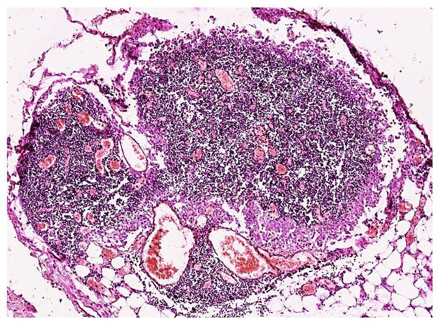 | Figure 1. A small image of the mesenteric lymph node shows signs of massive perfusion in all blood vessels. A massive decrease in lymphocytes in the stroma, foci of delymphatization in the cortical layer. Coarsely dispersed pink homogeneous deposits are detected in the subcapsular area. Staining GE. Size 4x10 |
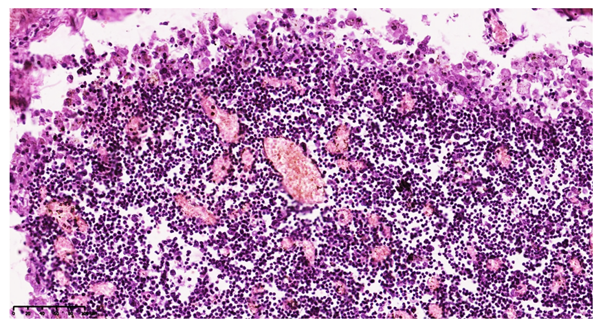 | Figure 2. Mesenteric lymph node. Macrophages and reticulocytes are abundantly accumulated and lymphocytes are sharply reduced. Paint G.E. The size is 40x10 |
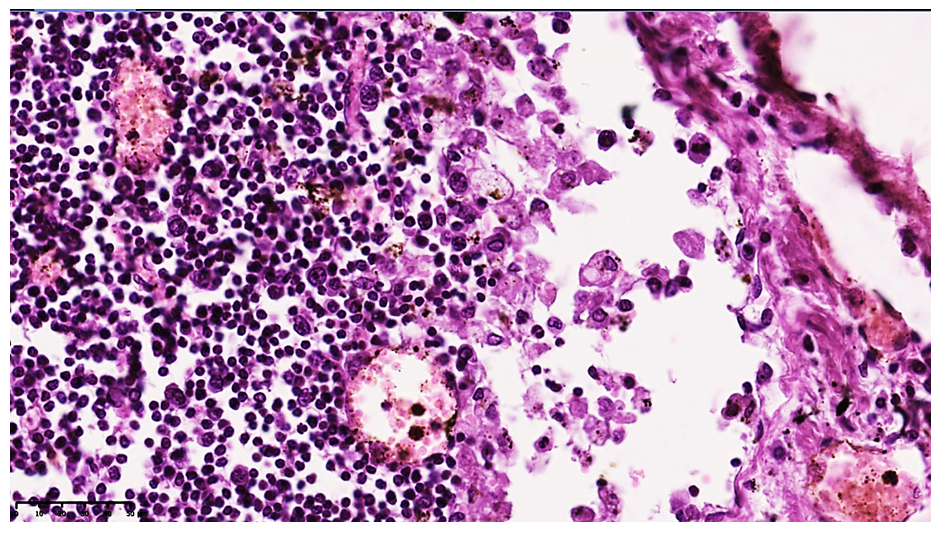 | Figure 3. Mesenteric lymph node. In the subcapsular space, macrophages and various tissue fragments, cell fragments are detected (1), pink homogeneous protein deposits and Motta cells are detected (2). Paint G.E. 10x10 |
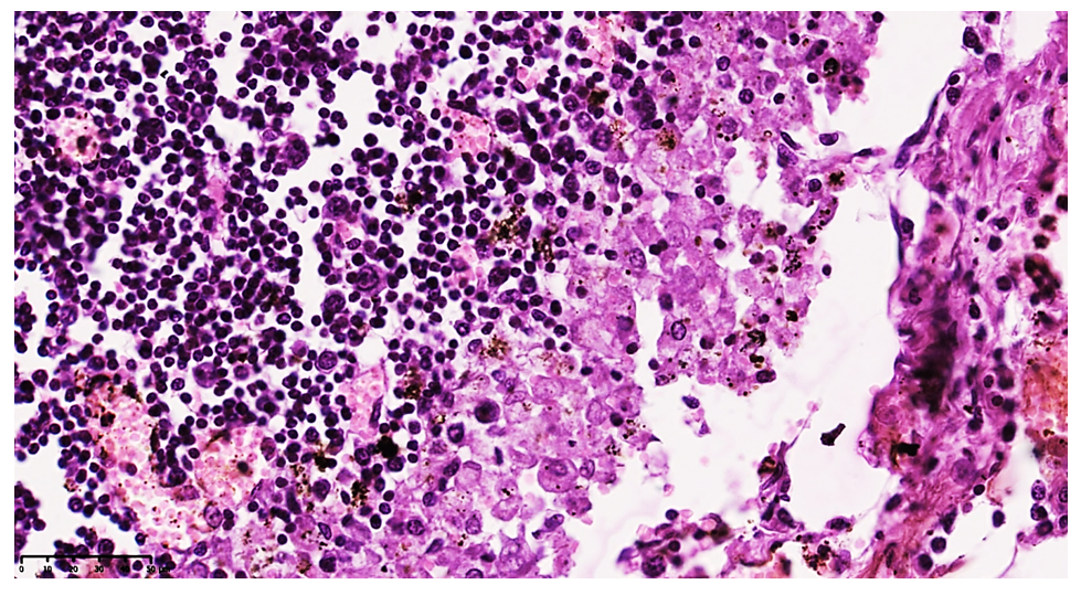 | Figure 4. Coarse homogeneous protein deposits, tissue fragments are detected in subcapsular spaces. Macrophages and Motta cells are detected (1). Paint G.E. The size is 40x10 |
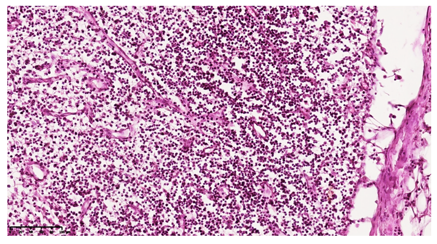 | Figure 5. Mesenteric lymph node. Macrophages and reticulocytes are abundantly accumulated and lymphocytes are sharply reduced. Paint G.E. Size 4x10 |
5. Conclusions
- Acute secondary peritonitis is the main source of morphological changes in the mesenteric lymph nodes in individuals aged 18–45 years, manifested by relaxation of the morphofunctional zones of the lymph nodes during the first 1–3 days of the disease. The development of lesions in the cortex and medulla is accompanied by the presence of lymphadenitis, various fragmented cell fragments, coarsely dispersed colloidal fluid, macrophages, a small number of neutrophils, and the appearance of lymphadenitis. At the same time, the morphological features are characterized by the presence of large macrophages undergoing incomplete phagocytosis in the reticular fibers of the subcapsular and medullary layers of the lymph nodes. It is precisely these macrophages that are found in the form of adhesions to the interdigitating cells of the medullary layer of the lymph node.Within the first 24 hours of peritonitis development, all antigen-specific differentiated lymphocytes in the regional lymph nodes migrate to the foci of inflammation. This is accompanied by a sharp decrease in T-lymphocytes in the paracortical areas and an increase in reticulocytes, macrophages, and a small amount of fibroblasts. In the medullary layer, a variable mixture of T and B lymphocytes in the medullary bands is observed, and the ratio of histiocytes and reticulocytes changes sharply. Mainly, an accumulation of reticulocytes, fibroblasts, histiocytes, and macrophages is detected. Intermediate tumors also develop in the medullary layer, which indicates the accumulation of various dispersed metabolites in large quantities and the development of acute morphofunctional disorders in the lymph nodes. The lymph nodes appear swollen, their capsule is tense, and the subcapsular spaces are filled with lymphatic fluid. Secondary lymphoid follicles are not detected, the germinal center is stable, and the active areas of the lymph nodes are less densely populated than usual. Foci of lymphocytes migrating toward the postcapillary venules are detected in the active areas of the cortex, and the lymphatic portal areas are edematous in the direction of the medullary cords.As a result of a sharp increase in the level of fluid drainage, massively exposed stroma is detected in the lymphoid follicles in the active areas of the lymph node. Lymphoid follicles are sharply blurred against the general background, the follicle texture is lost, and foci of congestion and stasis are detected in the postcapillary venules in the paracortical area. In the medullary layer, a variable admixture of other cell types, predominantly macrophages, and an increase in mast cells are detected in the direction of the cerebral striations. These changes are accompanied by the development of acute inflammation, vasoparalysis, and vasodilation of the vessels in response to inflammatory mediators, increased absorption of metabolites through paralysis of the vascular wall. Clinically, toxic substances cause toxic dystrophy in parenchymal organs and ultimately lead to endogenous intoxication and multiorgan failure. This is characterized by the rapid degranulation of mast cells and neutrophils and the release of large amounts of cytokines, leading to a "cytokine storm," which leads to a systemic vascular response and ultimately to multiorgan failure. Clinically, it is characterized by severe swelling of the organs, diapedesis, hemorrhages, the appearance of fibrous plaques, and foci of microabscesses on the serous membrane surfaces. At the same time, there is an increase in mast cells, an admixture of destroyed fibrous structures and tissue detritus, and histologically, dispersed debris formations are preserved. Lymphostasis in the lymphatic vessels and dilation of lymphatic capillaries are preserved. Thus, the morphological features of regional lymph node lesions in secondary acute peritonitis reflect the severity of the inflammatory process and play an important role in the development of endogenous intoxication and multiorgan failure, which requires the development of specific recommendations taking into account the specific morphofunctional aspects of each period of the disease.